| Journal of Clinical and Analytical Medicine
1
Thyroid Nodule in the Carotid Triangle
Güçlü Kaan Beriat, Hande Ezerarslan, Sinan Kocatürk, Şefik Halit Akmansu Kulak Burun Boğaz ABD, Ufuk Üniversitesi Tıp Fakültesi Dr. Rıdvan Ege Hastanesi, Ankara, Türkiye
Giant Thyroid Nodule Atypically Located in
The Carotid Triangle of The Neck: A Case Report
Boyun Karotid Üçgende Atipik Yerleşim
Gösteren Dev Tiroid Nodülü: Bir Olgu Sunumu
DOI: 10.4328/JCAM.586 Received: 17.01.2011 Accepted: 02.03.2011 Printed: 01.05.2016 J Clin Anal Med 2016;7(3): 402-4 Corresponding Author: Güçlü Kaan Beriat, Ufuk Üniversitesi Tıp Fakültesi Dr. Rıdvan Ege Hastanesi Kulak Burun Boğaz Anabilimdalı, Balgat, 06520, Ankara, Türkiye. T.: +90 3122044175 F.: +90 3122847786 E-Mail: [email protected]
Özet
Ektopik tiroid dokusu tiroid dokusunun anormal migrasyonuyla oluşan nadir görü-len bir tiroid patolojisidir. Yaklaşık olarak 100,000-300,000 doğumda bir görülür ve genellikle asemptomatiktir. Tiroid doku hiperplazisine bağlı olarak, asempto-matik ektopik tiroid dokusu büyüyerek boyun kitlesi şeklinde semptoasempto-matik hale gelebilir. Trakea önündeki normal lokalizasyonu dışında tiroid bezi, dil kökü ile tra-kea arasında genellikle orta hatta herhangi bir seviyede görülebilir. 71 yaşında erkek hasta, boyun sağ tarafında, sağ karotid üçgende 5x4x3 cm boyutlarında kitle ile kulak burun boğaz polikliniğimize başvurdu. Yapılan ince iğne aspirasyon biyopsisi sonucu tiroid dokusu olarak rapor edildi ve takiben kitle genel anestezi altında eksize edildi. Yapılan patolojik değerlendirmesi sonucunda kitlenin atipik olarak yerleşim gösteren ektopik yerleşimli tiroid nodülü olduğu anlaşıldı. Hastanın yapılan bir yıllık takibinde herhangi bir komplikasyonla karşılaşılmadı.
Anahtar Kelimeler
Ektopik Tiroid Dokusu; Karotid Üçgen; Tiroid Nodülü
Abstract
Ectopic thyroid tissue is a rare pathology of the thyroid that is formed by the abnormal migration of the thyroid tissue. It is seen once in about every 100.000 to 300.000 births and is generally asymptomatic. Asymptomatic ectopic thyroid tissue may grow larger and become symptomatic in the form of a neck mass due to thyroid tissue hyperplasia. The thyroid gland, apart from its normal localization before the trachea, may lie between the tongue root and the trachea generally on the middle line at any level. A 71-year-old male patient presented to our ear- nose and throat clinic with a mass of 5x4x3 cm located in the right carotid triangle. The thin-needle aspiration biopsy specimen was reported as thyroid tissue and sub-sequently the mass was excised under general anesthesia. The histopathological evaluation of the mass revealed that it was an atypically located ectopic thyroid nodule. No complications were seen within one year follow-up of the patient.
Keywords
Ectopic Thyroid Tissue; Carotid Triangle; Thyroid Nodule
| Journal of Clinical and Analytical Medicine
| Journal of Clinical and Analytical Medicine Thyroid Nodule in the Carotid Triangle
2
Introduction
The embryologic development of the thyroid gland begins on the 24th day of fetal life as an epithelial proliferation at the foramen cecum. The thyroid tissue reaches its final position be-fore the trachea during the 7th week of fetal life [1, 2].
Ectopic thyroid tissue (ETT) is a rare congenital anomaly and is generally asymptomatic. Its prevalence is about 1/100.000-1/300.000 [1].
ETT develops as a result of the incomplete migration of the thyroid gland and is generally located between the tongue root and the trachea mostly on the mid-line (90% of the cases) al-though it is sometimes found in various other localizations on the neck [3].
In this case report, we present a patient with an ectopic thyroid nodule located in the carotid triangle of the neck.
Case Report
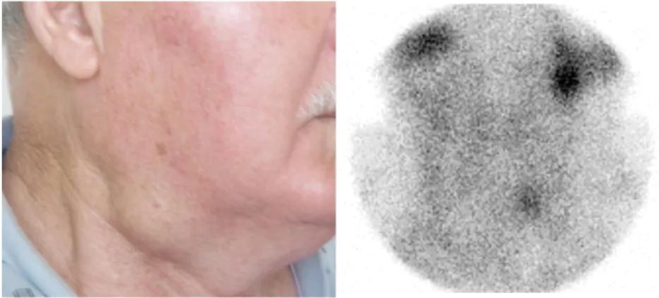
A 71-year-old male patient presented to our polyclinic with complaint of a mass that was present on the right half of his neck for the last twenty years and which gradually grew within the last three months. The patient had underwent total thyroid-ectomy because of multinodular goiter about thirty years ago at a different health center. The physical examination revealed a mass of 5x3 cm on the right side of the neck located on the anterior border of the upper 1/3rd sternocleidomastoid muscle. The mass had smooth borders, was semi-mobile and had a hard texture (Figure 1).
Within the scope of the pre- diagnosis the patient was thought to have a mass of unknown primary in the neck and no patho-logical findings were seen in his pan-endoscopy. Neck ultraso-nography (USG) revealed a solid lesion of about 5x4x3 cm with smooth borders that was homogenous and lobulated in the upper right jugular region inferior to the right submandibular gland. Color Doppler USG reported that the vascularity in this
region was very distinct and was equivalent to thyroid tissue. For a differential diagnosis of aberrant thyroid tissue, the pa-tient was subjected to thyroid scintigraphy. Thyroid scintigra-phy with 5mCi Tc-99 mm pertechnetate showed no involvement in the normal localization of the thyroid gland and reported an amorphous, intensively radioactive activity consistent with thy-roid tissue in the upper right jugular localization (Figure 2). The patient’s preoperative thyroid hormones and their biochemical values were within normal limits.
Thin-needle aspiration biopsy was performed in order to help preoperative diagnosis and its result revealed intensive colloid and mostly isolated rare thyrocid groups (Figure 3).
The patient’s ectopic thyroid tissue was totally excised by dis-secting it from the surrounding tissues under general anesthe-sia (Figure 4, 5).
No complications were seen postoperatively and the histopa-thology results indicated nodular goiter rich in colloid, showing different sizes, with follicles containing single file epithelium, and displaying no findings in favor of malignancy (Figure 6). Taking into consideration the possibility of postoperative hypo-thyroidism, the patient was administered thyroxin treatment by the department of endocrinology and metabolic diseases and was followed-up.
Discussion
Abnormalities in the embryologic development and migration of the thyroid gland may result in ETT. ETT can be seen at any localization on the middle neck line, from the tongue root to the mediastinum [4- 6]. Its prevalence is approximately 1/100.000-1/300.000. This rate is 1/4000-1/8000 for those suffering from a thyroid disease [1].
ETT is a rare thyroid pathology. It is more frequently seen in the female population than the male and it generally has an asymptomatic course [7]. However, dysphagia, dysphonia,
dys-pnea, cardiac failure, and bleeding can be seen based on the localization of the gland [4- 6]. Further, it has been reported to exert pres-sure on the surrounding tissues and to display malignant degeneration [4, 8].
Asymptomatic ETT may become symptomatic specifically during puberty and pregnancy, de-pending on the increase in the thyroid stimu-lating hormone (TSH) level and the hyperpla-sia of the thyroid tissue [4]. ETT is generally hypoactive, it rarely becomes hyperactive. The thyroid mass presented in our case, had
hy-perplasia with an increased TSH level follow-ing total thyroidectomy, possibly assumfollow-ing the hormonal task of the excised thyroid tis-sue.
Figure 3. Tyrocid clusters, single or in groups, looking benign on a colloid base (Giemsa x40). Figure 1. 5x4x3 cm mass on the right side of the neck located anterior to the upper 1/3 of the sternocleidomastoid muscle, over digastric muscle’s back belly.
Figure 2. Thyroid scintigraphy; activity involvement displaying an amorphous, intensively radioactive thyroid tissue in the upper right cervical region.
Figure 5. Complete and half macro-scopic images of the excised thyroid nodule.
Figure 4. Intraoperative image of the dissection area following the excision of the mass.
Figure 6. Thyroid follicules in different sizes. ( H&E, x10)
Journal of Clinical and Analytical Medicine | 403
| Journal of Clinical and Analytical Medicine Thyroid Nodule in the Carotid Triangle
3
ETT can even be seen in the mediastinal, intracardiac, gastro-intestinal tract, and intraperitoneal localizations besides along the normal migration route of the thyroid [2, 4]. ETT is mostly found in the sublingual position (90%). Our case with aberrant thyroid tissue is one of the rare cases in literature with its atyp-ical place in the right carotid triangle.
The etiology of ETT is not wholly known but it is argued that gene mutations might have an effect on it [4]. The mutation of “thyroid transcription factor 2” (TTF-2) is found to be related to thyroid agenesis, the mutation of the “Pax 8 gene” to the various forms of thyroid dysgenesis, and the gene mutation of TTF-1 to thyroid agenesis or dysgenesis. These gene mutations also give way to ectopic migration [4].
Seventy percent of ETT cases contain only the thyroid tis-sue, whereas others also house different cell groups in their surroundings. All diseases of the thyroid gland present in the thyroid tissue in its normal localization can also be seen in the ectopic thyroid tissue [6].
USG, scintigraphy, computerized tomography (CT), and magnet-ic resonance imaging (MRI) are radiologmagnet-ical methods that can be used in the diagnosis. Thyroid scintigraphy is both a sensi-tive and a specific method in determining whether the thyroid is in its normal location or not. Scintigraphy is a valuable method in the differential diagnosis of ETT from other mid-line cervi-cal masses such as thyroglossal duct cysts, lipoma, epidermoid cysts, lingual thyroid, etc. [1]. Thyroid scintigraphy enabled us to reach a differential diagnosis concerning other lesions that might be present in this region by showing that the mass, which was not on the mid-line, was thyroid tissue. USG, CT, and MRI are also useful in diagnosis but they are methods that have low levels of sensitivity and specifity. These methods are more valuable in the determination of the detailed localization and surroundings of masses.
ETT settled in lymph nodes and localized in the lateral neck region is very rare and many authors see it as the metastasis of papillary thyroid carcinoma. It was reported that 0.3-1.6 % of neck dissection material of cases operated for head-neck cancer, coincidentally contained thyroid tissue [9]. In our case, pathological evaluation revealed no lymph tissue and malignan-cy. On the contrary, a whole thyroid nodule with smooth borders was reported.
The treatment of ETT is either medical treatment (thyroid hor-mone suppression treatment) or surgical excision [1]. Therapeu-tic method is chosen depending on the size of the lesion, symp-toms of local pressure, patient’s age, presence of the thyroid gland and degeneration in the thyroid tissue (e.g. ulceration, hemorrhage, and cystic degeneration), suspicion or presence of malignancy, and the functional situation of the tissue.
If the ETT is the only thyroid tissue and if this tissue has a nor-mal function and is snor-mall and asymptomatic, the patient can be followed-up without any treatment or it can be repressed through the external introduction of thyroid hormone [9]. The mass can be reduced in size slowly but distinctively by radioac-tive iodine treatment. Through this therapy the volume can be reduced by 30-50 % in 4 to 6 months [3]. This treatment, how-ever, does not guarantee success and ablation with radioactive iodine is not appropriate for young patients [7].
Surgical treatment can be selected if there is pressure to sur-rounding tissues and organs or obstructive symptoms, if the pa-tient is nonresponsive to radioactive iodine treatment, if there is a suspicion or presence of malignancy, or if the patient pre-fers surgery [1]. Moreover, surgical treatment is a therapeutic
modality which is rapid, practical, and has low morbidity rates; and it also offers histological definitive diagnosis [3]. The dis-advantage of surgical treatment is the possibility of long term thyroid hormone replacement in patients because of postop-erative permanent hypothyroidism. Therefore, patients should be tested in regard to postoperative thyroid functions and hor-monal replacement treatment for regulation when necessary [3].
It is seen that in literature some authors suggest medical treat-ment even if there are no suspicion of malignancy and serious clinical symptoms. On the other hand, other authors suggest the excision of the thyroid tissue in the selection of treatment method regardless of the presence of normal thyroid tissue [1]. In the latter suggested method the patient’s condition improves and the possibility of a future development of malignant de-generation is eliminated. The fact that the lesion was large, the patient was old and the fact that the patient informed us about an enlargement in the last three months made us conclude that the risk of malignancy may be high. Besides, the insistence of the patient to get the mass surgically removed and the fact that he was living in a rural and remote area again made us move away from the option of medical treatment that would take a long time and would necessitate careful follow-up. It should be remembered that thyroid tissue can rarely be seen outside its normal localization and it should definitely be taken into consideration in the differential diagnosis. It should also be noted that, although it is rare and is most of the time a benign anomaly, ETT may cause serious problems based on its local mass effect and potency for malignant degeneration.
Competing interests
The authors declare that they have no competing interests. References
1. Chawla M, Kumar R, Malhotra A. Dual ectopic thyroid: case series and review of the literature. Clin Nucl Med. 2007;32:1.
2. Rinkel RN, Manni JJ, van der Beek JM. Ectopic thyroid tissue manifesting as a unique cause of an oropharyngeal mass. Otolaryngol Head Neck Surg. 2001 ;124:340
3. Zieren J, Paul M, Scharfenberg M, Menenakos C. Submandibular Ectopic Thyroid Gland. J Craniofac Surg. 2006 ;17:1194
4. Maino K, Skelton H, Yeager J, Smith KJ. Benign ectopic thyroid tissue in a cuta-neous location: a case report and review. J Cutan Pathol. 2004:31:195.
5. Rao PN, Pandit N, Kumar R, Upadhya IV, Vidya Sagar MS. Ectopic Functioning thyroid in the thyroglossal duct detected by radionuclide ımagıng. Clin Nucl Med. 2005;30:630.
6. Kumar R, Sharma S, Marwah A, Moorthy D, Dhanwal D, Malhotra A. Ectopic goi-ter masquerading as submandibular gland swelling. Clin Nucl Med. 2001;26:306. 7. Gallo A, Leonetti F, Torri E, Manciocco V, Simonelli M, DeVincentiis M. Ectopic lingual thyroid as unusual cause of severe dysphagia. Dysphagia. 2001; 16:220. 8. Shah BC, Ravichand CS, Juluri S, Agarwal A, Pramesh CS, Mistry RC. Ectopic thyroid cancer. Ann Thorac Cardiovasc Surg. 2007;13:122.
9. Leon X, Sancho FJ, Garcia J, Sanudo JR, Orus C, Quer M. Incidence and signifi-cance of clinically unsuspected thyroid tissue in lymph nodes found during neck dissection in head and neck carcinoma patients. Laryngoscope 2005;115:470. 10. Thomas G, Hoilat R, Daniels J. S, Kalagie W. Ectopic lingual thyroid: A case report. Int J Oral Maxillofac Surg. 2003;32:219.
How to cite this article:
Beriat GK, Ezerarslan H, Kocatürk S, Akmansu ŞH. Giant Thyroid Nodule Atypically Located in The Carotid Triangle of The Neck: A Case Report. J Clin Anal Med 2016;7(3): 402-4.
| Journal of Clinical and Analytical Medicine
404