E
FFECTS OF
C
RT
M
ONITOR-
E
MITTED
R
ADIATION IN
R
AT
T
ISSUES:
P
REVENTIVE
E
FFECT OF
V
ITAMIN C
Sibel Serin Kılıço lu MD,
1Selçuk Tabak MD,
2Aslıhan Avcı MD,
3Bülent Kılıço lu MD,
4Erdinç Devrim
MD,
3B. mge Ergüder MD,
3Ebru Gürleyik MD,
3Salih Celepli MD,
4Recep Çetin MD,
5lker Durak MD
31 Ufuk University Faculty of Medicine Department of Histology & Embryology, Ankara, Turkey 2 Adıyaman University Faculty of Medicine Department of Gynecologie & Obstetric, Adıyaman, Turkey 3 Ankara University Faculty of Medicine Department of Biochemistry, Ankara, Turkey
4 Ankara Training and Research Hospital, Department of General Surgery, Ankara, Turkey
5 Süleyman Demirel University Faculty of Medicine Department of General Surgery, Isparta, Turkey
ABSTRACT
Objective: To investigate the effects of CRT (cathode ray tube)-monitor-emitted radiation on the oxidant/ antioxidant status in kidney, liver, heart, brain tissues of rats and to observe the histopathological findings of these tissues, and to examine any protective role of vitamin C supplementation.
Material and Method: The study carried out on 40
Wistar albino adult female rats. There were 10 animals in each four group (control, vitamin C, computer, and computer plus vitamin C). The computer and computer plus vitamin C groups were exposed to computer monitors while the other groups were not. Vitamin C was administered 250 mg/kg/day orally. In the kidney, liver, heart, and brain tissues, malondialdehyde (MDA) levels and superoxide dismutase (SOD), catalase (CAT), and glutathione peroxidase (GSH-Px) activities were measured
spectrophotometrically. In addition, histopathological examination is carried out.
Results: In the kidney tissues, MDA levels significantly increased in the computer group compared with the computer plus vitamin C group and the control group (p<0.05). Histomorphologic changes were observed in the kidney and liver tissues of the computer group while there were no alterations in other tissues.
Conclusion: The results of this study suggest that
CRT-monitor-emitted radiation leads to oxidative stress and cellular changes in kidney and liver tissues and the antioxidant supplementation like vitamin C could prevent these possible oxidative effects.
Key Words: Histopathology, radiation, vitamin C, kidney, liver, antioxidants, oxidative stress. Nobel Med 2012;
Crt mOnitör kullanımı sOnuCu Oluşan raDyasyOnun sıçan DOkularına
etkileri: C vitamininin kOruyuCu etkisi
özet
amaç: Çalışmamızın amacı, CRT (katot ışınlı tüp)-monitör kullanımına bağlı oluşan radyasyonun çeşitli sıçan dokularında oksidan/antioksidan parametreleri ile histopatolojisi üzerine etkilerini ve C vitamininin oksida-tif strese karşı olası koruyuculuğunu değerlendirmektir.
materyal ve metod: Çalışmada 40 albino dişi rat (sı-çan) kullanılmış ve ratlar 4 grup (kontrol, C vitamini, bilgisayar, bilgisayar ile C vitamini) şeklinde ayrılmıştır. Bilgisayar ve bilgisayar ile C vitamini grupları monitör radyasyonuna maruz bırakılmıştır. C vitamini 250 mg/ kg/gün oral olarak uygulanmıştır. Hayvanların böbrek, karaciğer, kalp ve beyin dokularında malondialdehit (MDA) düzeyleri ve süperoksit dismutaz (SOD), kata-laz (CAT), glutatyon peroksidaz (GSH-Px) enzim ak-tiviteleri spektrofotometrik yöntemlerle ölçülmüştür. Ayrıca dokularda histopatolojik inceleme yapılmıştır.
Bulgular: Yalnızca monitör radyasyonuna maruz bı-rakılan bilgisayar grubu, monitör radyasyonu ile bir-likte C vitamini alan grup ve kontrol grubu ile kar-şılaştırıldığında, böbrek dokusu MDA düzeylerinde istatistiksel olarak anlamlı bir artış saptandı (p<0,05). Ayrıca bu grubun böbrek ve karaciğer dokularında be-lirgin histomorfolojik bulgular görülürken diğer doku örneklerinde anlamlı bir değişiklik saptanamadı. Bilgi-sayar monitörü radyasyonuna bağlı olarak böbrek ve karaciğer dokularında ortaya çıkan bu değişikliklerin C vitamini uygulamasıyla önlenebildiği bulundu.
sonuç: Çalışmanın sonuçları, CRT-monitör kulla-nımına bağlı oluşan radyasyonun oksidatif stresi ar-tırdığı ve böbrek ile karaciğer dokularında hücresel değişikliklere neden olduğunu desteklemektedir. C vitamini gibi antioksidanların kullanımı radyasyonun neden olduğu olası hücresel hasarı önleyebilir.
anahtar kelimeler: Histopatoloji, radyasyon, C vitamini, böbrek, karaciğer, antioksidanlar, oksidatif stres. Nobel Med 2012; 8(3): 81-85
ıntrODuCtıOn
Computer using is become popular in our offices, homes, hospitals etc. With the increased use of electric and electronic equipment such as personal computer (PC), notebook, cellular phones, wireless communication in our offices, our daily exposure to electromagnetic radiation (EMR) has increased. These devices use radio frequency waves which are the sources of radio frequency fields in our offices.1 As CRT
monitors are widely used in homes, offices and users are exposed to an electromagnetic field (EMF), many researchers have reported some disorders related to electromagnetic radiation (EMR). Free radicals (e.g., superoxide anion, nitric oxide, and hydroxyl radicals) and other reactive species (e.g., hydrogen peroxide, peroxynitrite, and hypochlorous acid) occur primarily as a result of aerobic metabolism. Malondialdehyde (MDA) is an oxidation product and generated by decomposition of lipid peroxides.2 Antioxidants
(vitamin E, vitamin C, beta-caroten, selenium, zinc) and antioxidant enzymes as superoxide dismutase (SOD), catalase (CAT), glutathione peroxidase (GSH-Px) exert synergistic actions in scavenging free radicals.3 Antioxidant agents transform free radicals
into less reactive species and limit their toxic effects. In the body, there is a delicate balance between the formation of reactive oxygen species and the endogenous protective mechanisms. These protective mechanisms could be insufficient because of some
diseases, genetic defects or various environmental factors like radiation, air pollution. When the balance between oxidants and antioxidants changes, that leads to oxidation and excessive production of reactive oxygen species damage the tissues. This may result in excessive production of reactive oxygen species, leading to damage of nuclear and mitochondrial DNA, cell membrane lipids and intracellular proteins, culminating in cell death.
The uncontrolled rise in computerization generates new dangerous source of electromagnetic fields (EMF), which is combined with deteriorating influence of personal computer (PC)-related overloads, acquires great importance and places stringent requirements. At the same time, the adverse impact of EMF of video displays terminals (VDT) and PCs have not been studied thoroughly although many researchers have reported the relation between electromagnetic radiation (EMR) and some disorders. And therefore the assessments are uncertain. Due to the fact that there are limited experimental data on oxidative stress caused by EMR, we aimed to determine the effects of CRT monitor-emitted EMR on oxidant/antioxidant status in tissues and to examine possible protective effects of vitamin C.
materıal and metHOD
the study. The study was approved by the Ethics Committee of Ankara Oncology Hospital according to the “Guide for the Care and Use of Laboratory Animals” criteria. The rats weighed 165±5 g and were 12 weeks old. Animals were divided into four groups (Control, Vitamin C, computer, computer plus Vitamin C) and in each group there were ten animals. All animals were fed a laboratory diet and water ad libitum.
In the computer group and computer plus vitamin C group, the rats were exposed to computer monitor released radiation system for 8 hours/day (from 9 am to 5 pm) for three weeks. Monitors used in the study were two cathode ray tube (CRT) type monitors (Tatung Model No: CM14UHR and Hyundai Model No: S450) and positioned face-to-face in 1.5 m2 areas
that the cages of each study group were between the monitors without a screen-saver. The rats were group-caged and were allowed free motion in their cages. In the computer plus vitamin C group, rats received vitamin C in the doses of 250 mg/kg/day for three weeks adding to laboratory diet.
At the end of the study period, the animals were sacrificed under ether anesthesia and their kidney, liver, heart, and brain tissues were removed and prepared for the biochemical and histopathological investigations.
Biochemical Examination: The tissues were first
washed with deionized water to remove the blood and then homogenized in a homogenizator for about three minutes. During tissue preparation process, the temperature was kept at +4 °C and, after the centrifugation at 6000 g for about 20 minutes; the supernatants were used in the analyses. Oxidant (MDA level) and antioxidant (SOD, CAT and GSH-Px, activities) parameters were measured in all tissues of the groups with spectrophotometric methods. MDA level was measured by thiobarbituric acid reactive
substances (TBARS) method.4 SOD activity was
measured as described.5 One unit for SOD activity was
expressed as the enzyme protein amount causing 50% inhibition in nitroblue tetrazolium (NBT) reduction rate. CAT activity was determined by measuring decrease of hydrogen peroxide (H2O2) absorbance at 240 nm as described.6 GSH-Px activity was measured
by following changes in NADPH absorbance at 340 nm as described.7 The protein levels of the samples
were measured by Lowry’s method.8 The results are
expressed as nmol/mg, U/mg, IU/mg, mIU/mg of protein respectively.
Histopathological Examination: The study was
carried out in the Department of Histology and Embryology, Ankara University, Faculty of Medicine.
For light microscope analyses, the samples were obtained from the tissues, fixed in 10% neutral buffered formaline solution for two days. They were washed in flowing water and were dehydrated with rising concentrations of ethanol (50%, 75%, 96%, and 100%). After dehydration, specimens were put into xylene to obtain the transparency and were then infiltrated with and embedded in paraffin. The embedded tissue is cut into sections of 5 µm thickness by Leica RM 2125 RT and then cut into sections of 3 µm for clear detailed examination. The sections were stained with hematoxylin and eosin. The histopathological examinations were performed and photographed by Nikon eclipse E 600.
The histopathological sections were evaluated by two blinded researchers and photographed.
statistical analysis
All statistical analyses were carried out using SPSS statistical software (SPSS for Windows, version 11.0, SPSS Inc., Chicago, IL, USA). The data were given as arithmetic mean ± standard deviation (mean ± SD). In the statistical evaluation of the results, the Student’s
t test was carried out. The statistical significance was
defined as p value lower than 0.05.
Figure 1. Haematoxylin and eosin staining with light microscope. The
glomerule (G), tubules (T), proximal tubule (pt), distal tubule (dt) in group I (Figure A), in group II (Figure B). Congestion (c), lymphocyte infiltration (*), mesangial cell nucleus (N), glomerular capillary (cp), hyperemia (h) and Bowman’s capsule (arrow) in group III (Figure C, C’) and in group IV (Figure D, D’). Scale bar: A, B, D: 20 µm, C: 40 µm, C’, D’: 4 µm
D C A D’ C’ B G G 20 µm 40 µm 20 µm 4 µm 20 µm 4 µm G G G C C G G G G G G N T T pt pt dt pt dt pt pt dt dt dt dt v v cp pt pt dt cp v h * C C N N G C C C C C G T T G G G G T T
results
Oxidant/antioxidant parameters
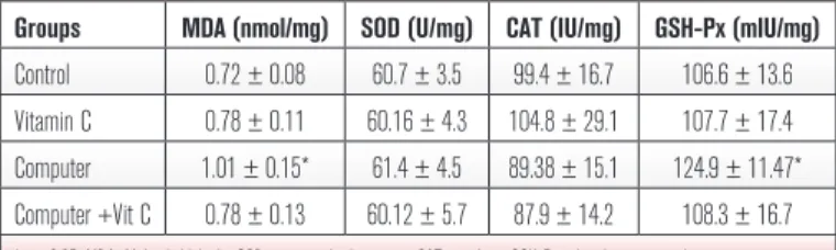
As seen in Table 1, in kidney tissue, there was a significant increase in MDA level in computer group compared with the other groups. However no significant differences were seen between any of the groups in terms of antioxidant enzyme activities. In addition, in the other tissues, there were no statistically significant differences in these parameters among the groups (Table 1-4).
Histopathological examination of the liver tissues:
The specimens of the control group presented original morphological feature(s) of the liver tissue. The hepatocytes were polyhedral normal in size with nucleus and cytoplasm. The intercellular space between the two adjacent hepatocytes was in normal ranges (Figure 2A, 2A’). In vitamin C group, the
tissues displayed some non-specific histopathological changes such as fading of hepatocyte cytoplasm and shrinkage of the whole tissue. The shrinkage of sinusoids besides the cellular shrinkage was remarkable (Figure 2B, 2B’). In computer group, sinusoidal dilatation and edema around vena centralis were observed. The cytoplasm and the nucleoplasm of hepatocytes were discolored. The pale cytoplasm was related to the cytoplasmic lipid vacuolization (Figure 2C, 2C’). When we evaluate the specimens of computer plus vitamin C group, we examined the existing paleness of the hepatocyte cytoplasm because of lipid vacuolization. The marked regression of the sinusoidal dilatation was observed. The cellular shrinkage of whole liver tissue was remarkable (Figure 2D, 2D’).
Histopathological examination of the kidney tissues:
The specimens of the control group presented original morphological feature of cortex and medulla of the renal tissue. Renal corpuscles and most of the cortical parenchyma surrounding them consist of proximal and distal convoluted tubules were seen. The cortex consists of mainly of proximal convoluted tubules lined by more eosinophilic epithelial cells, with smaller numbers of distal convoluted tubules and collecting tubules (Figure 1A). In vitamin C group, the tissues displayed no specific histopathological changes (Figure 1B). In the computer group, numerous renal corpuscles and tubules were seen in Figure 1C. Hyperemia and congestion were observed at the cortical parenchyma around the renal corpuscules. The tubules were become tight because of the shrinkage of the space between them. The lymphocyte infiltration was existed in the parenchyma tissue and in particular peritubullar mononuclear inflammation was present (Figure 1C). The glomerular cell proliferation was remarkable. The augmentation in the number of mesangial and endothelial cells was observed (Figure 1C’). When we evaluate the specimens of the computer plus vitamin C group, we examined the certain amount of shrinkage between the tubules. The glomerular cell proliferation was regressed (Figure 1D, 1D’).
Histopathological examination of the heart and brain tissues: The specimens from the whole groups
presented no alteration to the original morphological feature of cardiac muscle, and brain tissue.
DısCussıOn
EMFs are all around us. Rapidly increasing use of wireless communication systems has caused a growing public concern about possible health effects of EMFs.9
An increasing number of people report subjective
Figure 2. Haematoxylin and eosin staining with light microscope. The
hepatocytes surrounding vena centralis (vc) and liver sinusoids (*) in group I (Figure A), group II (Figure B), group III (Figure C) and group IV (Figure D). Scale bar: 10 µm. The hepatocytes (arrow head) and sinusoids (*) viewed in all groups (Figure A’, B’, C’, D’). Scale bar: 4 µm.
A B C D A’ B’ C’ D’ VC VC VC VC * * * * * * * * * * * * * * * * * * * * * * * * * * * * * * 1 µm 4 µm 4 µm 10 µm 10 µm 10 µm 10 µm 4 µm
symptoms and hypersensitivity to a wide variety of electromagnetic sources such as power lines, radio and TV broadcasting stations, cellular phones and their base stations, computer monitors (cathode ray tubes or CRTs) and electrical home appliances.10
Computer users suffer from some complaints such as eye strain, headaches, general malaise, and other visual and musculoskeletal problems.11 Additionally,
the frequent self-reported symptoms are fatigue, dizziness, concentration diffi culties, nausea, heart palpitation, digestive disturbances, and facial skin symptoms such as redness, tingling, and burning sensation.12,13 Provocative studies on EHS patients
have not shown any worsening of symptoms or physiological reactions related to laboratory exposure to EMF.14,15 Overall, the whole issue of EMF health
effects still remains controversial.16 In a study by
performed Balci et al., the effects of mobile-phone-emitted radiation were investigated on the oxidant/ antioxidant balance in corneal and lens tissues. They observed protective effects of vitamin C in this setting. For this aim, vitamin C (250 mg/kg/day) was given to albino Wistar rats. They found increased MDA level and CAT activity in the corneal tissue from the animals in the mobile phone group compared with the mobile phone plus vitamin C group and the control group (p<0.05). However, SOD activity was found to decrease signifi cantly in the cornea. (p<0.05). In the lens tissues, MDA mean level was found to increase signifi cantly in the mobile phone group relative to mobile phone plus vitamin C group and the control group (p<0.05). Their results suggested that mobile telephone radiation leads to oxidative stress in corneal and lens tissues and antioxidants like vitamin C could help prevent these effects.17
In another study performed by Erguder et al., possible effects of computer use were investigated on human saliva samples. They observed oxidant effect of computer use in saliva. They found increased MDA levels (an oxidation marker) in saliva at 2nd and 4th
hours. However, SOD (an antioxidant enzyme) activity was found to decrease. Their results suggested that computer-released radiation causes changes in enzymatic antioxidant defense system and, leads to oxidant stress in saliva samples from subjects.18
Many researchers shown interest in studying the mechanisms of the interaction between EMR and living organisms. Studies have shown that radiation may interfere with chemical reactions involving free radical generation.19 In the study performed by Balci
et al., in the lens tissue, signifi cantly increased MDA levels were found in the group exposed to radiation compared to the control group.20
In our study, we found that computer system based electromagnetic radiation (CSBER) caused oxidative stress in kidney tissues, and led to some histopathological changes in kidney and liver tissues that have high metabolic actives, of the computer group. In the liver, no oxidation was found but histopathologic changes were observed. May be any oxidative harmful effects did not occur in the liver tissues. It has been observed that vitamin C can prevent these changes signifi cantly. In the other tissues, there were no meaningful changes. Therefore, our results suggest that vitamin C supplementation could give benefi cial results to eliminate harmful effects of PC-monitor emitted radiation, particularly in the intensive computer users. Further studies are needed on the subject before reaching defi nite conclusions in this regard.
Table1: Oxidant/antioxidant parameters of the kidney tissues (Mean±SD)
Groups MDA (nmol/mg) SOD (U/mg) CAT (IU/mg) GSH-Px (mIU/mg)
Control 0.72 ± 0.08 60.7 ± 3.5 99.4 ± 16.7 106.6 ± 13.6
Vitamin C 0.78 ± 0.11 60.16 ± 4.3 104.8 ± 29.1 107.7 ± 17.4
Computer 1.01 ± 0.15* 61.4 ± 4.5 89.38 ± 15.1 124.9 ± 11.47*
Computer +Vit C 0.78 ± 0.13 60.12 ± 5.7 87.9 ± 14.2 108.3 ± 16.7
*: p<0.05, MDA: Malondialdehyde, SOD: superoxide dismutase, CAT: catalase, GSH-Px: glutathione peroxidase
Table 2: Oxidant and antioxidant parameters of rat liver tissue (Mean±SD)
Groups MDA (nmol/mg) SOD (U/mg) CAT (IU/mg) GSH-Px (mIU/mg)
Control 0.55 ± 0.084 144.4 ± 40.7 101.6 ± 4.3 58.6 ± 13.6
Vitamin C 0.50 ± 0.092 132.3 ± 15.7 99.9 ± 6.2 44.2 ± 10.7
Computer 0.56 ± 0.0075 105.9 ± 22.6 106.5 ± 6.2 57.1 ± 15.2
Computer +Vit C 0.43 ± 0.0082 140.17 ± 35.5 101.4 ± 10.2 61.3 ± 13.7
MDA: Malondialdehyde, SOD: superoxide dismutase, CAT: catalase, GSH-Px: glutathione peroxidase
Table 4: Oxidant and antioxidant parameters of rat heart tissues (Mean±SD)
Groups MDA (nmol/mg) SOD (U/mg) CAT (IU/mg) GSH-Px (mIU/mg)
Control 0.81 ± 0.10 58.49 ± 8.0 7.06 ± 1.7 53.96 ± 14.3
Vitamin C 1.00 ± 0.19 53.7 ± 6.2 6.8 ± 1.82 52.39 ± 24.9
Computer 0.86 ± 0.13 64.36 ± 9.4 11.8 ± 5.1 45.37 ± 10.29
Computer + Vit C 1.17 ± 0.23 66.96 ± 9.9 10.24 ± 4.6 43.67 ± 10.8
MDA: Malondialdehyde, SOD: superoxide dismutase, CAT: catalase, GSH-Px: glutathione peroxidase
Table 3: Oxidant and antioxidant parameters of rat brain tissue (Mean±SD)
Groups MDA (nmol/mg) SOD (U/mg) CAT (IU/mg) GSH-Px (mIU/mg)
Control 4.19 ± 0.93 23.3 ± 1.67 4.8 ± 0.95 40.5 ± 4.8
Vitamin C 2.68 ± 0.65 21.7 ± 1.08 3.59 ± 0.81 34.6 ± 3.9
Computer 2.3 ± 0.77 22.9 ± 2.16 3.2 ± 0.8 34.4 ± 3.75
Computer +Vit C 3.1 ± 0.63 22.7 ± 1.52 3.67 ± 0.78 36.5 ± 7.6
REFERENCES
1. Kim IN, Megeda EV. Impact of electromagnetic fields on a computer user.
Gig Sanit 2007; 1: 44-48.
2. Kahkonen MP, Hopia AI, Vuorela HJ, et al. Antioxidant activity of plant
extracts containing phenolic compounds. J Agric Food Chem 1999; 47: 3954-3962.
3. Kahkonen MP, Hopia AI, Heinonen M. Berry phenolics and their
antioxidant activity. J Agric Food Chem 2001; 49: 4076-4082.
4. Dahle LK, Hill EG, Hollman RT. The thiobarbituric acid reaction and the
autoxidations of polyunsaturated fatty acid methyl esters. Arch Biochem Biophys 1962; 98: 253-261.
5. Durak I, Canbolat O, Kacmaz M, et al. Antioxidant interference in
superoxide dismutase activity methods using superoxide radical as substrate. Clin Chem Lab Med 1998; 36: 407-408.
6. Aebi H. Catalase. In Bergmeyer HU, ed. Methods of Enzymatic Analysis
New York: Academic Press, 1974; 673-677.
7. Paglia DE, Valentine WN. Studies on the quantitative and qualitative
characterization of erythrocyte glutathione peroxidase. J Lab Clin Med 1967; 70: 158-169.
8. Lowry O, Rosenburg N, Farr L, Randall R. Protein measurement with
folin phenol reagent. J Biol Chem 1951; 182: 265-275.
9. Markova E, Hillert L, Malmgren L, Persson BR, Belyaev IY. Microwaves
from GSM mobile telephones affect 53BP1 and gamma-H2AX foci in human lymphocytes from hypersensitivite and healthy persons. Environ Health Perspect 2005; 113: 1172-1177.
10. Sandstrom M, Lyskov E, Berglund A, Medvedev S, Mild KH. 1997.
Neurophysiological effects of flickering light in patients with perceived electrical hypersensivity. J Occup Environ Med 1997; 39: 15-22.
11. Woods V. Musculoskeletal disorders and visual strain in intensive data
processing workers. Occup Med 2005; 55: 121-127.
12. Lyskov E, Sandstrom M, Mild KH. Neurophysiological study of patients
with perceived electrical hypersensitivity. Int J Psychoysiol 2001; 42: 233-241.
13. Hocking B. Preliminary report: Symptoms associated with mobile phone
use. Occup Med 1998; 48: 357-360.
14. Lyskov E, Sandstrom M, Mild KH. Provocation study of persons
with perceived electrical hypersensitivity and controls using magnetic field exposure and recording of electrophysiological characteristics. Bioelectromagnetics 2001; 22: 457-462.
15. Sandstrom M. Electromagnetic fields in offices. Int J Occup Saf Ergon
2006; 12: 137-147.
16. Maes A, Collier M, Vandoninck S, Scarpa P, Verschaeve L. Cytogenetic
effects of 50 Hz magnetic fields of different magnetic flux densities. Bioelectromagnetics 2000; 21: 589-596.
17. Balci M, Devrim E, Durak I. Effects of mobile phones on oxidant/
antioxidant balance in cornea and lens of rats. Curr Eye Res 2007; 32: 21-25.
18. Erguder IB, Durak I. Effects of Computer Use on Human Saliva Oxidant/
Antioxidant Status. Online J Biol Sci 2006; 6: 14-17.
19. Simko M. Cell type specific redox status is responsible for diverse
electromagnetic field effects. Curr Med Chem 2007; 14: 1141-1152.
20. Balci M, Namuslu M, Devrim E, Durak İ. Effects of computer
emitted radiation on oxidant/antioxidant balance in cornea and lens from rats. Mol Vis 2009; 15: 2521-2525.
CORRESPONDING AUTHOR: Bülent Kılıço lu MD Ankara Training and Research Hospital, Department of General Surgery, Ankara, Turkey [email protected]