Eastern Journal of Medicine 16 (2011)231-234
B. Ekici et al / Ruptured Sinüs Valsalva Aneurysm
Case Report
231
Asymptomatic female patient with ruptured
sinus valsalva aneurysm: a case report
Berkay Ekicia, Gültekin Günhan Demirb, Aycan Fahri Erkanb, Fatih Atikb , Musa Sahina
Haldun Karagozc, Sengül Çehrelib, Enver Ekicib
a
Department of Cardiology,Bitlis State Hospital Bitlis, Turkey
b
Department of Cardiology,Ufuk University Faculty of Medicine, Ankara, Turkey
c
Department of Cardiovascular Surgery, Güven Hospital, Ankara, Turkey
Abstract. Sinus of Valsalva aneurysms (SVA) are relatively rare lesions with a variable clinical presentation.
Rupture of a SVA (RSVA) often causes hemodynamic instability due to intracardiac shunting or cardiac tamponade, therefore immediate diagnosis and urgent treatment are required. In this case, we presented a 53-year-old asymptomatic female patient, who had a ruptured right SVA with a fistula from the right sinus of Valsalva to the right ventricle (RV) of unknown etiology. A 53-year-old female patient with a history of erythema nodosum (EN) and hypertension (HT) for ten years was referred to our clinic, from department of dermatology for detailed evaluation of HT and target tissue damage. She was asymptomatic and incidentally diagnosed with a ruptured right sinus of Valsalva aneurysm during the transthoracic echocardiography (TTE). SVA are relatively rare anomalies. SVA may be either congenital or secondary to such underlying diseases as syphilis, bacterial endocarditis, atherosclerosis, and aortic dissection. In case of rupture, biventricular failure may occur as a result of systemic-pulmonary shunting. Surgical repair has been the traditional treatment choice of these aneurysms. Our case is interesting because the diagnosis was a coincidental finding in an asymptomatic patient.
Key words: Sinus Valsalva aneurysm, rupture, incidentally
1. Introduction
Sinus of Valsalva aneurysms are relatively rare pathologies. The first case was described in 1839 (1). It is usually pathology of the right (76, %) or the noncoronary sinus (20,2%), rarely of the left sinus (3%) of Valsalva (2). This type of aneurysm is typically congenital and may be associated with heart defects. It is sometimes associated with Marfan syndrome or Loeys-Dietz syndrome, but may also result from Ehlers-Danlos syndrome, atherosclerosis, syphilis, cystic medial necrosis, chest injury, or infective endocarditis (3). If unruptured, this type aneurysm may be asymptomatic and therefore go undetected until
*Correspondence: Berkay Ekici
Bitlis State Hospital, Department of Cardiology, Bitlis, Turkey.
Fax no: +904342468427 Tel:+0905324000173 E-mail:[email protected]
Received: 16.10.2010 Accepted: 21.12.2010
symptoms appear or medical imaging is performed for other reasons.RSVA often causes hemodynamic instability due to intracardiac shunting or cardiac tamponade, therefore immediate diagnosis and urgent treatment are essential. We presented a case of asymptomatic middle-aged female with a ruptured right SVA to the RV of unknown etiology.
2. Case report
A 53-years-old female with a history of EN and HT for ten years was referred to our clinic, from department of dermatology for detailed evaluation of HT and target tissue damage. She had recently been diagnosed with type 2 diabetes and had been smoking a pack of cigarettes a day for 30 years. In examination, blood pressure was 120/80 mmHg, heart rate was 82 beats/minute; a 3/6 degree sistolo-diastolic murmur was heard, and it was best audible over the left third and fourth intercostal spaces and right sternal border,
B. Ekici et al / Ruptured Sinüs Valsalva Aneurysm
232
Fig. 1. Transesophageal echocardiography, midesophageal level, aortic valve long axis view demonstrated rupture of right coronary sinus of Valsalva aneurysm into the right ventricle with typical windsock appearance (shown by the arrow).
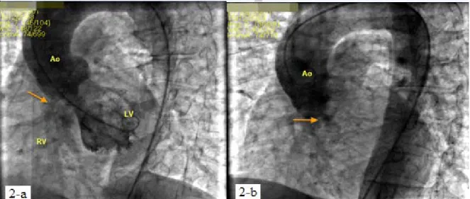
Fig. 2. Cardiac catheterization images demonstrated left to right shunt between right ruptured sinus of Valsalva aneurysm and the right ventricle (shown by the arrow).
radiating to the subxiphoid area. The patient’s functional capacity was class II according to the New York Heart Association classification. She had mild anemia (hemoglobin: 11.7 gr/dL) and occult blood in stool (OBS) was positive. Because of the EN and OBS, colonoscopy was offered to exclude inflammatory bowel disease but the patient refused this procedure. Detailed examination revealed no oral afts, genital ulcers or uveitis. Pathergy test was also negative, therefore Behçet’s disease was also ruled out. Cardiac and mediastinal shadows were normal on the chest X-ray and thorax computerized tomography. Electrocardiography showed sinus rhythm, normal axis and nonspecific ST segment and T wave changes. TTE revealed an enlarged
right SVA and color Doppler echocardiography showed mild mitral and tricuspid regurgitation and a left to right shunt between right SVA and the RV with normal right ventricular systolic function. Doppler interrogation revealed continuous flow from aorta to RV though the aneurysm with a peak gradient of 90 mmHg measured during diastole and Qp/Qs ratio was 2:1. Transesophageal echocardiography (TEE) performed to further delineation of the aortic valve pathology, and the ruptured SVA was clearly visualized (figure-1). At the end of the examination, we performed a cardiac catheterization and demonstrated a small shunt between the right SVA and the RV (figure-2). There were no atherosclerotic lesions or vessel
Eastern Journal of Medicine 16 (2011)231-234
B. Ekici et al / Ruptured Sinüs Valsalva Aneurysm
Case Report
233 abnormality in the coronary arteries. The patient underwent cardiac surgery and right SVA rupture was repaired by direct closure. At cardiac surgery she was found to have an in 6 mm diameter ruptured sinus of Valsalva aneurysm with classic windsock appearance. Postoperative TTE revealed no abnormality. The patient is currently on follow up with medical treatment.
3.Discussion
Sinus of Valsalva aneurysms occurs when aortic media is defective and there is absence of fusion between aortic media and the fibrous annulus of the aortic valve, with subsequent aneurismal enlargement at this weak point due to the high pressure at the root of the aorta (4). It is usually congenital but less commonly is associated with endocarditis, atherosclerosis, trauma, syphilis, aortic dissection, ulcerative colitis, Behçet’s disease, and Marfan’s syndrome (3). The male-to-female ratio is 3.5:1 and symptoms usually develop in the adult population (2). Coexisting cardiac lesions, especially a ventricular septal defect or aortic valve regurgitation, may both be present in about 30 to 40% of patients. Symptoms occur in 80% of patients, most commonly between 30 and 45 years of age. The severity of the shunt, the presence of associated lesions, and age at presentation determine the severity of symptoms (5). Symptoms associated with rupture are shortness of breath, chest pain, and fatigue (6). TEE has been regarded as the gold standard for the detection of SVA (7).
Surgical repair of SVA was found to be associated with an acceptably low incidence of both intraoperative and late adverse events and with an improved survival. Long-term survival after surgical treatment of RSVA is excellent. The risk for recurrent fistula or VSD is minimal in the current era. Late aortic insufficiency is still a risk, especially in right sinus of Valsalva-to-right ventricle fistula with associated subarterial VSD. Repair of RSVA through an aortotomy with or without cardiotomy permits inspection of the aortic root complex and facilitates aortic valve repair; this approach may reduce the incidence of late aortic insufficiency. Resection and repair of RSVA entails an acceptably low operative risk and yields long-term freedom from symptoms. Early, aggressive treatment is recommended to prevent endocarditis or lesional enlargement, which causes worse symptoms and necessitates more extensive repair (8). Surgery used to be the mainstay of treatment of RSVA, however, in the past few years several reports of transcatheter
closure of RSVA have come into light. Recently, transcatheter closure is reported as a feasible, effective, and safe technique with the Amplatzer devices (9). Definitely, it has the advantage of obviating open heart surgery but complete occlusion is mandatory to prevent hemolysis and infective endocarditis (10). As reported in the literature, surgery is the safest approach because of its lower association with both intraoperative and late adverse events, therefore the patient was referred to surgery.
Aortic aneurysm is an extremely rare complication of inflammatory bowel disease such as ulcerative colitis (11). Erythema nodosum is the most common skin abnormality seen in patients with inflammatory bowel diseases, like Crohn's and ulcerative colitis (12). The patient had mild anemia (hemoglobin: 11.7 gr/dL) and OBS was positive. Because of the EN and OBS, colonoscopy was offered to exclude inflammatory bowel disease but the patient refused this procedure.
We reported the case in order to impress the importance of ruptured SVA need prompt diagnosis and treatment in even the asymptomatic patients. Especially in the clinically silent situations suspicion, cautious examination and right tools for diagnosis are crucial.
References
1. Hope J. A treatise on the diseases of the heart and great vessels, (3rd ed.) Philadelphia: Lea & Blanchard, 1839, pp 466-471.
2. Reichert CL. Ruptured sinus Valsalva aneurysm, a rare cause of heart failure. Neth Heart J 2008; 16: 60-61.
3. Rothbaum DA, Dillon JC, Chang S, Feigenbaum H. Echocardiographic manifestation of right sinus of Valsalva aneurysm. Circulation 1974; 49: 768-771. 4. Wahab A, Khan HS, Wahab S. Rupture of
Sinus of Valsalva Aneurysm Presenting with Left Ventricular Failure. JIACM 2008; 9: 209-211
5. Moustafa S, Mookadam F, Cooper L, et al. Sinus of Valsalva aneurysms-47 years of a single center experience and systematic overview of published reports. Am J Cardiol 2007; 99: 1159-1164.
6. Weijerse A, van der Schoot MJ, Maat LP, et al. Cardiac tamponade due to a ruptured aneurysm of the sinus of valsalva. J Card Surg 2008; 23: 256-258.
7. Cancho ME, Oliver JM, Fernández MJ, et al. Transesophageal echocardiographic diagnosis of a ruptured sinus of valsalva aneurysm with right atrium fistula. Rev Esp Cardiol 2001; 54: 1236-1239.
B. Ekici et al / Ruptured Sinüs Valsalva Aneurysm
234 8. Van Son JA, Danielson GK, Schaff HV, et al.
Long-term outcome of surgical repair of ruptured sinus of Valsalva aneurysm. Circulation 1994; 90: 1120-1129.
9. Kumar P, Banerji A. Transcatheter closure of ruptured sinus of valsalva aneurysm. Catheter Cardiovasc Interv 2010; 76: 774-776.
10. Arora R, Trehan V, Rangasetty UM, et al. Transcatheter Closure of Ruptured Sinus of Valsalva Aneurysm. J Interven Cardiol 2004; 17: 53-58.
11. Karakurt C, Koçak G, Selimoğlu A, Ozen M. Aortic aneurysm: a rare complication of ulcerative colitis. Anadolu Kardiyol Derg 2007; 7: 461-462.
12. Larsen S, Bendtzen K, Nielsen OH. Extraintestinal manifestations of inflammatory bowel disease: epidemiology, diagnosis, and management. Ann Med 2010; 42: 97-114.