Introduction
Current modalities that are used to detect breast lesions include ultrasonography, mammography and magnetic resonance imaging (MRI). The sensitivity of MRI in detecting breast lesions is higher as compared to both ultrasonography and mammography (1). Breast MR examination has become an essential and integral component of breast imaging (2). MRI has a high sensitivity in investigating patients at high-risk for breast cancer, in follow-up of breast cancer patients, and in detecting ipsilateral or contralateral disease during preoperative evaluation (3-6). MRI can detect suspicious breast lesions that are neither palpable on clinical examination nor visible by mammography or ultrasonography (5, 7, 8).
Although MRI is a sensitive method for the early detection of breast cancer, its specifity is low despite recent technological advances (9). Suspicious MR-only visible breast lesions that are undetectable by inspection, ultrasonography and mammography should be histologi-cally confirmed. MR-guided needle biopsy, and MR-guided lesion marking followed by excisional surgical biopsy are used for evaluation of such lesions. MR-guided diagnostic biopsies are both less invasive and more suitable in terms of patient comfort as compared to surgi-cal excisional biopsy. According to the European Society of Breast Imaging guideline, MR-guided interventional procedures should be performed for clarification of MR-only visible questionable lesions (5). MR-guided interventional procedures include fine needle biopsy, core-needle biopsy and vacuum assisted biopsy. Vacuum assisted biopsy is superior to fine needle biopsy and core-needle biopsy in terms of providing more material (10). Core-needle biopsy and vacuum assisted biopsy are being increasingly used for histopathologic character-ization of suspicious lesions that cannot be detected by clinical examination and can be only monitored by MRI, as significant diagnostic
Magnetic Resonance Imaging Guided Vacuum Assisted
and Core Needle Biopsies
Fahrettin Kılıç
1, Abdulkadir Eren
2, Necmettin Tunç
3, Mehmet Velidedeoğlu
4, Selim Bakan
1, Fatih Aydoğan
4, Varol Çelik
4,
Ertuğrul Gazioğlu
4, Mehmet Halit Yılmaz
11Department of Radiology, İstanbul University Faculty of Medicine, İstanbul, Turkey 2Department of Radiology, İstanbul Medipol University, İstanbul, Turkey
3Clinic of Radiology, Memorial Hospital, Diyarbakır, Turkey
4Department of General Surgery, İstanbul University Cerrahpaşa Faculty of Medicine, İstanbul, Turkey
Address for Correspondence :
Fahrettin Kılıç e-mail: [email protected] Accepted: 26.10.2015Received: 30.09.2015
DOI: 10.5152/tjbh.2015.2769
25
ABSTRACT
Objective: The purpose of this study to present the results of Magnetic resonance imaging (MRI) guided cutting needle biopsy procedures of
suspi-cious breast lesions that can be solely detected on Magnetic resonance (MR) examination.
Materials and Methods: The study included 48 patients with 48 lesions which were solely be observed in breast MRI, indistinguishable in
ultrasonography and mammography, for MR guided vacuum-assisted cutting needle biopsy and 42 patients with 42 lesions for MR guided cutting needle biopsy for the lesions of the same nature. MR imaging was performed using a 1.5-Tesla MRI device. Acquired MR images were determined and biopsy protocol was performed using computer-aided diagnosis system on the workstation. Vacuum biopsies were performed using 10 G or 12 G automatic biopsy systems, cutting needle biopsy procedures were performed using fully automated 12 G biopsy needle.
Results: All biopsy procedures were finalized successfully without major complications. The lesions were 54 mass (60%), 28 were non-mass
con-trast enhancement (31%) and 8 were foci (9%) in the MR examination. Histopathological evaluation revealed 18 malignant (invasive, in-situ ductal carcinoma and lobular carcinoma), 66 benign (apocrine metaplasia, fibrosis, fibroadenomatoid lesion, sclerosing adenosis, fibrocystic disease and mild-to-severe epithelial proliferation) and 6 high-risk (atypical ductal hyperplasia, intraductal papilloma, radial scar) lesions.
Conclusion: Magnetic resonance guided vacuum and cutting needle biopsy methods are successful methods fort he evaluation of solely MRI
de-tected suspicious breast lesions. There are several advantages relative to each other in both methods.
methods with different advantages over each other. In our study, we aimed to present results of MR-guided vacuum-assisted biopsy and core-needle biopsy performed in our department.
Material and Methods
Patient selection and indications for biopsy
90 suspicious breast lesions that were detected with only MRI, in 90 patients at the İstanbul University Faculty of Medicine Department of Radiology between April 2011 and August 2013 were included in the study. Forty-eight patients underwent MR-guided vacuum biopsy and 42 patients underwent core-needle needle biopsy. The study was ap-proved by the Istanbul University Faculty of Medicine Ethical Board, all patients were informed about the procedure and informed consent was obtained from each patient.
Patients who were 18 years or older, with non-palpable suspicious le-sions that were only detected by MRI which were evaluated as BI-RADS 4 and 5 according to BI-BI-RADS criteria, and who consented to vacuum biopsy were included in the study. Patients younger than 18 years of age, those who did not consent to participate in the study, those with lesions detected by ultrasonography or mammography, those with palpable lesions, and those who withdrew their consent during the study period were excluded from the study.
Statistical analysis
The percentage, mean and median values of the data were evaluated by using SPSS (16.0; SPSS, Inc.; Chicago, IL, USA) software.
MR-Guided Breast Biopsy Techniques
Magnetic resonance imaging during biopsy procedures was performed by using a 1.5-Tesla MRI scanner. The patient was placed in the prone position, breast skin was disinfected with 10% Povidone-iodine at the site of the lesion, and imaging was performed with a 7-channel breast coil that contained compression plates. The compression was applied from both sides of the breast, medial and lateral aspects, in order to reduce artifacts by preventing motion and to enable accurate calcula-tions and stabilization of the breast during needle introduction. Since excessive compression may prevent contrast enhancement, the com-pression amount was carefully adjusted during stabilization. Bard Va-cora (10G) or Suros Atec (12G) biopsy systems were used for vacuum-assisted biopsy. Core-needle needle biopsy procedure was performed by using a 12-G fully automatic biopsy needle.
Interventional procedures began with MR sequences obtained for imag-ing. First, T1-weighted sagittal images were acquired to verify visibility of the reference points on the compression plates and if the lesion was accessible. Following these steps, T1A fast low angle shot (FLASH) 3D
sequence (TR / TE, 11 / 5.16, Gap 20, FOV 330, matrix 200x256, fre-quency direction R> L, bandwidth 150 Hz / Px) images were acquired once before and twice after bolus injection of 0.1 mmol/L per kg of body weight MR contrast agent that was administered via an intrave-nous catheter. The total imaging time was about 5 minutes.
The received MR images were further evaluated by a computer-aided diagnostic system (CAD; Dynacad, Invivo, Orlando, Florida, USA). The unenhanced images were simultaneously substracted from con-trast images by the workstation. After MR images were obtained following stabilization of the breast, localization of the lesion to be intervened was determined according to reference points within com-pression plates through the computer-aided diagnosis system. Lesion approach was planned prior to the intervention according to suitable
grid spacing, lesion depth and distance from the skin values provided by the computer, based on lesion localization (medial or lateral). The MR board was removed from the gantry, the patient was re-informed about the process, instructed not to move and the procedure was initi-ated. Local anesthesia was employed by infiltration of 4-6 cc Prilocaine HCl (Citanest) subcutaneously to the area of interest.
In both vacuum assisted and core-needle biopsy, both the needle and MRI compatible sheath were introduced into the breast together. The cutting needle was removed and a plastic cannula that prevents bleed-ing out was inserted through the sheath, and control T1A FLASH 3D axial MR images were obtained. The sheath and plastic cannula were visualized as thin hypointense artifacts on control MR images, and their localization was confirmed. After determination of appropriate localization, the biopsy procedure was performed with a MRI compat-ible vacuum biopsy needle that used an ignition system with single insertion, and 6-12 consecutive samples were obtained from the lesion at different points in a clockwise manner. In core-needle biopsy, sam-ples were obtained by 4-5 consecutive introductions with the ignition system. Samples underwent routine histopathologic examination. No major complications occurred during the procedures.
Control T1A FLASH 3D axial weighted MR images were obtained following biopsy to verify accuracy of the process, to assess if a second intervention is required, and to check for the presence of a hematoma (Figure 1-3). When the biopsy accuracy was verified, an MRI compat-ible marker was positioned to the biopsy site, and the procedure was terminated after obtaining control MR images for marker localization. The goal of leaving a marker was to localize the lesion for the surgeon if the pathology showed a malignant lesion, and to locate the lesion on follow-up MR images if the pathology result was benign.
Results
The study included MR-guided biopsies of 90 suspicious breast lesions that can only be detected with MR. Forty-eight patients underwent MR-guided vacuum biopsy and 42 patients underwent core-needle biopsy. The lesions were categorized as benign or malignant according to their histopathologic features on biopsy. The mean age of patients undergoing vacuum biopsy was 45.74 (26-69) years, and was 48.3 (35-56) years in those undergoing core-needle biopsy.
On MR examination, 54 of the 90 lesions were visualized as masses, 28 as non-mass contrast enhancement, and 8 as focus. The median size of the biopsied lesions was 9 mm (4 -15 mm) for those undergoing vacu-um-assisted biopsy, and was 15 mm (8-22 mm) for those undergoing core-needle biopsy. The median size of all lesions was determined as 12 mm (4-22 mm). The mean procedure duration was 38 minutes (24-69 min) for vacuum assisted biopsies, and was 41 minutes (28-58 min) for core-needle biopsies.
Histopathologic evaluation revealed 10 malignant (20.8%) (invasive and in-situ ductal carcinoma) and 38 benign (79.2%) (apocrine meta-plasia, fibrosis, fibroadenomatoid lesion, sclerosing adenosis, fibrocystic disease and mild-moderate-severe epithelial proliferation) lesions among vacuum assisted biopsy samples. Among core-needle biopsies; 8 lesions were malignant (in-situ ductal carcinoma, invasive ductal and lobular carcinoma) (19.4%), 6 were high-risk (atypical ductal hyperplasia, in-traductal papilloma, radial scar) (14%), and 28 were benign (fibrocystic changes, sclerosing adenosis, fibroadenoma) (66.6%) lesions.
Patients diagnosed with malignant and high-risk lesions were referred to medical and/or surgical treatment, and those with benign lesions were recommended to undergo a follow-up MRI or US at 6 months and 1 year. Malignant lesions were treated with breast conserving sur-gery after MR-guided lesion localization. The results of excisional bi-opsies of high-risk lesions were compatible with core-needle biopsy results, with no additional malignant findings.
Benign lesions did not show progression and these patients are being monitored by a routine annual follow-up program. A biopsy failure
was considered based on radiology-pathology discordance in 2 patients with core-needle biopsy and 1 patient with vacuum assisted biopsy. Excision was performed in these patients all of which revealed benign findings similar to their biopsy results.
All lesions were visualized on MRI sections during the biopsy dure. Since the routine breast protocol was not used, and the proce-dures were performed quickly with rapid decisions based on the CAD system, issues related to contrast wash-out and thus lesion disappear-ance on control MR cross-sections was not encountered.
27
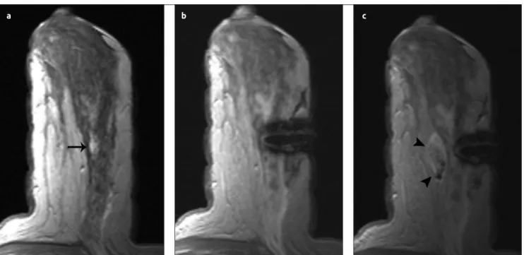
Figure 1. a-c. Core-needle biopsy of a lesion in the right breast of a 35-year-old woman (a) The view of an irregular shaped lesion 1cmin diameter that was suspicious for malignancy on subtracted axial enhanced image (arrow) (b) The lesion on axial contrast-enhanced T1-weighted image and the adjacent sheath (c) Post-biopsy image showing a barely distinguishable lesion and hypointense areas indicating hemorrhage posterior to the lesion. The biopsy revealed invasive ductal carcinoma
a b c
Figure 2. a-c. Core-needle biopsy of a lesion in the left breast of a 42-year-old woman (a) A 12mm in diameter, non-mass contrast
enhancement in the middle outer section of the left breast on axial contrast-enhanced T1-weighted image (black arrow) (b) Image of the lesion and the adjacent guiding sheath (c) Image of a 2cm hematoma just behind the lesion on control imaging after biopsy (arrowheads). The biopsy result showed an in-situ ductal carcinoma
Discussion and Conclusion
Biopsy of occult breast lesions can be performed by MR-guided lesion marking; wire-guidance, Technetium-99 m ROLL (Radio guided oc-cult lesion localization), and by MR-guided radiologic biopsy meth-ods. MR-guided biopsies are both less invasive and more comfortable for the patient as compared to lesion marking and surgical excisional biopsy (11). Obtaining a diagnosis by biopsy prior to lesion mark-ing and excision reduces the number of surgical interventions. If the biopsy result is benign and is consistent with radiologic findings then additional surgical treatment is not required. If the result is malignant then therapeutic procedures may be directly applied. The rate of de-tecting malignancy after surgical excision of BI-RADS 4-5 lesions var-ies between 15-40%, while this rate varvar-ies between 70-80% in surgical excision after detection of malignancy by radiologic sampling methods (12, 13). Meta-analyses including many studies state that radiologic biopsy methods should be preferred to surgical biopsy, if it can be performed (14, 15).
Vacuum assisted biopsy is less invasive and faster than surgical biopsy and does not cause deformity (16). It has lower risk and morbidity as compared to surgical biopsy, better accuracy than fine needle as-piration biopsy (17), and more sampled tissue than core-needle bi-opsy (18). It provides bigger and multiple samples in a single entry as compared to fine-needle aspiration and core-needle biopsy. Vacuum assisted biopsy also has a higher technical success rate as compared to fine needle aspiration biopsy.
Other advantages of vacuum biopsy are allowing quick intervention to small lesions (<10 mm) and providing more material for histopatho-logic evaluation. Core-needle biopsy is superior to fine needle biopsy in terms of providing more material and to vacuum biopsy by being less invasive (19). Carbognin et al. (20) stated that vacuum-assisted biopsy is an effective and reliable method for the diagnosis of MR-only lesions that are non-palpable and smaller than 1 cm. In a study evalu-ating the efficiency of vacuum-assisted biopsy system and core-needle
biopsy in removing microcalcifications, the failure rate in sampling was reported as 16% for 14G needle as compared to the rate of 4% in 14G vacuum biopsy, and 1% in 11G vacuum biopsy. This valuable study indicated that material sufficiency in vacuum biopsy increased parallel to an increase in needle diameter. The false negativity rate in core-needle biopsy was reported as 8%, while that of vacuum biopsy was determined as 0.67% (21).
Biggest drawback of vacuum biopsy is higher cost as compared to core-needle biopsy (16). The complication rate is higher in vacuum biopsy than other biopsy procedures. Core-needle biopsy is a quite good alternative method to vacuum-assisted biopsy, in case it cannot be performed, with much less complications (22). On the other hand, disadvantages of core-needle biopsy include requirement for multiple insertions for repetitive sampling, decrease in breast tissue with an in-crease in bloody samples in repeat specimens, and insufficiency in di-agnostic sensitivity of calcified lesions, atypical ductal hyperplasia and ductal carcinoma in situ.
Perlet et al. (23) evaluated the histopathologic characteristics of vac-uum biopsy on 538 patients, and reported 138 (27%) malignant, 17 (3%), atypical ductal hyperplasia, and 362 (70%) benign findings. To-zaki et al. (24) detected 34 (33%) malignant, 4 (4%) atypical ductal hyperplasia, 5 (5%) flat epithelial atypia, and 59 (58%) benign find-ings among 100 patients. In the study by Eby and Lehman, (10) 422 (25%) malignant and 1234 (75%) benign findings were detected in 1656 patients.
In our study of 90 patients, 18 (20%) lesions were reported as malig-nant, 66 (73.3%) as benign, and 6 (6.7%) as high-risk lesions. An in-dication for operation was detected in 26.7% of cases, and all of these lesions were removed with surgical excision for pathologic evaluation. Our results are comparable with other studies. One reason for the rela-tively low rate of malignancy in our study was successful utilization of second-look ultrasonography by experienced specialized physicians. Ultrasonography is performed by technicians in most centers except our country. The incidental MR lesions which could be detected by the
28
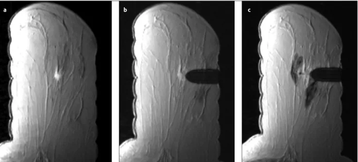
Figure 3. a-c. Vacuum-assisted biopsy for a lesion of the left breast in a 54-year-old woman (a) The view of an irregular bordered, round,
nodular lesion 5 mm in diameter on contrast-enhanced T1-weighted axial image of the left breast (b) Image of the lesion and the adjacent sheath. Pay attention to the slight contrast washout in the lesion (c) The lesion cannot be clearly viewed in control imaging after biopsy along with hypointense hemorrhagic areas around the lesion and the cannula. Biopsy result was reported as invasive ductal carcinoma
guidance of ultrasonography tends to be malignant, and are sampled under ultrasonography guidance (25). With effective use of second-look ultrasonography, the likelihood of ultrasound-guided sampling will increase and the number of cases with MR-guided malignant biopsy will change in favor of the more challenging MRI suspicious but histopathologically benign lesions. Nevertheless, the requirement for specific biopsy indications is evident. A biopsy failure was consid-ered in three patients and the surgical excisions revealed benign lesions similar to the biopsy findings.
The mean duration of vacuum assisted biopsy was reported as 65 min by Malhaire et al. (26), and as 35 min by Tozaki et al. (24). The mean duration of vacuum assisted biopsy was determined as 38 min (24-69 min) in our study, which was in concordance with previous studies. This duration was shortened gradually with increasing patient num-bers and experience. Additionally, most of the biopsy procedures were performed with a semi-automatic biopsy system in order to reduce cost, which prolonged duration of the biopsy procedure. The mean biopsy duration for core-needle biopsy was 41 min (28-58 min), and was similar to studies by Liberman et al. (27, 28).
In this study presenting our initial experience, promising results were obtained in computer-aided magnetic resonance imaging guided core-needle and vacuum-assisted biopsy of MR-only visible breast lesions. These methods can be used as an alternative to excisional biopsy for histological diagnosis of lesions detected by MRI, in appropriate cases. It is our opinion that with increasing expertise and accumulating data on follow-up of these patients, these methods will be used more ef-fectively.
Ethics Committee Approval: Ethics committee approval was received for this study.
Informed Consent: Written informed consent was obtained from pa-tients who participated in this study.
Peer-review: Externally peer-reviewed.
Author Contributions: Concept - M.H.Y.; Design - F.K., S.B.; Su-pervision - M.H.Y., V.Ç., E.G.; Materials - M.V., S.B.; Data Collec-tion and/or Processing - F.K., A.E., N.T.; Analysis and/or Interpreta-tion - F.K.; Literature Review - A.E., N.T.; Writing - F.K., A.E., F.A.; Critical Review - M.H.Y., V.Ç., E.G.
Conflict of Interest: No conflict of interest was declared by the au-thors.
Financial Disclosure: The authors declared that this study has re-ceived no financial support.
References
1. Kuhl C. The current status of breast MR imaging. Part I. Choice of tech-nique, image interpretation, diagnostic accuracy, and transfer to clinical practice. Radiology 2007; 244:356-378. (PMID: 17641361) [CrossRef]
2. Morris EA. Diagnostic breast MR imaging: current status and future directions. Magn Reson Imaging Clin N Am 2010; 18:57-74. (PMID: 19962093) [CrossRef]
3. Riedl CC, Ponhold L, Flory D, Weber M, Kroiss R, Wagner T, Fuchsjäger M, Helbich TH. Magnetic resonance imaging of the breast improves de-tection of invasive cancer, preinvasive cancer, and premalignant lesions during surveillance of women at high risk for breast cancer. Clin Cancer Res 2007; 13:6144-6152. (PMID: 17947480) [CrossRef]
4. Liberman L, Morris EA, Kim CM, Kaplan JB, Abramson AF, Menell JH, Van Zee KJ, Dershaw DD. MR imaging findings in the contralateral breast of women with recently diagnosed breast cancer. AJR Am J Roent-genol 2003; 180:333-341. (PMID: 12540428) [CrossRef]
5. Viehweg P, Bernerth T, Kiechle M, Buchmann J, Heinig A, Koelbl H, Laniado M, Heywang-Köbrunner SH. MR-guided intervention in wom-en with a family history of breast cancer. Eur J Radiol 2006; 57:81-89. (PMID: 16364583) [CrossRef]
6. van den Bosch MA, Daniel BL, Pal S, Nowels KW, Birdwell RL, Jeffrey SS, Ikeda DM. MRI-guided needle localization of suspicious breast le-sions: results of a freehand technique. Eur Radiol 2006; 16:1811-1817.(PMID: 16683117) [CrossRef]
7. Morris EA, Liberman L, Dershaw DD, Kaplan JB, LaTrenta LR, Abramson AF, Ballon DJ. Preoperative MR imaging-guided needle local-ization of breast lesions. AJR American journal of roentgenology 2002; 178:1211-1220. (PMID: 11959734) [CrossRef]
8. Orel SG, Rosen M, Mies C, Schnall MD. MR imaging-guided 9-gauge vacuum-assisted core-needle breast biopsy: initial experience. Radiology 2006; 238:54-61. (PMID: 16304093) [CrossRef]
9. Parkin DM, Bray F, Ferlay J, Pisani P. Estimating the world cancer burden: Globocan 2000. Int J Cancer 2001; 94:153-156. (PMID: 11668491)
[CrossRef]
10. Eby PR, Lehman CD. Magnetic resonance imaging--guided breast in-terventions. Top Magn Reson Imaging 2008; 19:151-162. (PMID: 18941395) [CrossRef]
11. Oxner CR, Vora L, Yim J, Kruper L, Ellenhorn JD. Magnetic resonance imaging-guided breast biopsy in lesions not visualized by mammogram or ultrasound. Am Surg 2012; 78:1087-1190. (PMID: 23025947) 12. Schueller G, Schueller-Weidekamm C, Helbich TH. Accuracy of
ul-trasound-guided, large-core needle breast biopsy. Eur Radiol. 2008; 18:1761-1773. (PMID: 18414872) [CrossRef]
13. Wallis M, Tardivon A, Helbich T, Schreer I. Guidelines from the Euro-pean Society of Breast Imaging for diagnostic interventional breast proce-dures. Eur Radiol 2007; 17:581-588. (PMID: 17013595) [CrossRef]
14. Bruening W, Fontanarosa J, Tipton K, Treadwell JR, Launders J, Schoelles K. Systematic review: comparative effectiveness of core-needle and open surgical biopsy to diagnose breast lesions. Ann Intern Med 2010; 152:238-246. (PMID: 20008742) [CrossRef]
15. Dahabreh IJ, Wieland LS, Adam GP, Halladay C, Lau J, Trikalinos TA. AHRQ Comparative Effectiveness Reviews. Core Needle and Open Surgi-cal Biopsy for Diagnosis of Breast Lesions: An Update to the 2009 Report. Rockville (MD): Agency for Healthcare Research and Quality (US); 2014. 16. Liberman L. Percutaneous image-guided core breast biopsy. Radiol Clin
North Am 2002; 40:483-500, vi. (PMID: 12117188)
17. Wald DS, Weinreb JC, Newstead G, Flyer M, Bose S. MR-guided fine needle aspiration of breast lesions: initial experience. J Comput Assist To-mogr 1996; 20:1-8. (PMID: 8576457) [CrossRef]
18. Chen X, Lehman CD, Dee KE. MRI-guided breast biopsy: clinical expe-rience with 14-gauge stainless steel core biopsy needle. AJR Am J Roent-genol 2004; 182:1075-1180. (PMID: 15039191) [CrossRef]
19. Philpotts LE, Hooley RJ, Lee CH. Comparison of automated versus vacu-um-assisted biopsy methods for sonographically guided core biopsy of the breast. AJR Am J Roentgenol 2003; 180:347-351. (PMID: 12540431)
[CrossRef]
20. Carbognin G, Girardi V, Brandalise A, Baglio I, Bucci A, Bonetti F, Pozzi Mucelli R. MR-guided vacuum-assisted breast biopsy in the management of incidental enhancing lesions detected by breast MR imaging. Radiolo-gia Med 2011; 116:876-885. (PMID: 21293942) [CrossRef]
21. Jackman RJ, Rodriguez-Soto J. Breast microcalcifications: retrieval failure at prone stereotactic core and vacuum breast biopsy--frequency, causes, and outcome. Radiology 2006; 239:61-70. (PMID: 16567483) [CrossRef]
22. Dershaw DD. Equipment, technique, quality assurance, and accredita-tion for imaging-guided breast biopsy procedures. Radiol Clin North Am 2000; 38:773-789, ix. (PMID: 10943277)
29
23. Perlet C, Heywang-Kobrunner SH, Heinig A, Sittek H, Casselman J, An-derson I, Taourel P. Magnetic resonance-guided, vacuum-assisted breast biopsy: results from a European multicenter study of 538 lesions. Cancer 2006; 106:982-990. (PMID: 16456807) [CrossRef]
24. Tozaki M, Yamashiro N, Sakamoto M, Sakamoto N, Mizuuchi N, Fu-kuma E. Magnetic resonance-guided vacuum-assisted breast biopsy: re-sults in 100 Japanese women. Jpn J Radiol 2010; 28:527-533. (PMID: 20799018) [CrossRef]
25. Spick C, Baltzer PA. Diagnostic utility of second-look US for breast le-sions identified at MR imaging: systematic review and meta-analysis. Ra-diology 2014; 273:401-409. (PMID: 25119022) [CrossRef]
26. Malhaire C, El Khoury C, Thibault F, Athanasiou A, Petrow P, Ollivier L, Tardivon A. Vacuum-assisted biopsies under MR guidance: results of 72 procedures. Eur Radiol 2010; 20:1554-1562. (PMID: 20119729)
[CrossRef]
27. Liberman L, Morris EA, Dershaw DD, Thornton CM, Van Zee KJ, Tan LK. Fast MRI-guided vacuum-assisted breast biopsy: initial experi-ence. AJR Am J Roentgenol 2003; 181:1283-1293. (PMID: 14573421)
[CrossRef]
28. Liberman L, Bracero N, Morris E, Thornton C, Dershaw DD. MRI-guid-ed 9-gauge vacuum-assistMRI-guid-ed breast biopsy: initial clinical experience. AJR Am J Roentgenol 2005; 185:183-93. (PMID: 15972421) [CrossRef]