ARAŞTIRMA/ORIGINAL ARTICLE
(*) GATA Haydarpaşa Eğitim Hastanesi, Ortopedi ve Travmatoloji Kliniği, İstanbul
(**) İstanbul Medipol Üniversitesi, Ortopedi ve Travmatoloji Kliniği, İstanbul (***) GATA Haydarpaşa Eğitim Hastanesi, Kardiyoloji Kliniği, İstanbul (****) GATA Haydarpaşa Eğitim Hastanesi, Kulak-Burun-Boğaz Kliniği, İstanbul
(*****) Şırnak Asker Hastanesi, Ortopedi ve Travmatoloji Kliniği, Şırnak
Ayrı Basım İsteği: Doç.Dr. Selami Çakmak GATA Haydarpaşa Eğitim Hastanesi Ortopedi ve Travmatoloji Kliniği Üsküdar/İstanbul [email protected]
Selami Çakmak(*), Osman Rodop(*), Mahir Mahiroğulları(**), Ömer Uz(***), Evren Erkul(****),
Kenan Keklikçi(*****)
ÖZET
Klippel Feil sendromlu erişkinlerdeki muhtelif organ anormalliklerinin farkında mıyız?
Klippel-Feil Sendromu (KFS) klasik bir üçleme ile karakterizedir (kısa boyun, düşük arka saç çizgisi, boyun hareketlerinde kısıtlılık). Asıl klinik tabloya servikal vertebranın segmentasyon problemleri neden olurken, iskelet sistemi dışındaki anomaliler de bildirilmiştir. Bu çalışmada KFS’lu hastalarda yaygın görülen klinik bulguların gözden geçirilmesi ve vurgulanması amaçlanmıştır. 2000-2011 yılları arasında yüz asimetrisi olan ve kısa boyunlu 120 kişi değerlendirilmiştir. 42 KFS’lu erişkin bu çalışmaya dahil edilmiştir. Hastalar iskelet sistemi ve iskelet dışı patolojiler açısından incelenmiştir. Hastaların 41’i erkek (%98) ve biri kadındır (%2). 42 hastanın 31’i tip I (%74), 9’u tip II (%21) ve 2’si (%5) tip III olarak sınıflandırılmıştır. Klasik klinik üçlü bulgu hastaların hepsinde saptanmıştır. Biri hariç tüm hastalarda skolyoz saptanmışken, Sprengel deformitesi 10 hastada (%24) gözlenmiştir. Eşlik eden diğer organ patolojilerinin %9,5’i ürogenital sistem, %23,8’i kardiyovasküler sistem ve %31’i işitme patolojileri idi. KFS’lu hastalardaki omurga ve diğer organ patolojileri sonuçta tıbbi tedavi gerektiren ciddi sorunlara yol açabilir. KFS’lu hastalarda olası bir sistemik problemin saptanması amacıyla detaylı bir inceleme veya yakın klinik takip yararlı olabilir.
Anahtar Kelimeler: Klippel-Feil Sendromu, konjenital, servikal vertebra, skolyoz,
anomaliler, malformasyonlar, füzyon, sınıflama SUMMARY
Klippel-Feil syndrome (KFS) is characterized by a classical triad (short neck, low posterior hair line, limitation of neck movements). Segmentation problems of cervical vertebra cause the main clinic in patients, and extraskeletal anomalies are also reported. The aim of this study is to review and emphasize common clinical findings of KFS patients. Between 2000 and 2011, 120 individuals with facial asymmetry and short neck were evaluated. Forty-two adult patients with KFS were included in this study. Patients were assessed for associated skeletal and extraskeletal pathologies. There were 41 male (98%) and 1 female (2%) individuals. Of forty-two patients, thirty-one (n=31; 74%) were classified as type I, nine (n=9; 21%) as type II, and two (n=2; 5%) as type III. Classical clinical triad was detected in all patients. Congenital scoliosis has been observed in all patients except one. Sprengel deformity was observed in 10 individuals (24%). Associated other system disorder ratios were 9.5% for urogenital problems, 23.8% for cardiovascular pathologies and 31% for audiological problems. Spinal and other multi-organ pathologies of patients with KFS may lead to serious problems which may eventually need medical treatment. Detailed assessment for possible systemic disorders and close follow-up may be helpful for KFS patients.
Key words: Klippel-Feil syndrome, congenital, cervical vertebrae, scoliosis, anomalies,
malformations, fusion, classification
Do we know multifarious organ abnormalities of adults
with Klippel-Feil syndrome?
Gülhane Tıp Derg 2016;58: 37-44 © Gülhane Askeri Tıp Akademisi 2016 doi: 10.5455/gulhane.207263
Introduction
Klippel-Feil syndrome was described for the first time in 1912 and characterized by the classic triad of short neck, low posterior hair line, and restricted neck movements (1). This classical clinical triad emerges in 40-50% of patients with KFS (2-4). The most common clinical finding is limited cervical movements, especially in rotation and lateral bending (4-7). KFS is a rare disorder and estimated to appear in every 1:40.000-50.000 births (3,4,6). It is characterized by the congenital fusion of two or more cervical vertebrae and caused by a failure in the segmentation period of cervical vertebrae during the 3 to 8 weeks of gestation. The causes of segmentation defects of cervical spine have not yet been explained exactly.
Diagnosis of KFS depends on the segmentation problem of at least one level of cervical vertebrae. Other skeletal and multi-systemic anomalies can be involved. The most common musculoskeletal anomalies are scoliosis, Sprengel deformity, torticollis, kyphosis, and congenital limb anomalies. Some other associated extra-skeletal anomalies have also been reported and these different anomalies are variable (3,8,9). Otological and ear anomalies, renal anomalies, cardiac problems, and neural tube disorders have been reported to occur in patients with KFS. According to Feil’s original classification, KFS is classified into three main types: Type I, fusion of multiple cervical vertebrae and/or upper thoracic vertebrae as a block; type II, fusion of two or limited number of cervical vertebrae, and type III, fusion of cervical vertebrae and concomitant lumbar vertebrae (8).
In previous study, senior author had reported the review of skeletal and non-skeletal findings in patients with KFS (10). The goal of this following study is to add new cases and highlight common clinical features of KFS that clinicians encounter in their regular practice and emphasize the importance of being awareness about possible serious disorders. We present a case series of 42 patients, who diagnosed and after assessed to look for any overlooked anomalies associated with KFS that may need follow-up or treatment.
Materials and methods
This observational study was a review of the adult individuals diagnosed with KFS at two main military hospitals in Istanbul and Ankara, Turkey. These adult individuals selected from our outpatient clinic patients who were admitted for routine health check-up (mostly recruitment of health screening programme) or other disorders.
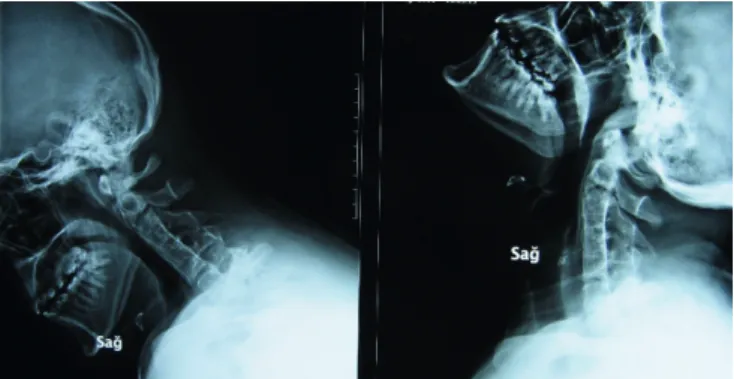
Between 2000 and 2011, individuals with short neck, decrease ROM of the neck and low posterior hairline were included the study and were sent for cervical anteroposterior and lateral radiographies to name the diagnosis as KFS. Adult patients with KFS were subjected to detailed orthopaedic physical examination, cervical flexion-extension radiographs (Figure 1) and lumbar anteroposterior and
lateral radiographies. Fusion was defined as bone bridging between segments and the loss of segmental motion on dynamic (flexion-extension) radiographies. Coronal alignment of spine (especially cervical) and Cobb angle are measured by a line starting from C1 vertebra and according to Samartzis method (11). The Cobb angle measurement is
started with determination of curve at apical vertebra which is most displaced from vertical axis. End vertebrae which are least displaced from vertical axis and mostly tilted are then marked through the curve proximally and distally. Two lines are needed: first line is drawn along the superior end-plate of proximal end vertebra and second line is drawn along the inferior end-plate of distal end vertebra. The measured angle between these lines is Cobb angle. If Cobb angle of lateral spinal curvature more than 10°, it is defined as scoliosis. All patients were asked about whether or not there are any family members who have had similar posture and signs for KFS. All clinical and radiographic assessment was performed by an orthopedic surgeon.
These patients were also evaluated by cardiovascular examination, echocardiography, electrocardiography, urologic examination, pelvic and renal ultrasound, ENT (Ear-Nose-Throat) examination, routine audiological tests (pure tone audiometry, speech reception threshold assessment and tympanometry), neurologic and psychiatric examination. Other associated anomalies were also recorded and patients were classified into Feil’s classification. All patients also questioned for any activity disabilities or restrictions which may cause working distress or psychological problem. An approval from Ethical Committee of GATA Haydarpasa Training Hospital was obtained. Detailed information about the syndrome and screening program were stated and all patients’ written informed consents were obtained for the publication of individual clinical details and accompanying clinical images.
Results
120 adult individuals with short neck and low posterior hairline were sent for cervical anteroposterior and lateral radiographies and 42 of them diagnosed as KFS. Detailed multi-clinical and radiographic data were obtained from 42 patients and these patients included this study. There were 41 male (98%) and 1 female (2%) individuals. Of forty-two (n=42) patients, thirty-one (n=31; 74%) were classified as type I, nine (n=9; 21%) as type II and two (n=2; 5%) as type III, according to Feil’s classification. The average age at assessment time was 21.7 years (range, 19-40). All patients except one reported that none of other family members have similar findings.
Low posterior hair line, short neck, and fusion of cervical vertebrae (at least two) have been observed in all patients (Figure 2 and 3). The most commonly fused segments were
Figure 1. Fusion of cervical vertebrae seen on lateral dynamic radiographies of cervical vertebrae in flexion and extension.
Figure 2. Short neck and slight fascial asymmetry can be seen in front view of patient.
Figure 3. Low posterior hair line and Sprengel deformity at left side can be seen in back view of patient
C2-C3, C3-C4 and C5-C6 (each of which was present in 64% of patients). Single-level fusion rate was %33, 2-level rate was %36, and 3-level rate was % 20. The fusion rate for 4-5 level was % 11. Congenital scoliosis has been observed in nearly all patients except one (98%) and cervical/cervicothoracic region was the most included site of spine (Figure 4). The mean Cobb angle of all scoliotic patients was 23,3°. (Cobb angles specific to KFS types: 24° in type 1, 18° in type II and 32° in type III. Scoliotic curves were single in 56% and double in 44%, right curved in 60% and left curved in 40%. There was no instability and/or hypermobility signs on dynamic cervical flexion-extension radiographies.
Neck movements were limited to more than 50% of flexion-extension arch, both lateral tilt and rotation in 20 patients (47%) and less than 50% in 11 patients (26%). Limitations of rotational movements especially were significant in patients with torticollis component (n=13, 31%). Total restriction of cervical movements was rare.
Sprengel deformity was observed in 10 individuals (24%). Fusion of lumbar vertebrae was recorded in two individuals (Type III KFS). In one patient there was a unilateral sacralization at posteroanterior radiography. Cervical costae were observed in five individuals (12%) with no neurovascular compression symptoms. Posterior fusion defects (spina bifida etc.) were detected in 3 patients. (One was localized at C6 and C7 level, one was at between C6 and T2 level and the other one was at two different localization; C6-C7 and T1-T2). Developmental disorders of upper limbs (bilateral radius and ulnar tilt, syndactyly) are also noted in one patient.
Among non-skeletal system anomalies; urogenital system anomalies were found in four of 42 individuals (9.5%); unilateral renal agenesis was determined in one patient and renal cysts (asymptomatic) were found in three patients. Cardiovascular problems were detected in 10 patients (23.8%). Asymptomatic diastolic dysfunction of left ventricle (grade 2) was found in 2 individuals. There was also mitral valve insufficiency in three patients, aortic valve insufficiency in one patient, and mitral valve prolapse in two patients. In another asymptomatic individual, concomitant cardiac problems (mitral valve insufficiency, mitral valve prolapse and aortic insufficiency) were detected. One patient had secundum type atrial septal defect (he had been operated for ASD) and mitral valve prolapse. Synkinesia (mirror movements) was observed in four patients (9.5%) (advanced level in one and mild level in three). Audiological disorders were determined in13 patients (31%), involving conduction type hearing loss (n=6), sensorineural type hearing loss (n=5) and mixt type hearing loss (n=2). In one patient with Type I KFS, multiple systemic anomalies (pectus excavatum, microcephaly, pes planovalgus, and serious thenar atrophy at both hands) were detected and he had also an operation for esophageal fistula before. Associated anomalies of KFS reported in literature and this study are shown in Table I. All patients’ demographic data are shown in Table II.
Patients were not reported any activity restriction or psychological distress during routine life and working life.
Discussion
KFS is divided into three main groups according to Feil’ classification; Type I shows large fusions involving multiple cervical and upper thoracic vertebrae, Type II shows fusion at just one or two intervertebral spaces and Type III shows lumbar vertebral fusions associated with cervical fusion. Thomsen et al reported the rates of subgroups as 40% for Type I, 47% for Type II and 13% for Type III (3). Samartzis reported the pediatric patients’ rates as 50% for Type II, 25% for Type I and 25% for Type III (12). Guille et al also found rates as 54.5% for Type I, 27.3% for Type III and 18.2% for type II in their study which contains adult individuals (5). In our study, we evaluated adult individuals and rates of Type I, II and III types were 74%, 21% and 5%, consecutively. Thomsen and Samartzis’ study groups were mostly evaluated pediatric population and shows a predominance of Type II. However Guille’ group was evaluated an adult group (mean age was 35 years) and reported a predominance of Type I like our study. In this condition we think that patterns of fused segments and types in KFS are not age or time dependent issue. This contradictory result may strengthen the opinion of the need for new, molecular-based flow charts and classifications on pediatric-based and adult-based ones separately. Several other authors also recommended different classification schemes based on cervical fusion, genetic patterns, mobility of non-fused segments, or neurologic injury risk (3,5,8,12-14). Because of they have not been used widely in literature, we did not assess our individuals with them.
In literature, sex-type distribution of KFS individuals reported in a slight female predominance with 57-70% rates (3,5,12,14). In recent and our previous study, there is no correlation with respect to the reported literature rates (10). Nearly all of our patients were male. But here is a special case. Our both hospital are the final medical centers for candidates who were sent for assessment of health status for military service. In Turkey, there is a compulsory military service and we always perform fully medical examination for all male candidates at almost 20 years old. All of our male patients are the individuals who were diagnosed in this medical examination period. One female individual is the relative of one of male individuals. While reading the sex-type distribution rates in this study, one should consider our difference in rates in lights of all these information. However our male predominance of patients may
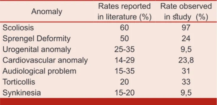
Table I. Various anomalies observed in individuals with KFS.
Anomaly in literature (%)Rates reported Rate observed in study (%)
Scoliosis 60 97 Sprengel Deformity 50 24 Urogenital anomaly 25-35 9,5 Cardiovascular anomaly 14-29 23,8 Audiological problem 15-35 31 Torticollis 20 33 Synkinesia 15-20 9,5
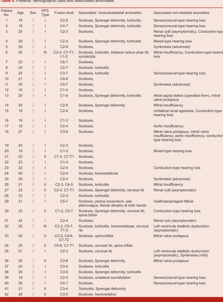
Table II. Patients’ demographic data and associated anomalies.
Patient
No. Age Sex TypeKFS Fusion level Associated musculoskeletal anomalies Associated non-skeletal anomalies 1 19 ♂ I C2-5 Scoliosis, Sprengel deformity, torticollis Sensoryneural-type hearing loss 2 19 ♂ I C4-7 Scoliosis, Sprengel deformity, torticollis Sensoryneural-type hearing loss
3 20 ♂ I C2-3 Scoliosis, Renal cyst (asymptomatic), Conduction-type
hearing loss
4 20 ♂ I C2-4 Scoliosis, Sprengel deformity, torticollis Mixed-type hearing loss
5 20 ♂ I C2-6 Scoliosis, Synkinesia (advanced)
6 20 ♂ III C2-4, C7-T1,
L1-2 Scoliosis, torticollis, bilateral radius-ulnar tilt, syndactyly Mitral insufficiency, Conduction-type hearing loss
7 23 ♂ I C5-7 Scoliosis,
8 20 ♂ I C2-7 Scoliosis, torticollis
9 20 ♂ I C3-7 Scoliosis, torticollis Sensoryneural-type hearing loss
10 21 ♂ I C4-6 Scoliosis,
11 20 ♂ I C5-7 Scoliosis, Synkinesia (advanced)
12 19 ♂ I C1-4 Scoliosis,
13 20 ♂ I C1-6 Scoliosis, Sprengel deformity, torticollis Atrial septal defect (operated from), mitral valve prolapsus
14 20 ♂ I C2-6 Scoliosis, Sprengel deformity Mitral insufficiency
15 19 ♂ I C2-4 Scoliosis, Unilateral renal agenesis, Conduction-type
hearing loss
16 19 ♂ I C1-2 Scoliosis,
17 19 ♂ I C2-4 Scoliosis, Aortic insufficiency
18 27 ♂ I C3-6 Scoliosis, Mitral valve prolapsus, mitral valve
insufficiency, aortic insufficiency, conduction-type hearing loss
19 20 ♂ I C2-3 Scoliosis,
20 19 ♂ I C1-3 Scoliosis, Mixed-type hearing loss
21 23 ♂ II C1-3, C7-T1 Scoliosis,
22 23 ♂ I C1-4 Scoliosis,
23 22 ♂ I C2-4 Scoliosis, Conduction-type hearing loss
24 40 ♂ I C2-4 Scoliosis, hemivertebrae
25 20 ♂ I C2-4 Scoliosis, Synkinesia (advanced)
26 21 ♂ II C2-3, C4-5 Scoliosis, torticollis Mitral insufficiency 27 23 ♂ II C2-4, C7-T1 Scoliosis, Sprengel deformity, cervical rib Renal cyst (asymptomatic)
28 23 ♂ I C2-3 Scoliosis, torticollis
29 21 ♂ I C5-7 Scoliosis, pectus excavatum, pes
planovalgus, thenar atrophy at both hands. Gastroesophageal fistula 30 23 ♂ II C1-2, C5-7 Scoliosis, Sprengel deformity, cervical rib,
spina bifida Conduction-type hearing loss
31 24 ♂ I C2-4 Scoliosis, Renal cyts (asymptomatic)
32 25 ♂ III C2-3, C5-7,
T1-3 Scoliosis, torticollis, hemivertebrae, cervical rib Left ventricule diastolic dysfunction (asymptomatic)
33 20 ♂ II C2-3, C4-6,
C7-T2 Scoliosis, spina bifida Mitral valve prolapsus 34 20 ♂ II C5-6, C7-T1 Scoliosis, cervical rib, spina bifida
35 21 ♂ I C2-3 Scoliosis, cervical rib Left ventricule diastolic dysfunction (asymptomatic), Synkinesia (mild) 36 20 ♂ II C3-6 Scoliosis, Sprengel deformity Mitral valve prolapsus
37 24 ♂ I C3-4 Scoliosis, torticollis
38 20 ♂ I C3-5 Scoliosis, Sprengel deformity, torticollis
39 19 ♂ I C2-5 Scoliosis, unilateral sacralization Sensoryneural-type hearing loss
40 35 ♀ I C4-7 Scoliosis, Sensoryneural-type hearing loss
41 21 ♂ II C3-4 Torticollis, Sprengel deformity
be helpful to discuss predominance of sex-type in terms of KFS types. Samartzis et al reported female predominance (57%) in harmony with literature and also mentioned a greater male predominance with increasing classification type. Authors also suggested that if males may be more at risk for type III (12). However in our studies (males evaluated) we did not found any male predominance specific to type III. Patterns of fused segments and Feil’s types may not be associated with sex-type.
Rates of classic clinical triad of KFS (short neck, low posterior hair line, and fusion of cervical vertebrae) are reported before as between 34-74% (3,9,14). In our study, all patients had classic clinical triad. This is one of the privileged sides of our study because when individuals were seen in outpatient clinic, physical appearance was the most determinative finding being first step to diagnose KFS. Facial asymmetry and torticollis were also among the first signs helps to diagnose KFS. Facial asymmetry and torticollis may be initial indicators for congenital cervical deformities. These anomalies are observed in 20% of patients with KFS (9). Ballock and Song evaluated the nonmuscular causes of torticollis and analyzed 288 patients’ data. 53 patients had nonmuscular causes for torticollis and of these patients, KFS rate was 30% (15). In our study, torticollis rate was 33% (14 patients). Both clinical appearances (classical triad and facial asymmetry) may canalize physicians to the way of KFS diagnose.
Skeletal pathologies
Scoliosis which is the most common associated anomaly has been determined in more than 50% of individuals of KFS. Hensinger reported a high incidence of 60% and Pizzutillo noted the rate of congenital scoliosis as 78% (9,14). We detected scoliosis in all patients except one (97%) and deformity was mostly located at cervicothoracic region in our study. Samartzis et al especially studied for rate of cervical scoliosis in KFS and reported the cervical scoliosis rate as 53.5% (12). On the other hand some authors investigated the incidence of KFS rate in scoliotic patients. In a retrospective study, Winter et al reviewed the radiographies of 1,215 patients with scoliosis and kyphosis and found that 298 of patients (25%) had segmentation defects located at cervical spine (16). However Xue at el reported lower incidence (5,42%) of KFS in patients with congenital scoliosis (17).
Underlying mechanism of the formation of both Sprengel deformity and KFS arise from intervention during gestation. It’s during week 8 that scapula descends to normal position and simultaneously segmentation of cervical vertebrae occurs. Our rate (24%) was similar with the Sprengel deformity rates reported at literature (26%-30%) (3). In a study evaluated the existence of Sprengel deformity in young patients (mean age 13,5 years) with KFS, authors reported the rate as %16,7 and no significant association of sex-type, classification-type, specific fused patterns and degree of scoliosis with the presence of Sprengel deformity (18). Tassabehji et al reported 50% percent of Sprengel deformity in individuals of a 5 generation family with KFS (19).
Neurologic Pathologies
Neurologic problems mostly related with KFS may come from spinal nerve stenosis, cord compression or/with instability. Patients with KFS commonly have nonspecific pain localized at neck, shoulder, or back but they rarely need medical treatment for neurologic problems or instability of cervical vertebrae (12). According to some authors, neurologic symptoms are independent from the cervical fusion levels and patterns (20). Rouvreau et al also found out that only five of 19 patients with KFS complained from neurologic complications during follow-up period of 12.5 years (21). However some other studies suggest that cervical fusions change the natural mobility patterns of the cervical spine and this process lead to some degenerative cervical problems, hypermobility and instability (5,22-24). Pizzutillo et al also conducted a study for assessment of cervical range of motion kinematically and reported that neurologic problems occur in patients with increased mobility at upper cervical vertebrae (14).
On other hand, in some conditions, radiographic evaluation of cervical radiographies for cervical instability or hyper mobility may be confused at childhood. For example pseudosubluxation of C2 on C3 and C3 on C4 may be a normal finding in a child younger than 8 years old (25). When instability occurred in accordance with neurologic symptoms, some studies recommend cervical fusion (4,14,26) while some others do not (27-30). Guille et al evaluated individuals with KFS and reported no sign of cervical instability (31). Similarly, no instability or hypermobility was seen in our study and all patients were asymptomatic neurologically. However, we agree with authors who advocate fusion, because risk of neurologic complications at future life may be related to cervical instability or hypermobility at mobile cervical levels. KFS patients should be informed to be careful against any trauma even following minor one and assessed periodically to rule out any neurological radiculopathy and/or myelopathy.
Although Ulmer has underlined that spine-related problems may be seen at adulthood, Ritterbusch and Rouvreau reported stenosis of cervical cord and neurologic symptoms at childhood (21,23,24). However, we did not detect neurologic symptoms like radiculopathy or myelopathy in our adult individuals. They had nonspecific neck pains but not clinically significant. As mentioned above, literature has some confusing reports in terms of being symptomatic or asymptomatic and the management way of these patients specific to age-groups (pediatric and adult). Magnetic resonance imaging can be useful to determine the pathologic anatomy and if cervical disorders have any involvement with spinal cord or nerves in individuals with significant symptoms (14,23).
Other organ pathologies
Association of congenital multiple organ anomalies with KFS (or other segmentation problems of cervical vertebrae) have been showed in literature before (9). These multisystem anomalies may result from mutations of genes responsible for segmentation process (13). Detection of exact causes of genetic deficiencies is a complicated process. Heterogeneity
of individuals with KFS makes it complex to identify classes. Increasing information with genetic advances and completion of the human genome sequencing project will reorganize the classification of KFS and make a descriptive link between genetic bases and clinical appearance of this multisystemic disease. Clarke et al reported a special family, in which most of the members had fusion of cervical vertebrae on radiographies (32). Paracentric inversion of chromosome 8q was demonstrated in all family members. There is a judgement that this inversion corrupts the functionality of SGM1 gene. In light of this information, this family can lead and encourage further genetic studies. Tassabehji et al also further followed up this family (now in 5th generation) and added new clinical features (19). Mutations in PAX genes family and HOX complexes may also play important roles in the segmental disorders at cervical vertebrae (33,34).
Lack of genetic analysis is one of the limited points of this study. Further genetic studies are needed to exhibit exact, underlying causes of KFS. Complex interactions between different genes must be clarified and then it may trigger new attempts for re-classification of KFS patterns as in the Clarke et al study (13). Secondly we did not assess the base of skull on radiographies and we did not evaluated basilar invagination. This was the second lack point of this study.
Now we know that there are well-known multi-organ anomalies associated with KFS including mainly cardiovascular, otological, urologic and other systems. Many sporadic cases are also being reported in time.
Cardiovascular anomalies associated with KFS have been reported in literature. The most frequently seen anomaly was ventricular septal defect (14). Morrison reported the rate of cardiovascular anomalies as 4.2% and Thomsen et al reported as 3.5% (3,35). Hensinger stated that the cardiac problem ratio was 14% (9). In our study 23.8% of individuals had cardiovascular anomalies, but most of them were asymptomatic and found at detailed echocardiographic assessment. Some of the other anomalies in association with KFS previously reported are agenesis of internal carotid artery, coarctation of aorta, atrial septal defect, and dextrocardia (36-38). Evaluation of adult KFS patients in our study may be speculated as one of the reasons for our patients’ lacking of these life-threatening serious cardiovascular anomalies.
Otological and audiological problems in patients with KFS may cause sensory or conductive hearing loss and may occur in one third of patients (9,39). Yildirim et al conducted a study and reported audiological abnormalities as 60% in individuals with KFS (40). Rate of otological and audiological disorders (31%) in our study was comparable with reported rates in literature. McGaughran et al reported the rate of audiologic abnormalities as %80 in KFS patients (n=45). Sensorineural deafness was the most common audiologic pathology (n=15) (41). Rate of conduction type deafness (46.1%, 6/13) was most the common type in our study, followed by sensorineural type (38.4%, 5/13) and mixed type (15.3%. 2/13). Our patients with audiological disorders were not aware of their problems and they were treated and began to follow-up. This point also
shows the clinical importance of the evaluation of patients with KFS for additional other organ anomalies.
During the fourth and eighth week of gestational life, any intervention may result with segmentation defects of cervical somites and urogenital anomalies. These anomalies have been determined as 30% of individuals with KFS (9). Moore et al evaluated genitourinary anomalies associated with KFS and found that 64% of patients had significant genitourinary system anomalies and unilateral agenesis of kidney was most common among them (42). In our study there is only one case with unilateral kidney agenesis. The other three cases had simple asymptomatic renal cysts.
Mirror movement (or synkinesia) is defined as a motor control disorder in which patient voluntarily moves one limb, the opposite limb involuntarily mirrors the same movements. It may be normal in patients less than 5 years of age and may be seen at 20% of KFS patients (9). The exact patho-physiologic mechanism of synkinesia is still unknown but, Gunderson et al reported the autopsy report of a KFS patient and revealed incomplete crossing of the pyramidal tracts (43). We detected the rate of synkinesia as 9.5%. In comparison with literature, our lower rates can be explained by a hypothesis that it becomes more muted with advancing age and is rarely found after 20 years of age.
In conclusion, comprehensive assessment of other organ system anomalies associated with musculoskelatal anomalies in individuals with KFS is indicated to find any existence of diseases which may be in need of medical treatment. Long term and close follow-up of KFS patients and suggestions for complete check-up should be kept in mind. Awareness of this rare syndrome is important for possible cervical spine instability and hypermobility which can cause severe neurological problems. Understanding of underlying etiology of KFS can be achieved by further broad and multi-center based genetic researches. Improvements on the field of study and new data about links between genetic background and the phenotypic appearance of KFS patients will be able to explain the heterogeneity of this syndrome and to lead up re-classification of KFS.
References
1. Klippel M, Feil A. The classic: A case of absence of cervical vertebrae with the thoracic cage rising to the base of the cranium (cervical thoracic cage). Clin Orthop Rel Res 1975; 109: 3–8.
2. Patel PR, Lauerman WC. Maurice Klippel. Spine 1995; 20: 2157-2160.
3. Thomsen MN, Schneider U, Weber M, Johannisson R, Niethard FU. Scoliosis and congenital anomalies associated with Klippel-Feil syndrome types I-III. Spine 1997; 22: 396-401.
4. Tracy MR, Dormans JP, Kusumi K. Klippel-Feil syndrome: clinical features and current understanding of etiology. Clin Orthop Relat Res 2004; 424: 183-190.
5. Guille JT, Miller A, Bowen JR, Forlin E, Caro PA. The natural history of Klippel-Feil syndrome: Clinical, roentgenographic, and magnetic resonance imaging findings at adulthood. J Pediatr Orthop 1995; 15: 617-626.
6. Shen FH, Samartzis D, Herman J, Lubicky JP. Radiographic assessment of segmental motion at the atlantoaxial junction in the Klippel-Feil patient. Spine 2006; 31: 171-177.
7. Van Kerckhoven MF, Fabry G. The Klippel-Feil syndrome: a constellation of deformities. Acta Orthop Belg 1989; 55: 107-118.
8. Gunderson CH, Greenspan RH, Glaser GH, Lubs HA. The Klippel-Feil syndrome: genetic and clinical reevaluation of cervical fusion. Medicine (Baltimore) 1967; 46: 491-512.
9. Hensinger RN, Lang JE, MacEwen GD. Klippel-Feil syndrome: a constellation of associated anomalies. J Bone Joint Surg Am 1974; 56: 1246-1253.
10. Mahiroğullari M, Ozkan H, Yildirim N, Cilli F, Güdemez E. [Klippel-Feil syndrome and associated congenital abnormalities: evaluation of 23 cases]. Acta Orthop Traumatol Turc 2006; 40(3): 234-239.
11. Samartzis D, Kalluri P, Herman J, Lubicky JP, Shen FH. Cervical scoliosis in the Klippel-Feil patient. Spine (Phila Pa 1976) 2011; 36(23): E1501-1508.
12. Samartzis DD, Herman J, Lubicky JP, Shen FH. Classification of congenitally fused cervical patterns in Klippel-Feil patients: epidemiology and role in the development of cervical spine-related symptoms. Spine 2006; 31: E798-804.
13. Clarke RA, Kearsley JH, Walsh DA. Patterned expression in familial Klippel-Feil syndrome. Teratology 1996; 53: 152-157.
14. Pizzutillo PD, Woods M, Nicholson L, MacEwen GD. Risk factors in Klippel-Feil syndrome. Spine 1994; 19: 2110-2116.
15. Ballock RT, Song KM. The prevalence of nonmuscular causes of torticollis in children. J Pediatr Orthop 1996; 16: 500-504.
16. Winter RB, Moe JH, Lonstein JE. The incidence of Klippel-Feil syndrome in patients with congenital scoliosis and kyphosis. Spine 1984; 9: 363-366. 17. Xue X, Shen J, Zhang J, et al. Klippel-Feil syndrome
in congenital scoliosis. Spine (Phila Pa 1976) 2014; 39(23): E1353-1358.
18. Samartzis D, Herman J, Lubicky JP, Shen FH. Sprengel’s deformity in Klippel-Feil syndrome. Spine (Phila Pa 1976) 2007; 32(18): E512-516.
19. Tassabehji M, Fang ZM, Hilton EN, et al. Mutations in GDF6 are associated with vertebral segmentation
defects in Klippel-Feil syndrome. Hum Mutat 2008; 29: 1017-1027.
20. Theiss SM, Smith MD, Winter RB. The long-term follow-up of patients with Klippel-Feil syndrome and congenital scoliosis. Spine 1997; 22: 1219-1222.
21. Rouvreau P, Glorion C, Langlais J, Noury H, Pouliquen JC. Assessment and neurologic involvement of patients with cervical spine congenital synostosis as in Klippel-Feil syndrome: study of 19 cases. J Pediatr Orthop B 1998; 7: 179-185.
22. Epstein NE, Epstein JA, Zilkha A. Traumatic myelopathy in a seventeen-year-old child with cervical spinal stenosis (without fracture or dislocation) and a C2-C3 Klippel-Feil fusion. A case report. Spine 1984; 9: 344-347.
23. Ulmer JL, Elster AD, Ginsberg LE, Williams DW 3rd. Klippel-Feil syndrome: CT and MR of acquired and congenital abnormalities of cervical spine and cord. J Comput Assist Tomogr 1993; 17: 215-224.
24. Ritterbusch JF, McGinty LD, Spar J, Orrison WW. Magnetic resonance imaging for stenosis and subluxation in Klippel-Feil syndrome. Spine 1991; 16: S539-541.
25. Dormans JP, Drummond DS, Sutton LN, Ecker ML, Kopacz KJ. Occipitocervical arthrodesis in children. A new technique and analysis of results. J Bone Joint Surg Am 1995; 77: 1234-1240.
26. Elster AD. Quadriplegia after minor trauma in the Klippel-Feil syndrome. A case report and review of the literature. J Bone Joint Surg Am 1984; 66: 1473-1474. 27. Chaumien JP, Rigault P, Maroteaux P, Padovani JP,
Touzet P. [The so-called Klippel-Feil syndrome and its orthopedic incidences]. Rev Chir Orthop Reparatrice Appar Mot 1990; 76: 30-38.
28. Hardy JR, Pouliquen JC, Penneçot GF. [Posterior arthrodeses of the upper cervical spine in children and adolescents. Apropos of 19 cases]. Rev Chir Orthop Reparatrice Appar Mot 1985; 71: 153-166.
29. Herring JA, Bunnell WP. Klippel-Feil syndrome with neck pain. J Pediatr Orthop 1989; 9: 343-346.
30. Nagashima H, Morio Y, Teshima R. No neurological involvement for more than 40 years in Klippel-Feil syndrome with severe hypermobility of the upper cervical spine. Arch Orthop Trauma Surg 2001; 121: 99-101.
31. Guille JT, Sherk HH. Congenital osseous anomalies of the upper and lower cervical spine in children. J Bone Joint Surg Am 2002; 84: 277-288.
32. Clarke RA, Singh S, McKenzie H, Kearsley JH, Yip MY. Familial Klippel–Feil syndrome and paracentric inversion inv(8)(q22.2q23.3). Am J Hum Genet 1995; 57: 1364–1370.
33. Krumlauf R. Hox genes in vertebrate development. Cell 1994; 78: 191-201.
34. McGaughran JM, Oates A, Donnai D, Read AP, Tassabehji M. Mutations in PAX1 may be associated with Klippel-Feil syndrome. Eur J Hum Genet 2003; 11: 468-474.
35. Morrison SG, Perry LW, Scott LP 3rd. Congenital brevicollis (Klippel-Feil syndrome) and cardiovascular anomalies. Am J Dis Child 1968; 115: 614-620.
36. Braga M, Pederzoli M, Beretta S, et al. Agenesis of the right internal carotid artery and Klippel-Feil syndrome: case report. Spine 2009; 34: E740-742.
37. Falk RH, MacKinnon J. Klippel-Feil syndrome associated with aortic coarctation. Br Heart J 1976; 38: 1220-1221.
38. Kaya E, Kayar AH, Ozyurek S, Dursun M. Klippel-Feil Syndrome and Dextrocardia: a case report. Turk J Rheumatol 2009; 24: 163-165.
39. Jarvis JF, Sellars SL. Klippel-Feil deformity associated with congenital conductive deafness. J Laryngol Otol 1974; 88: 285-289.
40. Yildirim N, Arslanoglu A, Mahirogullari M, Sahan M, Ozkan H. Klippel-Feil syndrome and associated ear anomalies. Am J Otolaryngol 2008; 29: 319-325. 41. McGaughran JM, Kuna P, Das V. Audiological
abnormalities in the Klippel-Feil syndrome. Arch Dis Child 1998; 79: 352-355.
42. Moore WB, Matthews TJ, Rabinowitz R. Genitourinary anomalies associated with Klippel-Feil syndrome. J Bone Joint Surg 1975; 57: 355-357.
43. Gunderson CH, Solitare GB. Mirror movements in patients with the Klippel-Feil syndrome: neuropathologic observations. Arch Neurol 1968; 18: 675-679.