Clinical Cases in Mineral and Bone Metabolism 2018; 15(2):300-303 300
Mehmet Akif Çaçan1 Salih Karaca2 Aslı Çakır3 Bekir Yavuz Uçar1
1Orthopaedics & Spine Surgery, Istanbul Medipol University Medipol Mega Hospital Complex, Istanbul, Turkey
2Medical Student, Istanbul Medipol University Medical School, Istanbul, Turkey
3Pathology, Istanbul Medipol University Medipol Mega Hospital Complex, Istanbul, Turkey
Address for correspondence: Bekir Yavuz Uçar
Orthopaedics & Spine Surgery
Istanbul Medipol University Medipol Mega Hospital Complex Istanbul, Turkey
E-mail: [email protected]
Summary
Background. We report a misdagnosed pediatric case of
osteoid osteoma in sacrococcygeal region. Previously it was said by general surgery that the source of pain was the pilonidal sinus and was therefore operated. A CT scan was taken and showed that the patient had sacro-coccygeal osteoid osteoma which nidus was very close the right S4 spinal root. The lesion was therefore marked on CT scans and resected by performing an open sur-gery.
Methods. This report reviewed the patient’s medical
re-cords, imaging studies, treatment, and related literature.
Results. Our patient, who had a continuous pain for 2
years, applied to our orthopedic clinic due to aches and pain at sacrococcygeal region. The characteristic clini-cal symptom was nocturnal pain especially night that decreases or resolves completely with salicylates. Previ-ously it was said by general surgery that the source of pain was the pilonidal sinus and was therefore operated. Our patient who had no decrease pain after surgery was advised to use salicylate which resulted in dramatic res-ponses. A CT scan was taken and showed that our pati-ent had sacrococcygeal osteoid osteoma which nidus was very close the right S4 spinal root. The lesion was therefore marked on CT scans and resected by perfor-ming a mini-open surgery.
Conclusion. Osteoid osteoma is a benign tumor of bone,
especially in children and young adults. Often seen in the lower bones of the lower extremity, the sacral ver-tebra is very rare and causes severe localized pain at night. When such a chart is encountered in children and young adults, osteoid osteoma should be considered ca-refully in the differential diagnosis.
KEY WORDS: osteoid osteoma; misdiagnosis.
Introductıon
Osteoid osteoma is a benign tumor of the bone that was first described by H.L. Jaffle in 1935 (1), usually seen in children and young adults. Osteoid osteomas with a frequency of approximately 10% seen in benign bone tumors are more common in males than females (2). The size is characterized by nidus that contains calcification in variable amounts not exceeding 1.5-2 cm. Especially at night, it is a clinical chart which is manifested by pain that is exacerbated by salicylate and nonsteroidal anti-inflammatory agents. It is usually seen in long bones and may occur in any skeletal structure, even though it is more localized to the lower extremities. The spi-ne is an area where osteoid osteomas are rarely localized. The vertebrate site is mostly in the lumbar vertebrae and at least in the sacrum (3). We present a rare case of osteoid osteoma in sacrum.
Case presentation
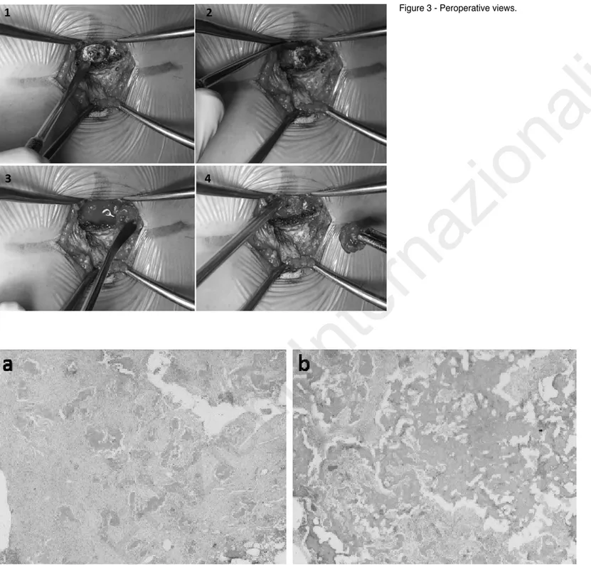
A 7-year-old female patient presented to our clinic with pain in the sacrococcigeal region. Our patient, who had pain for 2 years, had been operated due to the diagnosis of piloni-dal sinus at another center. But postoperative pain still did not pass. The pain was especially present at night. She suffered insomnia due to the pain mostly occurring during night hours, and started having psychological problems and treatment for it. Osteoid osteoma was considered for our patient and salicylate 100 mg was initiated and a dramatic response was obtained. On this, our patient had sacral CT scan and right S4-level posteriorly located osteoid osteoma was detected (Figure 1). CT-guided Kirschner wire marking was done under general anesthesia (Figure 2). Afterwards, the nidus in our patient who was taken to the operating ro-om was removed with open surgery (Figure 3). Histologi-cally, hematoxylin and eosin staining demonstrated diffe-rentiated osteoblasts lining the osteoid, and interconnected trabeculae of woven bone, which was compatible with oste-oid osteoma (Figure 4). Our patient’s pain just passed after surgery, and was discharged 1 day later. Our patient did not have any complaints. Insomnia and the psychiatric tre-atment were terminated.
Dıscussıon
Osteoid osteoma (OO) with a frequency of 2-3% seen among all bone tumors can be found in any skeletal structure (4). It is frequently localized to the long bones of the lower extremity. Spinal OO is rare (10%) (5), and present more of-ten in the posterior spinal elements. The region is most
com-A rare case of misdiagnosis: sacrococcygeal osteoid
osteoma that was treated surgically as a pilonidal
sinus in a pediatric patient
Case report
CCMBM 2 2018-4b XP-2018.qxp_- 05/10/18 17:52 Pagina 300
©
CIC
Edizioni
monly seen in the lumbar vertebrae (59%) followed by the cervical vertebrae (27%) in the thoracal vertebrae (12%) and at least in the sacrum (2%) (3). Only 1 of the 19 cases repor-ted in the literature on OO in sacrum was posteriorly locarepor-ted (6). In our case, the lesion is posteriorly located. The fact
Clinical Cases in Mineral and Bone Metabolism 2018; 15(2):300-303 301
A rare case of misdiagnosis: sacrococcygeal osteoid osteoma that was treated surgically as a pilonidal sinus in a pediatric patient
that the events reported in sacrum are mostly anterior regi-on, unlike other vertebrae’s , suggests that it is due to its uni-que shape (7). In clinical reflection, pain is an important fac-tor in the differential diagnosis of OO. Makley and Dunn (8) reported that the pain caused by OO was caused by abun-Figure 1 - Sacral CT scan of right S4-level pos-teriorly located osteoid osteoma (axial, sagittal and coronal views).
Figure 2 - CT-guided kirschner wire marking. CCMBM 2 2018-4b XP-2018.qxp_- 05/10/18 17:52 Pagina 301
©
CIC
Edizioni
dance of nodustic nerve fibers and excessive prostaglandin synthesis. In a study involving a large number of cases, it was found that 95% of the cases of indole was aggravated at night and that prostaglandins caused by pain were increased (9). Our patient had localized pain that became very severe at night. This caused our patient not to sleep at night and to suffer deformities such as redness, burning, swelling in the eyes from insomnia. The insomnia state was making the pa-tient restless and depressed by affecting the psychological state of our patient.
The operative treatment of the OO is the complete resection of the nidus. The CT guided resection was first reported by Voto et al. (10) who reported that this technique is useful for localization and removal of the nidus, and nowadays CT
gui-Clinical Cases in Mineral and Bone Metabolism 2018; 15(2):300-303 302
M. A. Çaçan et al.
dance is applied with treatment options such as trephine per-cutaneous excision (11) and CT guided perper-cutaneous radio-frequency ablation (RFA) (12). It has been reported that 16% recurrence is caused by inadequate ablation of RFA (6). Another deficiency in RFA is the lack of adequate tissue retrie-val for histological and pathological diagnosis (13). The use of RFA in lesions close to the nerve structure is limited in its use because it can cause the nearby nerve tissue to become war-med and thus damaged (13, 14). 15 of the 19 OO cases repor-ted in the sacrum were trearepor-ted with open resection. 4 cases were treated with resection in CT guideline (6). RFA was not considered in our case because it was very close to right S4 spinal root. It is appropriate to have resection treatment under the guidance of CT which gives good results.
Figure 3 - Peroperative views.
Figure 4 - a) Anastomosing, irregular trabeculae rimmed by single layer of osteoblasts in loose, fibrovascular stroma (HEx40); b) Sclerotic nidus of woven bone (HEx40).
CCMBM 2 2018-4b XP-2018.qxp_- 05/10/18 17:52 Pagina 302
©
CIC
Edizioni
Clinical Cases in Mineral and Bone Metabolism 2018; 15(2):300-303 303 A rare case of misdiagnosis: sacrococcygeal osteoid osteoma that was treated surgically as a pilonidal sinus in a pediatric patient
References
1. Jaffe HL. Osteoid osteoma: a benign osteoblastic tumor composed of os-teoid and atypical bone. Arch Surg. 1935;31:709-728.
2. Etemadifar MR, Hadi A. Clinical Findings and Results of Surgical Re-section in 19 Cases of Spinal Osteoid Osteoma. Asian Spine J. 2015 Jun;9(3):386-393.
3. Patnaik S, Jyotsnarani Y, Uppin SG, Susarla R. Imaging features of pri-mary tumors of the spine: A pictorial essay. Indian J Radiol Imaging. 2016 Apr-Jun;26(2):279-289.
4. Hakim DN, Pelly T, Kulendran M, Caris JA. Benign tumours of the bone: A review. J Bone Oncol. 2015 Mar 2;4(2):37-41.
5. Zenmyo M, Yamamoto T, Ishidou Y, Komiya S, Ijiri K. Osteoid osteoma near the intervertebral foramen may induce radiculopathy through tu-morous inflammation. Diagn Pathol. 2011 Jan 19;6:10.
6. Fukuda S, Susa M, Watanabe I, Nishimoto K, Horiuchi K, Toyama Y, Morioka H. Computed tomography-guided resection of osteoid osteoma of the sacrum: a case report. Journal of Medical Case Reports. 2014;8:206.
7. Schmitz A, Diedrich O, Schmitt O. Sacral osteoid osteoma-a rare cause
of back pain in childhood and adolescence. Klin Pediatr. 2000 May-Jun;212(3):110-112.
8. Makley JT, Dunn MJ. Prostaglandin synthesis by osteoid osteoma. Lancet. 1982 Jul 3;2(8288):42.
9. Cohen MD, Harrington TM, Ginsburg WW. Osteoid osteoma: 95 cases and a review of the literature. Semin Arthritis Rheum. 1983;12:265-281. 10. Voto SJ, Cook AJ, Weiner DS, Ewing JW, Arrington LE. Treatment of
os-teoid osteoma by computed tomography guided excision in the pediatric patient. J Pediatr Orthop. 1990 Jul-Aug;10(4):510-513.
11. Alemdar C, Çaçan MA, Dusak A, Özkul E, Atiç R, Kapukaya A. A com-parison of percutaneous trephine excision and open surgery in the treatment of osteoid osteoma. Int Orthop. 2016 Jul;40(7):1481-1487. 12. Garge S, Keshava SN, Moses V, Koshy G, Ahmed M, Mammen S, Mad-huri V. Radiofrequency Ablation of Osteoid Osteoma in Common and Technically Challenging Locations in Pediatric Population. Indian J Med Paediatr Oncol. 2017 Jul-Sep;38(3):302-305.
13. Ghanem I. The management of osteoid osteoma: updates and contro-versies. Curr Opin Pediatr. 2006 Feb;18(1):36-41.
14. Hadjipavlou AG1, Lander PH, Marchesi D, Katonis PG, Gaitanis IN. Min-imally invasive surgery for ablation of osteoid osteoma of the spine. Spine (Phila Pa 1976). 2003 Nov 15;28(22):E472-477.
CCMBM 2 2018-4b XP-2018.qxp_- 05/10/18 17:52 Pagina 303
©
CIC
Edizioni
Internazionali
View publication stats View publication stats