CASE REPORT
Progressive supra-aortic stenosis in a young adult
with the
findings of Singleton Merten Syndrome
Arda Ozyuksel, Cihangir Ersoy, Emir Canturk, Atif Akcevin
Department of Cardiovascular Surgery, Medipol University, Istanbul, Turkey Correspondence to Dr Arda Ozyuksel, [email protected] Accepted 15 August 2014 To cite: Ozyuksel A, Ersoy C, Canturk E, et al. BMJ Case Rep Published online: [ please include Day Month Year] doi:10.1136/ bcr-2014-205985
SUMMARY
Singleton Merten Syndrome is an autosomal dominant disorder of unknown origin. Patients often present with muscular weakness, failure to thrive, abnormal dentition, glaucoma, psoriatic skin lesions, aortic calcification and musculoskeletal abnormalities. In this case, we present a young girl with a history of aortic root replacement, who had an unusual progressive supra-aortic stenosis managed with urgent surgery during the course of the syndrome. Cardiovascular involvement needs special attention, since it is the major cause of mortality along with rhythm disturbances in the course of Singleton Merten Syndrome.
BACKGROUND
Singleton Merten Syndrome (SMS) is a genetically
inherited syndrome of unknown origin.1 It is an
autosomal dominant disorder with variable expres-sion of the symptoms with an onset during child-hood. In this case, we would like to present a
young girl withfindings of SMS who had a history
of operation for valvular aortic stenosis 3 years prior to admission to our clinic. We performed the replacement of the ascending aorta for severe and
progressive calcification leading to critical
supra-aortic stenosis.
CASE PRESENTATION
An 11-year-old girl was admitted to our hospital with recurrent aortic stenosis and syncope attacks. For the past 3 days, she had been experiencing attacks of dizziness, vertigo, deviation of the eyes and a presyncope-like situation with jerking and contraction of her arms, which lasted for a couple of minutes. Three years prior to her admission,
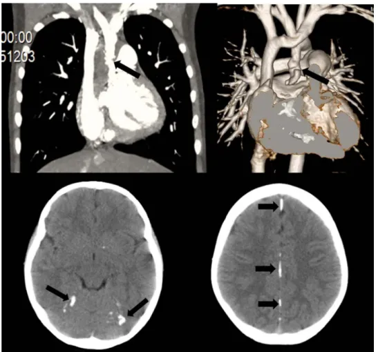
Figure 1 Critical supra-aortic stenosis at the ascending aorta (upper left side, arrow) at CT. Irregularity of the aortic
wall and a possible haematoma or dissection formation can be identified at the MRI (upper right side, arrow).
Scattered calcifications at the tentorium cerebelli and falx cerebelli at cranial CT are demonstrated in the lower images
(arrow heads).
Ozyuksel A, et al. BMJ Case Rep 2014. doi:10.1136/bcr-2014-205985 1
anterior aortic root enlargement procedure (Konno-Rastan operation) and replacement of the aortic valve with a 17 mm mechanical valve had been performed. The aortic valve had been reported to be at bicuspid morphology at that time. She
had been operated for glaucoma five times. Her parents
reported easy cavity formation in her teeth. Her incisor teeth were abnormally developed with double line including caries. Her physical examination revealed systolic murmur at the right side of the sternum. Neurological examination was normal except for vertical nystagmus at the left eye and weakness of the
lower extremity muscles. Transthoracic echocardiography
revealed a supravalvular critical aortic stenosis with a gradient of maximum 90 mm Hg and a mean of 55 mm Hg. Mechanical aortic valve was functioning normally with a mild stenosis.
Mitral valve leaflets were calcified and a mild mitral
insuffi-ciency and stenosis were noted. Cardiac MRI and CT were
per-formed and severe calcification at the ascending aorta leading to
a critical stenosis with a 7 mm passage at the narrowest point
was noted (figure 1). Cranial CT was performed, which revealed
severe calcifications at tentorium and falx cerebelli and bilateral
punctate calcifications at the basal ganglia (figure 1). Her total
and ionised blood calcium levels were 9.6 mg/dL and 1.16 mmol/
L respectively. An urgent operation was scheduled.
Re-sternotomy was performed following femoral arterial cannula-tion. Cardiopulmonary bypass (CPB) was established and low flow antegrade cerebral perfusion was initiated at 20°C. Aortotomy was performed at the ascending aorta, which was
heavily calcified with a pin hole antegrade flow (figure 2). The
ascending aorta was replaced with a 22 mm graft. The patient was weaned successfully from CPB on sinus rhythm. The post-operative course was uneventful, but episodes of severe
bradycar-dia attacks without any finding of convulsions were observed.
Although the patient was on sinus rhythm, on the 14th post-operative day a transvenous permanent pacemaker was implanted. The patient was subsequently discharged and was clinically doing
well, without any symptoms, in thefirst year after surgery.
OUTCOME AND FOLLOW-UP
The patient is free of symptoms in thefirst year after surgery.
DISCUSSION
SMS is a very rare clinical entity that is thought to be a disorder
of the calcium metabolism within a disturbed immune system.2
At its first description, the symptoms of abnormal dentition,
distal limb osteoporosis, marked calcification and intimal
weak-ening of the aortic arch and valve were mentioned, which were all present in our patient. Besides these landmarks of the syn-drome, our patient had glaucoma, psoriasis in the lower extrem-ities, muscle weakness and atypical facial appearance. Vertical nystagmus and preoperative convulsions were probably related
to central nervous system involvement, which included calci
fica-tions at the basal ganglion and tentorium cerebelli. Cardiac involvement and impaired cardiac function had been mentioned in a patient with aortic root replacement using pulmonary
auto-graft in the literature.3To the best of our knowledge, this is the
only case in the literature with aortic root enlargement due to severe valvular stenosis, followed by graft interposition of the ascending aorta for critical supra-aortic stenosis diagnosed
2 years after the first surgical intervention in a patient with
SMS. The development of severe supra-aortic stenosis and recent central nervous system involvement may imply the pro-gression of the disease in the puberty period.
Learning points
▸ Genetically inherited disorders of calcium metabolism may present with severe cardiovascular involvement.
▸ Urgent cardiovascular surgical interventions may be necessary in the natural course of these diseases.
▸ Multidisciplinary management of the acute exacerbations is mandatory in order to control the clinical outcome of complications.
Competing interests None. Patient consent Obtained.
Provenance and peer review Not commissioned; externally peer reviewed.
REFERENCES
1 Singleton EB, Merten DF. An unusual syndrome of widened medullary cavities of the metacarpals and phalanges, aortic calcification and abnormal dentition. Pediatr Radiol 1973;1:2–7.
2 Feigenbaum A, Müller C, Yale C, et al. Singleton-Merten Syndrome: an autosomal dominant disorder with variable expression. Am J Med Genet A 2013;161A:360–70. 3 Valverde I, Rosenthal E, Tzifa A, et al. Singleton Merten Syndrome and impaired
cardiac function. J Am Coll Cardiol 2010:56:1760.
Figure 2 Operative view indicating the severe calcifications and
dissection formation at the ascending aorta (white arrow). The previously replaced mechanical valve was functioning normally (black arrow).
2 Ozyuksel A, et al. BMJ Case Rep 2014. doi:10.1136/bcr-2014-205985
Copyright 2014 BMJ Publishing Group. All rights reserved. For permission to reuse any of this content visit http://group.bmj.com/group/rights-licensing/permissions.
BMJ Case Report Fellows may re-use this article for personal use and teaching without any further permission. Become a Fellow of BMJ Case Reports today and you can:
▸ Submit as many cases as you like
▸ Enjoy fast sympathetic peer review and rapid publication of accepted articles ▸ Access all the published articles
▸ Re-use any of the published material for personal use and teaching without further permission For information on Institutional Fellowships contact [email protected]
Visit casereports.bmj.com for more articles like this and to become a Fellow
Ozyuksel A, et al. BMJ Case Rep 2014. doi:10.1136/bcr-2014-205985 3
Rare disease
View publication stats View publication stats