REPUBLIC OF TURKEY BEZMIALEM VAKIF UNIVERSITY INSTITUTE OF HEALTH SCIENCES
INVESTIGATION OF DELAYED TYPE
HYPERSENSITIVITY RESPONSE AGAINST
CRIMEAN-CONGO HEMORRHAGIC FEVER
VIRUS NUCLEOPROTEIN
Nesibe Selma ÇETİNMASTER THESIS
Department of Biotechnology
SUPERVISOR
Prof. Dr. Mehmet Ziya Doymaz
TÜRKİYE CUMHURİYETİ BEZMİALEM VAKIF ÜNİVERSİTESİ
SAĞLIK BİLİMLERİ ENSTİTÜSÜ
KIRIM KONGO KANAMALI ATEŞİ VİRÜSÜ
NÜKLEOPROTEİNİNE KARŞI OLUŞAN
GECİKMİŞ TİP HİPERSENSİTİVİTE CEVABININ
ARAŞTIRILMASI
Nesibe Selma ÇETİN YÜKSEK LİSANS TEZİ
Biyoteknoloji Anabilim Dalı
DANIŞMAN
Prof. Dr. Mehmet Ziya DOYMAZ
Bu araştırma Bezmialem Vakıf Üniversitesi Bilimsel Araştirma Birimi tarafından desteklenmiştir.
ONAY
Kurum : Bezmialem Vakıf Üniversitesi Sağlık Bilimleri Enstitüsü Programın seviyesi : Yüksek Lisans ( ) Doktora ( )
Anabilim Dalı : Biyotoknolojı Tez Sahibi : Nesibe Selma Çetin Tez Başlığı : Kırım Kongo Kanamalı Ateşi Virüsü Nükleoproteinine Karşı Oluşan Gecikmiş Tip Hipersensitivite Cevabının Araştırılması
İmza
Jüri Bşk. (Danışman) Prof. Dr.Mehmet Ziya Doymaz ………
Bezmialem Vakıf Üniversitesi
Üye ... ... ……… Üye ... ... ……… Üye ... ... ……… Üye ... ... ………
Bu tez, Bezmialem Vakıf Üniversitesi Lisansüstü Eğitim ve Öğretim Yönetmeliği’nin ilgili maddeleri uyarınca yukarda belirtilen jüri üyeleri tarafından uygun görülmüş ve Enstitü Yönetim Kurulu’nun ……/……/……tarih ve ……/…… sayılı kararıyla kabul edilmiştir.
Prof. Dr. Mustafa Taşdemir Sağlık Bilimleri Enstitüsü Müdürü
APPROVAL
Institute : Bezmialem Vakif University, Institute of Health Sciences Level of Programme: Master ( ) Doctorate ( )
Department : Biotechnology Student : Nesibe Selma Cetin
Title of the Thesis: Investıgatıon Of Delayed Type Hypersensıtıvıty Response Agaınst Crımean-Congo Hemorrhagıc Fever Vırus Nucleoproteın
Signature
President of the Jury Prof. Mehmet Ziya Doymaz ………
Member: ... ... ……… Member : ... ... ……… Member: ... ... ……… Member: ... ... ………
This thesis was approved by the jury stated above in accordance with the related rules of the Postgraduate Education and Training Guide of Bezmialem Vakif University, and approved by Administrative Board with the decision dated ---/---/--- and numbered ---/---.
Prof. Dr. Mustafa Taşdemir Director of Institute of Health Sciences
BEYAN
Bu tezin kendi çalışmam olduğunu, planlanmasından yazımına kadar hiçbir aşamasında etik dışı davranışımın olmadığını, tezdeki bütün bilgileri akademik ve etik kurallar içinde elde ettiğimi, tez çalışmasıyla elde edilmeyen bütün bilgi ve yorumlara kaynak gösterdiğimi ve bu kaynakları kaynaklar listesine aldığımı, tez çalışması ve yazımı sırasında patent ve telif haklarını ihlal edici bir davranışımın olmadığını beyan ederim.
İmza İsim ve soy isim Tarih
ACKNOWLEDGMENT
I would like to express my gratitude to my supervisor, Prof. Mehmet Ziya Doymaz whose expertise, generous guidance, understanding and support in this study.
I am also grateful to my collegues, Msc. Elif Karaaslan, Msc. Merve Yazici and Sevde Hasanoglu whose support and contribution made this thesis real and to my family for their everlasting encouragment and moral, emotinal and financial support during my whole life.
I would like to dedicate this thesis to Yasin, my beloved husbund whose patience and sacrifices helped me to complete this work and to Yunus, my baby waiting to be born.
ÖZET
Kırım Kongo Kanamalı Ateşi Virüsü (KKKAV) bir arbovirüstür ve ixodid kenelerinde bulaşmaktadır. Kırım Kongo Kanamalı Ateşi Virüsünün yol açtığı Kırım Kongo Kanamalı Ateşi (KKKA) ise. Afrika, Asya, Güneydoğu Avrupa ve Ortadoğu’yu kapsayan çok geniş bir coğrafyada, %5 ile %50 arasında değişen fatalite oranına sahip bir hastalıktır. KKKAV Bunyaviridae ailesinin bir üyesi olup, Nairovirus cinsi altında sınıflandırılmaktadır. Virüs yaklaşık 90-100 nm çapında sferik viriona sahiptir. KKKAV genomu large (L), medium (M), and small (S) olarak adlandırılan üç tane negative sarmallı RNA segmentlerinden oluşmaktadır. RNA’ya bağlı viral RNA ploymerazı L segment tarafından üretilmektedir. S segment nükleoproteinleri, M segment ise Gn ve Gc proteinlerini kodlamaktadır. Viral replikasyonun ve transkripsiyonun konakçı hücrede başlayabilmesi için tüm segmentlerin nücleoproteinler (NP) tarafından sarmalanması gerekmektedir. KKKAV S segmenti yaklaşık 1.7 kb uzunluğunda olup 53 kDa ağırlığındaki nükleoproteini kodlamaktadır. KKKA genellikle hafif ve spesifik olmayan ateşli bir hastalık olarak rapor edilmiştir ancak bazı vakalarda çok şiddetli hemorajik hastalık gelişebilmektedir. Hastalığın 5 ile 14. günleri arasında devam eden hemoraj, çoklu organ bozukluğu ve şok ile sonuçlanabilmektedir. Hücre aracılı immünite kronik, kalıcı ve latent virüs enfeksiyonların seyrinde hayati önem taşımaktadır. Bu yüzden KKKAV enfeksiyonlarının hücresel immünite üzerindeki rolünü aydınlatmak oldukça önemlidir. Bu amaçla, çalışmamızda hayvan modellerinde KKKAV’ye karşı oluşacak DTH reaksiyonunun varlığının araştırılması hedeflenmiştir. Böylelikle KKKAV NP (1449 bç), NP’nin N-terminal parçası, NPNT (387 bç) ve NP’nin C-terminal parçası, NPCT (513 bç) rekombinant olarak prokaryotik ekspresyon sistemlerinde üretilerek DTH üzerindeki etkileri araştırılmıştır. Fare immunizasyon çalışmalarının ardından üç farklı dozda (50 µg/µl, 100 µg/µl, 200 µg/µl) antijen fare ayak tabanlarına enjekte edilmiş ve ayak tabanındaki şişme elektronik kumpas aleti ile ölçülmüştür. 24. saatte oluşan DTH cevabı her bir protein için en yüksek değeri göstermiş ve 72. saate kadar azalmıştır. Antijen enjekte edilen sağ ayak tabanındaki şişlik, kontrol grubuna kıyasla istatistiksel olarak anlamlı bulunmuştur (p<0,05). DTH reaksiyonu oluşturmak için 100 µg/µl ideal doz olarak saptanmıştır ve oluşan reaksiyonlar kıyaslandığında NP nin sırasıyla NPNT ve NPCT’ye göre daha antijenik olduğu gösterilmiştir.
ABSTRACT
Crimean–Congo hemorrhagic fever virus (CCHFV) is an arbovirus and transmitted both vertically and horizontally by ixodid ticks. Crimean–Congo hemorrhagic fever (CCHF) caused by CCHFV is reported in a wide geographic range including Africa, Asia, Southeast Europe and Middle East with a fatality rate from 5 to 50 %. CCHFV is a member of the family Bunyaviridae and classified in Nairovirus genus. Virus has spherical virions and a diameter of approximately 90-100 nm. CCHFV genome is composed of tripartite single-stranded negative RNA segments, called large (L), medium (M), and small (S) segment. RNA dependent viral RNA ploymerase is produced by L segment, S segment encodes nucleoproteins and M segment expresses Gn and Gc. To initiate the viral replication and transcription in the host cell, the segments are needed to be encapsidated by nucleoproteins (NP) .The S segment of CCHFV is approximately 1.7 kb long and encodes 53 kDa nucleoprotein (NP) made up of globular domain with a prominent arm. CCHFV infection is generally reported as a mild, nonspecific febrile illness but in some cases, severe hemorrhagic disease is also developed. Persisting hemorrhage, multi-organ failure and shock result in fatality on day 5-14 of illness. Cell mediated immunity (CMI) is of the essence resulting in chronic, persistent and latent virus infections. Therefore it is essential to clarify the role of cellular immunity in CCHFV infection. In this study, we aimed to investigate the presence of DTH reactions against CCHFV in animal model. Therefore, CCHFV NP (1449 bp), N-terminal part of NP, NPNT (387 bp) and C-N-terminal part of NP, NPCT (513 bp) were recombinantly produce in procaryotic expression systems and investigated their effect on DTH. Following immunization steps, antigens were injected in three different dosage (50 µg/µl, 100 µg/µl, 200 µg/µl) and footpad swelling was measured by SPI External Electronic Caliper Gages. For each protein DTH response has peaked at 24 hr and decreased by 72 hr. Footpad swelling was statistically significant on right hind pad injected with antigen for each groups, compared to negative control (p<0,05). 100 µg/µl was found to be optimum dose for DTH response and considering to DTH response NP was more antigenic than NPNT and NPCT, respectively.
TABLE OF CONTENTS
COVER... i INNER COVER... ii ONAY ... iii APPROVAL ... iv BEYAN... v ACKNOWLEDGMENT ... vi ÖZET... vii ABSTRACT... viiiLIST OF ABBREVATIONS AND SYMBOLS...xi
LIST OF FIGURES... xiii
LIST OF TABLES ... xv
1. INTRODUCTION... 1
1.1. Viral Hemorrhagic Fever ...1
1.2. Crimean-Congo Hemorrhagic Fever Virus ...1
1.2.1. History...2
1.2.2. Classification...3
1.3. Structure and genome...3
1.3.1. S segment...4 1.3.2. M segment ...5 1.3.3. L segment...6 1.4. Life cycle ...13 1.5. Transmisson of CCHFV ...14 1.6. Pathogenesis...16
1.7. Clinical features and treatment ...18
1.8. Treatment ...19
1.9. Delayed Type Hypersensitivity Response ...20
2. MATERIALS AND METHODS...21
2.1. Plasmid Constructions and Bacterial Hosts...21
2.1.1. Cloning vector for CCHFV NP...21
2.1.2. Preparation of competent cell...23
2.2. Transformation ...24
2.2.1. Analyzing positive transformants ...24
2.3. Construction Of Plasmids Expresssing CCHFV NP...26
2.3.1. TA cloning ...26
2.3.2. Directional cloning...27
2.4. Plasmid Isolation ...29
2.5. Expression...29
2.5.1. SDS-PAGE ...29
2.5.2. Scaling-up expression for purification ...30
2.6. His-tagged Protein Purification ...30
2.6.1. Protein concentration determination...31
2.8. Delayed Type Hypersensitivity...31
3. RESULTS ... 32
3.1. Production Of Recombinant NPs of CCHFV ...32
3.1.1. pET SUMO expression system...32
3.1.2. Purification of proteins tagged with 6xHis-SUMO ...33
3.1.3. SUMO cleavage...33
3.1.4. Expression of CCHFV NP proteins using pET28b vector...34
3.1.5. Purification of proteins tagged with 6xHis...35
3.2. Detection of Specific Antibodies Against Recombinantly Produced NPs...35
3.3. Recombinant CCHFV NPs Displays Cell Mediated Delayed Type Hypersensitivity In Mice...36
LIST OF ABBREVATIONS AND SYMBOLS
aa : Amino acids
APC : Antigen presenting cells
BCG : Bacille Calmette–Guérin
BSL-4 : Biosafety level 4
CCHF : Crimean–Congo hemorrhagic fever CCHFV : Crimean–Congo hemorrhagic fever virus
CFA : Complete Freund Adjuvant
CHF : Crimean hemorrhagic fever
CMI : Cell mediated immunity
DHF : Dengue hemorrhagic fever
DIC : Disseminated intravascular coagulation DTH : Delayed type hypersensitivitiy
DUGV : Dugbe virus
HAZV : Hazara virus
HFVs : Hemorrhagic fever viruses
hpi : Hours post infection
hRSV : Human respiratory syncytial virus HSV-1 : Herpes simplex virus type-1 ICAM1 : Leukocyte adhesion molecule 1
IFA : Incomplete Freund Adjuvant
IFN-γ : Interferon gamma
IFNs :Interferons
ISG15 : Interferon stimulated gene 15
KO : Knock out
LASV : Lassa virus
LB : Luria Bertani
MAbs : Monoclonal antibodies
MAVS : Mitochondrial antiviral-signaling protein MHC : Major histocompatibility complex
moDC : Monocyte-derived dendritic cells
MV : Measles virus
NP : Nucleoprotein
NS : Non-structural proteins
NSD : Nairo sheep disease
NSVs : Negative strand RNA viruses
ORF : Open reading frame
OTU : Ovarian tumor
RdRp :RNA- dependent RNA polymerase
RIG-I :Retinoic acid-inducible gene I
RNP : Ribonucleoprotein complexes
RVFV : Rift Valley Fever Virus
s.c. : Subcutaneous
SKI-1/S1P : Serine protease subtilisin-kexin isoenzyme-1/site-1-protease STAT-1 : Signal transducer and activator of transcription 1
SUMO : Small ubiquitin-related modifier
TCR : T-cell receptor
Th1 : Type 1 T-helper
Treg : Regulatory T cells
Ub : Ubiquitin
VCAM1 :Vascular cell adhesion molecule 1
LIST OF FIGURES
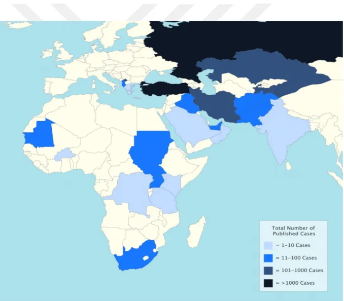
Fig 1-1. Number of reported cases of CCHF by country...1
Fig.1-2. Geographic distribution of CCHF according to WHO...2
Fig.1-3. Structure of a CCHFV virion ...4
Fig.1-4. CCHFV segment and their encoded proeins...4
Fig.1-5. Ribbon model of the CCHFV NP protein ...5
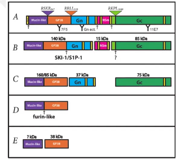
Fig. 1-6. Proteolytic process of CCHFV M segment...6
Fig. 1-7. OTU domain of CCHFV L protein deconjugates Ub and ISG-15 from cellular target proteins as a viral immune evasion mechanism ...7
Fig. 1-8. Sequence of CCHF L RNA segment-encoded proteins...9
Fig. 1-9. Life cycle of CCHFV. ...14
Fig.1-10. Geographic distribution and relatively incidence rate of Hyalomma spp. ticks ...15
Fig. 1-11. Routes of transmisson for CCHFV during the life cycle of Hyalomma spp. ticks ...16
Fig. 1-12. Pathogenesis of CCHF...17
Fig. 1-13. Escape mechanism of CCHFV from type I IFN response. ...17
Fig. 1-14. Clinical and laboratory course of CCHF ...19
Fig. 1-15. Development of DTH response on site of infection...20
Fig.1-16. DTH is directed by chemokines, cytokines and cytotoxins released by sensitized TH1 cells...21
Fig. 3-1. Plasmid constructs expressing CCHFV NPs...26
Fig. 3-1. Gel electrophoresis after PCR following transformation of plasmids into One Shot® Mach1TM-T1R competent E. coli...33
Fig 3-3. SDS page analysis after purification...33
Fig. 3-4. Unefficient SUMO cleavage of proteins with up to 4 units of SUMO protease .34 Fig. 3-5. Analysis of inserted CCHFV NP genes into pET28b plasmids by PCR...35
Fig. 3-6. Restriction analysis of isolated plasmids expressing NP...35
Fig. 3-7. Western blot analysis...36
Fig.3-8. DTH measurement of antigen injected footpad swelling ...37
LIST OF TABLES
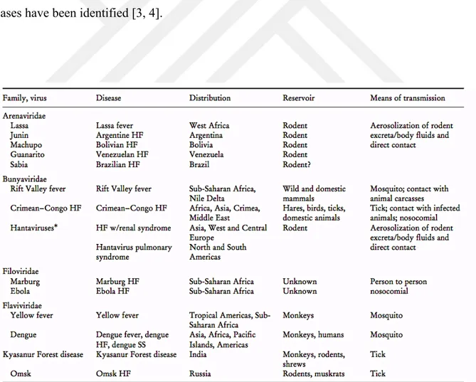
Table 1-1. The hemorrhagic fever (HF) viruses, their distribution, and principal mode
of transmission ...1
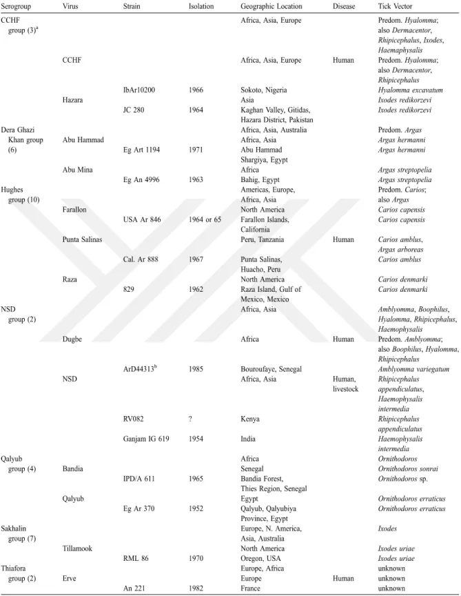
Table 1-2. Nairovirus serogroups and their host tick relationships ...4
Table 2-1. Time and temperature set up for PCR...24
Table 2-2. Primers used for TA cloning ...25
Table 2-3. Primers used in directional cloning ...27
1. INTRODUCTION
1.1. Viral Hemorrhagic Fever
The term of viral hemorrhagic fever (VHF) describes a virus-induced acute febrile, hemorrhagic disease reported from wide areas of the world. This terminology is first used by Russian and Japanese scientists in the 1930's to define typical clinical symptoms including fever and varying degrees of hemorrhage occurring in the Manchurian-Russian-Korean triangle of East Asia [1]. Hemorrhagic fever viruses (HFVs) are enveloped, single-stranded RNA viruses consisting of 14 viruses belong to four viral families: Arenaviridae, Bunyaviridae, Filoviridae and Flaviviridae (Table 1-1) [2]. Crimean Congo Hemorrhagic Fever Virus (CCHFV) belonging to Bunyaviridae family has become more of an issue for Turkey since when the first case of CCHF was announced in 2002 and until now over 6300 cases have been identified [3, 4].
1.2. Crimean-Congo Hemorrhagic Fever Virus
Crimean–Congo hemorrhagic fever virus (CCHFV) is an arbovirus and transmitted both vertically and horizontally by ixodid ticks, especially Hyalomma spp. ticks and some vertebrates like hares, hedgehogs, rodents, and birds serve as a reservoir for viral replication without displaying any symptoms. Crimean–Congo hemorrhagic fever (CCHF) is caused by CCHFV and occurs sporadically throughout much of Africa, Asia, Southeast Europe and Middle East with up to 50 % fatality rate (approximately %30) [5]. CCHFV is the most geographically disseminated tick-born virus among others and since the last decades the incidence (Fig.1-1) and geographic distribution (Fig. 1-2) of CCHF cases have remarkably increased [5, 6].
Fig 1-1. Number of reported cases of CCHF by country.The darkness of the coloration increases with case numer as defined by the key (5)
Even though CCHFV has very wide range of genetic variability with 20% sequence differences for the S segment and 31% for the M segment among virus isolates, viruses showing close genotypes can be present in far distinct regions and also distinct subtype of viruses can be found in the same region. The widespread dispersion of CCHFV could be explained by ticks carried on migratory birds. Indeed, birds migrating between Russia and Africa were counted as one of the reasons behind the Turkish epidemics and the other being the international livestock trade [7]. Moreover, broad host range of virus might cause the genetic variability by reassortment of genetic segments during co-infection of ticks or vertebrates, which enables to appearance of novel viruses[5].
Fig.1-2. Geographic distribution of CCHF according to WHO. Countries in red report more than 50 human cases, and those in orange report fewer than 50 cases. Countries in yellow have not reported human cases, but CCHFV has been isolated, or its presence has been inferred from serologic studies, and a transmission-competent tick vector is also present [5]
1.2.1. History
In 12th century, a persian physician Ismail Jorjani described a hemorrhagic disease caused by a louse or tick parasitizing a blackbird and in his book, Zakhireye Khwarazmshahi he called the disease as “kara khalak“ [8]. Hyalomma ticks are common vector for CCHF and frequently found on blackbirds. Currently, this disease considered to be CCHF according to his clinical and epidemiological descriptions [9]. In Uzbekistan, CCHF has also been denominated as khungribta (blood taking), khunymuny (nose bleeding), or karakhalak (black
death) for ages [10, 11] since 16th and 17th centuries the term ‘black dead’ entered European literature to refer to plague [12].
In modern terminology, CCHF has two historical background; firstly Crimean hemorrhagic fever (CHF) described in 1944-1945 when about 200 Soviet soldiers were infected in Crimea [13, 14]. In 1967, Chumakov and his colleagues in Moscow first isolated CHFV from newborn white mice [15, 16]. Then, isolation of virus by this method from a patient (Drosdov) in Astrakhan Drosdov led to use as a prototype strain for much experimental work all around the world. The researches working on viruses causing hemorrhagic fever discovered that CCHFV was antigenically indistinguishable from the Congo virus (Casals, 1969; Chumakov et al. 1969), originally isolated in 1956 from Congo [17, 18]. Afterwards, this virus began to be called CHF–Congo virus and later it was replaced with Crimean–Congo hemorrhagic fever virus [11].
1.2.2. Classification
CCHFV is a member of the family Bunyaviridae and classified in Nairovirus genus. Bunyaviridae also contains other 4 genus comprising over 350 arthropod-borne viruses; Orthobunyavirus, Phlebovirus, Hantavirus and Tospovirus [19]. The nairoviruses consist of 34 predominantly tick-borne viruses and are distinguished from other bunyaviruses due to size of their L segments [5]. They are divided into seven serogroups (Table 1-2) and predominantly cause disease in human [20, 21]. CCHFV and Hazara virus (HAZV) are listed in the CCHF serogroup. HAZV was firstly isolated from ticks on wild rodents in Pakistan and until now, there is no report about HAZV to cause disease in humans [22]. Because CCHFV is highly pathogen and require biosafety level 4 (BSL-4) laboratories for experimental works, HAZV can pave the way of research with antiviral agents against CCHFV [23].
1.3. Structure and genome
CCHFV has spherical virions and a diameter of approximately 90-100 nm [24].Viral envelope has two glycoproteins Gn and Gc in the lipid bilayer and they are recognized by cellular receptors to attach to the host cell surface (Fig 1-3.) [5]. CCHFV genome is composed of tripartite single-stranded negative RNA segments, called large (L), medium (M), and small (S) segment. These segments encode structural proteins: RNA dependent viral RNA ploymerase is produced by L segment, S segment encodes nucleoproteins and M
segment expresses Gn and Gc (Fig. 1-4). Each of the 3 segments contains single open reading frame (ORF) and terminal complementary sequences at 5’-UCUCAAAGA and at 3’AGAGUUUCU. These terminal sequences are conserved in all nairoviruses [25, 26]. This highly conserved ends might
Table 1-2. Nairovirus serogroups and their host tick relationships (81)
basebairs which provides functional promoter region for the binding of viral polymerase, and form of non-covalent closed circular structure of segments [5, 25, 27]. To initiate the viral replication and transcription in the host cell, the segments are needed to be encapsidated by nucleoproteins (NP) and by the RNA- dependent RNA polymerase (RdRp) [5].
Fig.1-3. Structure of a CCHFV virion which is spherical and approximately 90-100 nm in diameter. Virion consists of three single stranded negative RNA genome segment surrounded by nucleoproteins (NPs) complexed with the RNA dependent RNA polymerase (RdRp) and it is encapsidated by glycoproteins (Gn and Gc) in the lipid bilayer [5]
Also one or two non-structural proteins (NS) are expressed from some viruses in Bunyaviridae which called NSs and NSm encoded by S and M segment, respectively. They can act as an interferon antagonist, a regulator of replication and determinant of host range [28].Previously it was thought that, unlike other bunyaviruses CCHFV does not express any NSs or NSm but recent studies reports that NSm is cleaved during maturing process of glycoproteins encoded by M segment of CCHFV [29-32]. However, the function of this non-structural protein NSm in CCHFV is unkown [33]. Besides that S segment of CCHFV encodes also a non-structural protein NSs in positive sense [34]. A recent study of Barnwal et al. indicates that NSs induces apoptosis as activated by caspases due to mitochondrial membrane permeabilization [35].
Fig.1-4. CCHFV segment and their encoded proeins [5]
1.3.1. S segment
The S segment of CCHFV is approximately 1.7 kb long and encodes 53 kDa nucleoprotein (NP) made up of globular domain with a prominent arm comprising two long alpha helices containing a conserved caspase-3 cleavage site (Fig. 1-5) [35, 36]. The structural function of NP is the encapsidation of viral RNA due to homo-oligomerization to form ribonucleoprotein complexes (RNP). In addition to this, globular region of NP with viral polymerase (L protein) is critical for viral replication [5, 31]. Besides, the CCHFV polymerase activity is enhanced during viral vRNA transcription by the disruption of the caspase-3 cleavage site [37]. NP interacts also with actin which provides perinuclear localization of NP [38]. However, it is known that the interaction of the viral proteins with is actin filaments enhance replication of
the virus as is the case in paramyxoviruses such as Newcastle disease virus and Sendai virus [31, 39]
Fig.1-5. Ribbon model of the CCHFV NP protein. Helices are coloured as gray for N-terminal portion, dark blue for C-terminal portion of globular domain and purple for protruding arm. The red sphere indicates the N terminus, and the gold sphere indicates the C terminus. Yellow arrow shows the caspase-3 DEVD cleavage motif [35]
Interestingly, after structural analysis for CCHFV NP, it is concluded that CCHFV NP resembles Lassa virus (LASV) NP more than other bunyaviruses [35]. Furthermore, it is exhibited by phylogenetics analysis of L and NP sequences among segmented negative-strand RNA viruses (sNSVs) that nairoviruses are more closely related to arenaviruses than to any other bunyaviruses and as a result revision of current sNSV taxonomy might be essential [35].
1.3.2. M segment
As with other Bunyaviridae, CCHFV M segment is approximately 5.4 kb long [40] and translated into a single polypeptide of 1689 amino acids (aa) in length which is cotranslationally cleaved by the signal peptidase and post-transcriptionally modified in Golgi to form the type I transmembrane glycoproteins Gn (37 kDa) and Gc (75kDa), named for their relative proximity to the respective ends of the polyprotein, and a nonstructural protein NSm [29, 33]. Generation of the mature glycoproteins occur with a series of very complex
endoproteolytic events. Maturing process of 225 kDa M polyprotein begins with its cotranslational cleavage into the glycoprotein precursors PreGn (140 kDa) and PreGc (85 kDa) presumably by signal peptidase [29, 33]. N terminal of both Gn and Gc is generated by the serine protease subtilisin-kexin isoenzyme-1/site-1-protease (SKI-1/S1P) cleavage in the ER/cis-Golgi [41, 42]. After that, mucin-GP38 domain, named as GP85 when it is in monomer form and as GP160 when alternatively secreted as dimer, is cleaved by furin into GP38 and a mucin like domain [29, 41, 42] (Fig. 1-6). Mucin like domains are very unique for genera Nairovirus and notably hypervariable and rich in serine, threonine, and proline amino acids; heavily O glycosylated, which is similar to the mucin-like domain of the Ebola virus glycoprotein [43-45].
Fig. 1-6. Proteolytic process of CCHFV M segment. A) M polyprotein domains are coloured yellow for potential transmembrane domains; purple for mucin-like domain; orange for GP38;blue for Gn; pink for NSm and green for Gc. Signal peptidase cleavage sites are indicated by black arrows and furin-like (RSKR247), SKI-1/S1P (RRLL519), and SKI-SKI-1/S1P-like (RKPL1040) cleavage sites are illustrated by inverted triangles. B) PreGn and PreGc is cleaved by SKI-1/S1P and PreGc convertase (indicated arrows) the early secretory pathway. C) Generation of a nonstructural mucin-like GP38 protein of either 160 or 85 kDa, and the structural
glycoproteins Gn (37 kDa) and Gc (75 kDa). D) The mucin-like GP38 domain. E) Furin-like enzyme cleavage of GP85/GP160 into GP38 glycoprotein (38 kDa) and a mucin-like protein of unknown mass (? kDa) [29]
Secretion of Gn and Gc in trans-golgi is an interdependent process. Gn is indispensible for transportation of Gc from ER to golgi due to presumably having golgi localization signal on it [46-48] and also has a chaperone-like function for correctly folding of Gc [49]. However, Bertolotti-Ciarlet et al. has showed that only monoclonal antibodies (MAbs) against Gc, but not against Gn, were able to neutralize virus in vitro, suggested to be more important for infection [46]. Interestingly, a recent research has indicated that CCHFV Gn contains dual CCHC-type zinc finger motif on its C- terminal cytoplasmic tail, which can bind viral RNA [5, 50]
1.3.3. L segment
Like all nairoviruses, CCHFV L segment is about 12.2 kb in length, aproximately twice the size of those of other bunyaviruses. CCHFV L segment has single open reading frame comprising of 12164 nucleotides and encodes 3944 amino acid polyprotein, viral RNA-dependent RNA polymerase (RdRp), which has 62% nucleotide and amino acid identity with nairovirus Dugbe virus (DUGV) [40, 51]. L segment contains a high conservative ovarian tumor (OTU)-like cysteine protease domain domain (residues 35 to 152), a zinc finger type C2H2 domain (residues 606 to 632) and a leucine zipper motif (residues 1386–1407), suggests that CCHFV L polyproteins may have other functions than being a polymerase. In addition to this, a sequence in the CCHFV L is indicated as highly similar to a eukaryotic DNA topoisomerase I active site motif, like in DUGV L at amino acid position 76-94 (Fig. 1-8)[51].
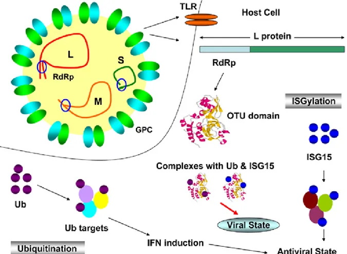
Viral OTU domains are present in eukaryotic, viral and bacterial proteins and functions as a core element of a cystein protease which can hydrolyze ubiquitin (Ub) and interferon stimulated gene 15 (ISG15) conjugates form target protein [52, 53]. Viruses capable to use this activity can evade two different cytokine pathways, IFNαβ and TNFα, in innate immunity as well as adaptive cellular immunity during MHC I and II antigen presentation [54, 55], TLR/IL1 signaling [56] and induction of type I IFN by the cellular viral sensor retinoic acid-inducible gene I (RIG-I) [53, 57]. Predictably, a study of Frias-Staheli et al. is reported that CCHFV L protein can deconjugate Ub and ISG-15 from cellular target proteins as a viral immune evasion mechanism (Fig. 1-7) [53]. Nevertheless, a minigenom replication assay is indicated that OTU domain containing CCHFV L protein does not
provide any evidence of autoproteolysis to generate additional protein products and OTU protease activity is dispensible for virus RNA replication [52].
Fig. 1-7. OTU domain of CCHFV L protein deconjugates Ub and ISG-15 from cellular target
Fig. 1-8. Sequence of CCHF L RNA segment-encoded proteins. The upper panel represents the predicted location of various sequence motifs within the entire CCHF L segment. In the lower panel, the alignment of the amino terminus of the L encoded protein of CCHF, Dugbe, and NSD (partial sequence) viruses are compared. Conserved positions are coloured in yellow. The highly conserved D, C, and H residues (blue) believed to constitute the OTU-like protease core amino acid
motif. The boxed region within the DUG virus sequence indicates the position of the predicted topoisomerase I active site motif which is also seen in both CCHF and NSD viruses [51]
1.4. Life cycle
The knowledge about cellular entry mechanism of CCHFV is currently deficient but Xiao and his collegues has identified that 180-300 amino acid residues of Gc has significant receptor binding capacity and the human cell surface nucleolin as a putative CCHFV entry factor (Fig. 1-9a) [59]. Simon and his collegues has indicated that CCHFV utilizes clathrin-dependent endocytosis, requires low pH for productive infection like other Bunyaviruses [60] and cholesterol in events following CCHFV binding and internalization [61]. Internalization of virus within the clathrin-coated vesicles occurs by fusing with endosomes and then lysosomes. Acidic environment in lysozyme induces conformational changes, which allow the virus to fuse with endosomal membranes (Fig. 1-9b) [62]. Moreover, Simon and his collegues has found that virus internalization was dependent on intact microtubules, and depolymerization of microtubules inhibited the expression of CCHFV RNA following progeny virus production. Within the first hour after infection, CCHFV is delivered by microtubules to the cellular sites where the viral transcription and replication will take place [63].
Replication starts with production of mRNA from negative sense viral RNA segment by virion associated viral polymerase in cytoplasm of host cell [64]. According to the study of Simon et al. about the kinetics of CCHFV RNA expression and synthesis in vitro, CCHFV positive sense RNA (mRNA and copy RNA ) are principally increased during the first 6 h of infection while negative sense RNA (vRNA) is kept constant till vRNA levels increased on 16 h post infection (hpi) in Vero E6 cells [63].
Bunyaviruses utilize generally same cap snatching mechanism for transcription with those of other arenaviruses and orthomyxoviruses [65] through approximately 11-15 nt capped RNA fragments used as primer to start transcription of nonpolyadenylated mRNA on the three viral RNA segments [37, 66]. CCHFV NP has unique endonuclease activity unlike other members of family. Even so, monomer form of CCHFV NP shows extremely low or no cap-binding affinity in vitro, compared to LASV NP which has highly structural smilarity with CCHFV [67]. Although cap-snatching mechanism of CCHFV is still unknown, it is reported that CCHFV NP superhelical polymer presents these RNA primer fragments, which leads to conformational change of subunits following liberation of monomeric NP. Thus, vRNA would be prepared for transcription and replication [37].
It is convinced that NP plays major role in viral replication. In order to start CCHFV viral replication, both interaction of NP with viral polymerase and enough NP should be present to encapsidate viral mRNA and genomic RNA to prevent degredation [68]. During microtubule dependent replication of CCHFV, actin filaments are involved in viral NP internalization to perinuclear region [38].
During the maturation of virions in the family of Bunyaviridae, the S, M, and L RNPs are packaged intracellularly into budding virions through cisternae within the Golgi structure [68-70]. In infected cells, N and L are translated on free ribosoms in cytosol and internalized to a non-Golgi perinuclear compartment via actin dependent mechanism [38, 71]. On the other hand, CCHFV NP proteins are found in Golgi at the late stage of viral assembly [68]. Syntesis of CCHFV glycoproteins is a multistep process. Primarily, a polyprotein from whole M segment is produced in ER. After SKI-1/S1P cleavage process, Gn is transported to Golgi complex by its Golgi localization signal in hydrophobic region of cytoplasmic tail while Gc is retained in ER until Gn is co-expressed, suggesting that the proper transport of two virion-forming glycoproteins is important for virus replication [29, 47]. Final assembled proteins in Golgi generate mature virions which are transported to the cell surface and released by secretory pathway toinfect other cells (Fig. 1-9j) [64]. Involvement of microtubules affects CCHFV entry, replication, assembly and egress [63].
Fig. 1-9. Life cycle of CCHFV. A)Viral attachment to surface receptor (unknown) B) Internalization through clathrin-dependent, receptor-mediated endocytosis. C) Fusion between the envelope and endosomal membranes by reduced pH in endosome D) Dissociation of the nucleocapsids and generation of mRNA and cRNA by RdRp. E) Translation of viral proteins and genomic vRNA production following new nucleocapsids formations. F) M polyprotein production in ER. G) Cleavage of olyprotein into Gn and Gc precursor forms transported to the Golgi complex H) Maturing process of glycoproteins, I) Formation of new virions. J) Virion egress [5]
1.5. Transmisson of CCHFV
CCHFV is an arthropod-borne virus and causes zoonosis despite circulating in an enzootic tick–nonhuman vertebrate–tick cycle and developing only a transient viremia without apparent of illness [5, 11]. At least 31 ticks species and one biting midge (Culicoides spp.) have been identified as CCHFV reservoirs [9, 11, 72]. But in fact, not all of these reservoirs serve as CCHFV competent vectors. Virus can be simply transmitted by a recent blood meal from a viremic host into them [9]. Hyalomma as a member of family Ixodidae is the predominant vector of CCHFV and posses a rigid shield or scutum unlike Argasidae containing soft ticks [40, 73]. Because of its wide geographic distrubition extending over southern Europe, a part of Middle East and Central Asia, H. marginatum is the most important vector for CCHFV [74] and commonly collected from humans and animals in endemic regions of Turkey (Fig. 1-10) [75]. H. marginatum rufipes (mainly in Africa) and H. anatolicum anatolicum are also common vector for CCHFV. The maintance of CCHFV can be achived by transstadially (from larva to nymph to adult) and interseasonally in several tick species and transovarially to the F1 generation (in some cases to F2) in Hyalomma marginatum marginatum, H. marginatum rufipes, Dermacentor marginatus and Rhipicephalus rossicus [11].
Transmission of CCHFV to tick vectors is occured principally during the spring and summer months, and is supported by viral replication in different host tissues reaching the highest titers in the salivary glands and reproductive organs during metamorphosis of competent tick vectors feeding on host blood [76]. CCHFV is vertically transmitted by adult females producing eggs and by adult males to females during copulation [5, 77-80].
All species maintain their life on at least two different hosts: larvae and nypmphs feed on the same individual host while feeding and molting or until dropping off, but adults feed on second host, particularly mammals. As an example, immature H. m. marginatum and H. m. rufipes feed on birds, hares and hedgehogs and adults on cattle and other large mammals, while H. a. anatolicum feed on domesticated mammals through their entire lifecycle [81]. Furtermore, some species feed on more than two host which means they drop off their host each molt. Hyalomma ticks are hunting ticks and their larvae and nymphs quest aggressively to feed on several hosts, including hedgehogs, hares and ground-feeding birds while the adults actively seek out sheep, cattle and other large animals including humans [5, 11].
Fig.1-10. Geographic distribution and relatively incidence rate of Hyalomma spp. ticks [82]
Like other naioroviruses, co-evolution of CCHFV with several tick species which have themselves evolved along with the vertebrates as sources of blood meals may contribute to the wide genetic diversity of the virus [5, 83].
Humans are actually not a main component of tick life cycle but actually a dead-end host. They are bitten occasionally by infected ticks when entered in enzootic area, which leads to CCHF in humans while other animals develop only asymptomatic transient viremia. Because of this, farmers, livestock owners and herders, abattoir workers, and veterinarians carry the highest risk of contracting CCHFV [7, 84-87]. CCHF can also caused by direct contact with the blood of infected animals and by contact with the virus-containing body fluids of a patient during the first 7–10 days of illness (Fig. 1-11) [5].
Fig. 1-11. Routes of transmisson for CCHFV during the life cycle of Hyalomma spp. ticks. Blue arrows indicate the course of the tick life cycle. After hatching, larvae feed on first hosts containing small mammals and birds. Some tick larvae and nyhmphs feed on two host while other species dropp off each molt (three-host ticks). This transmission is indicated by asteriks. Beside that, virus can be spread by ticks through co-feeding (dashed arrow). Then adult ticks take blood from large animal and after copulation virus is transmitted to eggs. Viral transmission to human can occur in different ways like infected ticks bites and direct blood contact with infected animals or humans, which is therefore marked as solid red arrows. The thickness of the red arrow indicates the efficiency of transmission [5].
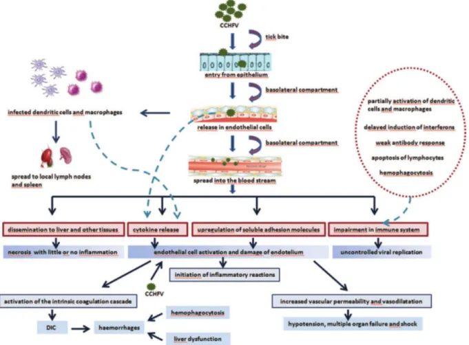
1.6. Pathogenesis
CCHFV pathogenesis is poorly understood because of necessity of BSL-4 laboratory conditions and lack of animal models. Blood analyses, autopsies, and liver biopsies of patients provide limited knowledge about pathogenesis [9].
Virus overcomes epithelium as a first barrier through tick bite. Basolateral membran reserving viral attachment proteins enable viral entry and viral release into the bloodstream, which causes systemic dissemination [88]. Internalization of the virus into host cells is mediated by the clathrin-, pH- and cholesterol-dependent pathway [61] by binding of the envelope glycoprotein Gc to cell-surface-associated receptors suggested human cell-surface nucleolin [59]. Upon activation of endothelial cells both directly and indirectly by cytokines like IL-1β, IL-6, and TNF-α released from dendritic cells, inflammatory responses is initiated and leukocytes are recruited [89, 90]. Upregulation of the leukocyte adhesion molecules ICAM1, vascular cell adhesion molecule 1 (VCAM1), and E-selectin increases vascular permeabilization [91]. Expression level of soluble ICAM1 (sICAM1) is correlated with elevated CCHFV titers and disease severity [92, 93]. After the viral proliferation in tissue
resident macrophages and dendritic cells, virus can be transmitted to local lymph nodes, spleen, and finally to systemic circulation of the host [88]. Excessive release of the cytokines named as cytokine storm have toxic effects on the endothelium causing to increased vascular
permeability, vasodilatation, multiple organ failure, and shock (Fig. 1-12) [94].
Fig. 1-12. Pathogenesis of CCHF. Endothelial cells are crossed over by CCHFV upon tick bite and are activated either directly by the virus or indirectly by virus-induced soluble mediators. Endothelial cell activation leads to the inflammatory reactions,increase of vascular permeability, and activation of the intrinsic coagulation cascade which then causes haemorrhages, hypotansion, multiple ogan failure and shock. CCHFV can be replicated uncontrollably by blocking several immune mechanism. DIC, disseminated intravascular coagulation [94]
According to animal studies, CCHFV replicates in blood on the first day of infection, in liver and spleen on the following day and then spreads systemically to lung, kidney, and finally to brain as a result of increased vascular permeability causing disruption of the blood– CSF barrier as seen on other viral hemorrhagic fevers [4, 95-98].
Connolly-Andersen and her colleagues reported that monocyte-derived dendritic cells (moDCs) and macrophages (MPs) were permissive for CCHFV replication following release of IL-6, Il-10 and TNF-α [99, 100]. TNF-α and IL-6 is highly correlated with disease
severity [101, 102]. Supernatants of infected DCs activated endothelial cells by up-regulating ICAM-1 expression [100]. Additionally, CCHFV-infected DCs also partially matured to an antigen-presenting cell as a result of inadequate upregulation of major histocompatibility
complex II (MHC II) priming of naive T cells [94, 99]. Hyperactivation of monocytes and macrophages leads to cytopenias, an excessive phagocytosis of blood cells [103, 104].
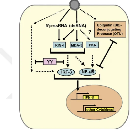
Fig. 1-13. Escape mechanism of CCHFV from type I IFN response. The monophosphorylate 5’ ends of the CCHFV ssRNA is recognized byRIG-I unlike other bunyaviruses. PKR is presumably activated,
either by trace amounts of dsRNA or by cell stress resulting from infection. IRF-3 is triggered by an unknown mechanism. CCHFV L protein containin OTU protease antagonizes the ntiviral effects of ISG15 and inhibits NF-kB-dependent signaling [105].
As a member of innate immune system, interferons (IFNs) are very important for a rapid and efficient antiviral response by limiting the spread of the infection. Upon viral
replication, secretion of type I IFNs (IFN-α and β) and subsequently upregulation of interferon-stimulated genes (ISGs) are induced [106, 107]. Therefore, they can regulate
inhibiton of viral protein expression, cell proliferation and apoptosis. However, IFN treatment at 1 hr after infection has no significant impact against CCHFV replication but it is effective to apply before infection [105]. MxA, a component of IFN-pathway is responsible for the inhibition of primer virus replication, which co-localize and interact with CCHFV nucleocapsid protein in the perinuclear region of infected cells [108, 109]. It is of interest to investigate, wheter CCHFV counteracts IFN signalling by several independent strategies. For example, CCHFV introduce viral ssRNA carrying 5’ monophosphate, not 5’ triphosphate triggering RIG-I signaling pathway in other bunyaviruses. This evasion mechanism can be explained by a possible phosphate cleavage during genome replication or by an anti-IFN factor like NSs of Rift Valley Fever Virus (RVFV) and La Crosse virus [110, 111]. Although triphospate on 5’ end is a characteristic motif for recognition by RIG-I, a recent study has reported that RIG-I recognizes CCHFV RNA, resulting in downstream type I IFN antiviral signaling and subsequent ISG stimulation [112]. On the other hand, the CCHFV OTU would block signaling by RIG-I and mitochondrial antiviral-signaling protein (MAVS) due to its deubiquitinase and deISGylase activities, which is almost seen in other nairoviruses targeting activated RIG-I to control innate immune signaling [113]. The OTU domain is a superfamily of ubiquitin (Ub)-deconjugating proteases and cleaves Ub and interferon stimulated gene product 15 (ISG15) from covalently conjugated proteins mediating innate antiviral responses. Frias-Staheli and her collegues showed that the CCHFV OTU protease deconjugate Ub and ISG15 from cellular target proteins and antagonizes the antiviral effects of ISG15 thereby inhibits NF-kB-dependent signaling ( Fig. 1-13) [53]. Beside its effect on NFkB signaling, protein ubiquitination plays also a critical role in the induction of adaptive cellular immune system such as MHC class I and II antigen presentation [54, 55, 114]. Deubiquitination might also affect other cellular processes subverted by viruses for their own advantage, such as the proteasome-mediated protein degradation system, multiple signal transduction events, or cell cycle progression[53].
1.7. Clinical features and treatment
CCHFV infection is generally reported as a mild, nonspecific febrile illness but in some cases, severe hemorrhagic disease is also developed. Clinical course of infection could be explained by four phases: incubation, prehaemorrhagic, haemorrhagic, and convalescence periods (Fig. 1-14) [11]. Although incubation period take 3-7 days following tick bite, period
of time can be decreased or increased depending on the route of exposure and viral load. The prehaemorrhagic period range from 1 to 7 days and reveals itself in fever (39–41°C) persisting for 4-5 days, headache, myalgia, and dizziness [7, 9, 11, 85, 115-117]. Diarrhoea, nausea, and vomiting are also reported in some cases [116, 117]. Hyperaemia of the face, neck, and chest, congested sclera, and conjunctivitis are also commonly observed in this period of disease. In case of asymptomatic course, RT-PCR would be performed for diagnosis [5, 6]. The haemorrhagic phase begins usually on day 3-5 and takes 2-3 days[6]. Common haemorrhagic manifestations are petechiae, conjuntivitis and large cutaneous ecchymoses as well as gastrointestinal, gingival, cerebral, vaginal, abdominal and urinary tracts bleeding [117]. About 30% of patients are subject to hepato- and splenomegaly [11]. Persisting
hemorrhage, multi-organ failure and shock result in fatality on day 5-14 of illness [5]. The convalescence period begins about 10–20 days if patients could survive CCHF but disease can take its effect up to a year [6]. However, such health problems after convalescent period does not reported by recently published articles from Turkey and Iran [85, 118].
Fig. 1-14. Clinical and laboratory course of CCHF DIC=disseminated intravascular coagulation [6]
1.8. Treatment
A number of studies have been reported on treatment of CCHF. Accordingly, a fully effective treatment is not attenable at the moment [119]. Because its initial nonspecific symptoms resemble myriad of other infections, CCHF patients can not be treated properly [120, 121]. Patients developing hypotension and hemorrhage are usually hospitalized for monitoring and supportive therapy including the administration of thrombocytes, fresh frozen plasma, and erythrocyte preparations. These supportive therapies are accompanied by laboratory tests such as complete while blood cells count, serum electrolytes and transaminases, renal function tests, and coagulation parameters [120, 122]. Initially, it is reported that a combination of high-dose methylprednisolone, intravenous immunoglobulin and fresh-frozen plasma was effective to cure CCHF [5, 123].
In recent publications, ribavirin is debated as an effective antiviral agent and widely administered [5]. Furthermore, its inhibitor effect on CCHFV replication has been noted in a minigenome system [52], in virus-infected cells [124-126], in newborn mice [127] and in signal transducer and activator of transcription 1 (STAT-1) KO mice [4]. These effects were noted in a concentration-dependent manner both in vivo and in vitro [5]. Detailed reviews about ribavirin therapy concluded that ribavirin is beneficial in CCHFV infections despite the lack of inscrupulous evidence [120, 128]. For example, a meta analysis has found no significant correlation between survival rate, length of hospitalization or need for blood perfusion and ribavirin treatment [129].
Passive immunotherapy has been carried out by injecting of anti-CCHFV antibodies prepared from convalesent patients, intramuscularly or intravenous for prophylactic and therapeutic purposes, particularly for high-risk patients [130-132].
1.9. Delayed Type Hypersensitivity Response
During the initial course of viral haemorrhagic fever infections, immune system mount a significant inflammatory response leading to disseminated intravascular coagulation following haemorrhage, multiple organ failure and septic shock [133].
The delayed type hypersensitivitiy (DTH) response is known to occur during the repeated exposure to the antigens. Consequently, the role of DTH response played during the
initial encounter with CCHFV is currently unknown. However, an essential arms of protective immunity against a microbial agent usually contain an effective DTH response. Furthermore, DTH reactions is usually helpful in gauging the breadth of T cell responsiveness [134-136].
Delayed type hypersensitivity response is primarily a function Th1 type response where CD4+ T lymphocytes are activated. This response sometimes could be to the detriment of the host and there are many example of viral, bacterial and parasitic infections where the DTH response against microbial antigens trigger tissue destruction causing harm to the host [137]. For this intial response to role played during granulomas, edemas, and exudates are formed after induction of DTH mediators and in case of intensive response not only tissues and small vessels but also larger vessels are damaged, which causes hemorrhages [137].
In this study, we aimed to investigate the presence of DTH reactions against CCHFV in animal model.
Fig. 1-15. Development of DTH response on site of infection. Following secondary exposure to antigen, TH1 cells recognizes processed antigen by APCs and release cytokines acting on vascular endothelium. Recruiting an inflammatory cell infiltrate causes the accumulation of fluid and protein which forms visible lesion [138]
Delayed type hypersensitivity (DTH) is initially described by Koch as tuberculin test used to determine whether an individual has previously been infected with Mycobacterium tuberculosis [138]. Following exposure, mycobacterial antigen is processed by antigen presenting cells (APC) and introduced by major histocompatibility complex (MHC) class II molecules to CD4+ lymphocytes and subsequently circulating sensitized memory T cells are generated [139]. After intradermal injection of small amounts of tuberculin derived from M. tuberculosis, a local T cell-mediated inflammatory reaction will be develop over 24–72 hours for individuals exposed to the bacterium, either by infection with the pathogen or by immunization with BCG, an attenuated form of M. tuberculosis [138]. This response is mediated by TH1 cells and several inflammatory cytokines are secreted to recruit and activate macrophages and other non-specific inflammatory cells (Fig. 1-16) [140]. Stimulated expression of adhesion molecules on endothelium leads to increase local blood vessel permeability, allowing plasma and accessory cells to enter the site; this causes a visible swelling (Fig. 1-15) [138]. Each of these phases takes several hours and so the fully developed response appears only 24–48 hours after challenge.
Fig.1-16. DTH is directed by chemokines, cytokines and cytotoxins released by sensitized TH1 cells [138]
2.
MATERIALS AND METHODS
2.1. Plasmid Constructions and Bacterial Hosts 2.1.1. Cloning vector for CCHFV NP
CCHFV Kelkit’06 NP (1449 bp) retrieved from GenBank (Accession no. GQ337053) and their estimated antigenic sequences by Kolaskar Tongaonkar (Kolaskar ve Tongaonkar, 1990; Immune Epitope Database and Analysis Program- http://tools.immuneepitope. org/tools/bcell/ iedb_ input0), NP124-507 (390 bp) and NP886-1392 (513 bp) were optimized for bacterial expression, synthesized and cloned into pUC19 by GenScript®. They were transformed into chemically competent DH5 E.coli cells.
1) NP (1449 bp) sequence
5’ atg gaa aac aaa att gaa gtg aat agc aaa gat gaa atg aac aaa tgg ttc gaa gaa ttt aaa aaa ggc aac ggt ctg atg gat acg ttt acc aac agc tat tct ttc tgc gaa aac gtt ccg aat ctg gat aaa ttt gtc ttc cag atg gca agt gct acg gat gac gcc caa aaa gac agc att tat gcg tct gcc ctg gtg gaa gcc acc aaa ttt tgc gca ccg att tac gaa tgt gcg tgg gtg agc agc acc ggc atc gtt aaa aaa ggt ctg gaa tgg ttc gaa aaa gat agt ggc acc att aaa tcc tgg gac gaa aac tat gct gaa ctg aaa gtg gat gtt ccg aaa atc gaa cag ctg gca aat tac cag caa gcg gcc ctg aaa tgg cgc aaa gat att ggc ttt cgt gtt aac gcc aat acc gca gct ctg agc aac aaa gtc ctg gca gaa tat aaa gtg ccg ggt gaa atc gtc atg agc gtg aaa gaa atg ctg tct gat atg att cgt cgc cgt aac ctg atc ctg aat cgc ggc ggt gac gaa aat ccg cgc ggt ccg gtt tca cgt gaa cat gtc gaa tgg tgt cgt gaa ttt gtg aaa ggc aaa tac att atg gcc ttc aac ccg ccg tgg ggt gat att aat aaa agt ggc cgc tcc ggt atc gca ctg gtt gct acc ggc ctg gcg aaa ctg gcc gaa acg gaa ggc aaa ggt gtc ttt gat gaa gcg aag aaa acc gtg gaa gcg ctg aat ggt tat ctg gat aaa cat cgc gat gaa gtg gac aaa gca tca gct gac tcg atg att acg aac ctg ccg aaa cac atc gcg aaa gcc cag gaa ctg tac aaa aat agt tcc gca ctg cgt gct cag ggc gcg caa atc gat acc ccg ttt tca tcg ttc tat tgg ctg tac aaa gcg ggt gtt acg ccg gaa acc ttt ccg acg att tct cag ttt ctg ttc gaa ctg ggc gaa caa ccg cgt ggc acc aag aaa atg aaa aaa gcg ctg ctg tca acg ccg atg aaa tgg ggc aaa aaa ctg tat gaa ctg ttt gcc gat gac tcg ttc cag caa aac cgc atc tac atg cat ccg gct gtg ctg acc gcg ggt cgt att agt gaa atg ggc gtg tgc ttc ggt acg atc ccg gtt gca aac ccg gat gac gca gca cag ggc tca ggt cac acc aaa tcg att ctg aat ctg cgt acc agc acc gaa acc aac aat ccg tgt gcg aaa acc att gtc aaa ctg ttt gaa atc cag gaa acg ggc ttc aat att caa gat atg gac atc
gtt gca agc gaa cat ctg ctg cac cag agt att gtc ggc aaa cag tcc ccg ttt caa aac gcg tat aat gtt aag ggt aac gcc acc tct gca aat att atc taa 3’
2) NP124-507 (390 bp) sequence
5’ atg aac ctg gat aaa ttc gtg ttc caa atg gcc tcc gca acc gat gac gct caa aaa gac tca atc tac gcc tca gcc ctg gtc gaa gcg acc aaa ttt tgc gcc ccg att tat gaa tgt gcg tgg gtt agc tct acg ggc atc gtc aaa aaa ggt ctg gaa tgg ttc gaa aaa gat agt ggc acc att aaa tcc tgg gac gaa aac tat gcg gaa ctg aaa gtg gat gtt ccg aaa atc gaa cag ctg gcc aat tac cag caa gcg gcc ctg aaa tgg cgt aaa gac att ggc ttt cgc gtc aac gca aat acg gca gct ctg agc aac aaa gtg ctg gct gaa tac aaa gtt ccg ggt gaa atc gtc atg agc gtg aaa gaa atg taa 3’
3) NP886-1392 (513 bp) sequence
5’ atg gcc ctg cgt gct caa ggc gct caa atc gac acc ccg ttc tca tca ttc tat tgg ctg tat aaa gct ggc gtg acc ccg gaa acc ttt ccg acg att tcc cag ttt ctg ttc gaa ctg ggc gaa caa ccg cgt ggc acc aag aaa atg aaa aaa gcg ctg ctg tca acc ccg atg aaa tgg ggc aaa aaa ctg tat gaa ctg ttt gcc gat gac tcg ttc cag caa aac cgt att tac atg cat ccg gca gtt ctg acg gct ggt cgc att agc gaa atg ggc gtg tgc ttc ggc acc atc ccg gtt gca aac ccg gat gac gca gca cag ggc agc ggt cac acc aaa tct atc ctg aat ctg cgc acc agc acg gaa acc aac aat ccg tgt gca aaa acg att gtc aaa ctg ttt gaa atc cag gaa acc ggc ttc aat att caa gat atg gac atc gtc gct tct gaa cat ctg ctg cac cag agt atc gtg ggt aaa caa tcc taa 3’
2.1.2. Preparation of competent cell
All the cells used for transformation for this assay were plated on Luria Bertani (LB) agar (1.0% Tryptone, 0.5% yeast Extract, 0.5% sodium chloride, 1.5% agar ) and a single colony from overnight grown culture were inoculated into 25 ml LB (1.0% tryptone, 0.5% yeast Extract, 0.5% sodium chloride) in a 250 mL bottle and incubated at 37°C for 4-6 hours. Cells were chilled for 10 minutes and harvested at 4100 rpm for 10 minutes at 4°C. Pellets were gently resuspended in 10 ml cold 0.1M CaCl, incubated on ice for 20 minutes and centrifuged at 4100 rpm for 10 minutes at 4°C. After resuspension in 5mL cold 0.1MCaCl/15% glycerol, cells were dispensed in microtubes (300μL/tube) and frozen in -80°C for storage.
2.2. Transformation
One µl isolated plasmid DNA (100 pg) or 5 µl ligation mixture were gently added into competent cells and incubated on ice for 10 minutes. Cells were heat shocked at 42°C for 2 minutes, immediately transfered on ice and incubated for 10 minutes. 250 µl S.O.C medium 2% tryptone, 0.5% yeast extract, 10 mM sodium chloride, 2.5 mM potassium chloride, 10 mM magnesium chloride, 10 mM magnesium sulfate and 20 mM glucose) was added to the mixture and the cells were incubated at 37°C with shaking 220 rpm for 1 hour. Following incubation, different amount of culture was spread on a prewarmed LB agar plate supplemented with relative antibiotics and incubated overnight at 37°C.
2.2.1. Analyzing positive transformants
Antibiotic selection
Transformed cells were plated on LB agar with appropriate antibiotic. For the cells containing pUC19 vectors, ampicilin were used to a final concentration of 100µg/ml while 50µg/ml kanamycin were added to LB agar to select the cells containing pETSUMO and pET28b expression vectors.
Polymerase Chain Reaction
Isolated plasmids (see Subheading 2.4) were used as template for CCHFV NP specific PCR. The reaction was conducted in Veriti® 96-Well Thermal Cycler (Applied
Biosystem) using cycling conditions as outline in Table 2-1. Two and half µl isolated plasmid was mixed with 2.5 µl 1 µM primers (Table 2-2) and 50 µM dNTPs in 1 µl (D7295, SIGMA), 5 µl 10X PCR Buffer (P2192, SIGMA), 0.5 µl 1u Taq polymerase (D6677, SIGMA) and 1.5 µl 25mM MgCl2 (M8787, SIGMA) were added into 50 µl PCR mixture. Tm was calculated using OligoAnalyzer3.1 (Integrated DNA Technologies, Inc, Coralville, IA, USA).
Table 2-1. Time and temperature set up for PCR
Step Number of cycles Phase Duration Temperature
1 1 Initial melt 5 min 95 °C
2 25 Melt 30 s 95 °C
3 25 Annealing 30 s 52 °C
4 25 Extension 30 s 72 °C
5 1 Final extension 10 min 72 °C
Table 2-2. Primers used for TA cloning
Name Sequence Target gene
F1 5’atg gaa aac aaa att gaa gtg 3’
R1 5’ tta gat aat att tgc aga ggt g 3’
NP
F2 5’ atg aac ctg gat aaa ttc gtg ttc c 3
R2 5’ tta cat ttc ttt cac gct cat gac 3’
NPNT
F3 5’ atg gcc ctg cgt gc 3’
R3 5’ tta gga ttg ttt acc cac g 3’
Restriction analysis
Analysis of inserts directionally cloned into pET28b were performed by using restriction enzymes. To cut 1µg plasmid DNA, 0.2 µl XhoI (10 u/µl), 0.2 µl NcoI (10 u/µl) and 10X Buffer (CutSmart® Buffer, New England Biolabs, Inc.) were mixed in a total volume of 20 µl. The mixture was incubated at 37°C for 1 hour and enzymes were inactivated at 80°C for 20 minutes.
Sequencing
Before starting to expression all positive transformants were analyzed by sequencing performed by Prof. Dr. Ali Osman Kilic at Karadeniz Technical University.
2.3. Construction Of Plasmids Expresssing CCHFV NP
To investigate CCHFV NP induced DTH response, 3 complete NP: 1449 bp, N-terminal part of NP, NPNT: 387 bp and C-N-terminal part of NP, NPCT: 513 bp) were separately amplified by PCR and cloned into Champion™ pET SUMO expression vector (Thermo Fisher Scientific). The cloning was TA based cloning system, which allows to express gene of interest as a fusion to N-terminal polyhistidine (6xHis) tag and SUMO protein for increased expression and solubility of recombinant fusion proteins and generation of native protein following cleavage by SUMO Protease. CCHFV NP and its both N- and C-terminal parts were also cloned into pET28b (kindly provided by Asst. Prof. Esen Bakhautdin, Fatih University, Istanbul) by using restrictiction enzymes in a directional cloning strategy.
Fig. 3-1. Plasmid constructs expressing CCHFV NPs. Gene of interests were inserted A) downstream of SUMO protein following N-terminal 6XHis tag into pETSUMO expression vector via TA cloning B) by using
restriction enzymes XhoI and NcoI before C-terminal 6XHis tag into pET28b vector via directional cloning.
2.3.1. TA cloning
To add single deoxyadenosine (A) to the 3 ́ends of PCR products for properly TA cloning, HOT FIREPol® DNA polymerase (Solis Biodyne) with proofreading activity was used primarily among the extension period during repeated cycles, then a Taq polymerase lacking 3’→ 5’ exonuclease activity (D1806, SIGMA) and100 µM adenine were added and incubated at 72C for 10 minutes in final extension. Primers used in PCR are listed in Table 4. PCR products were directly ligated with pET SUMO vector to avoid degradation of the single 3 A-overhangs over time. For ligation, 1:3 molar ratio of vector:insert was mixed with 10X Ligation Buffer and 4 Weiss units T4 DNA Ligase (both included with kit, K300-01, Thermo Fisher Scientific) and incubated at 15C for overnight. Afterwards, all ligation reaction was transformed as described in subheading 2.2 into One Shot® Mach1TM-T1R Chemically Competent E. coli cells (C8620-03, Thermo Fisher Scientific) used only for plasmid propagation. After cloning, plasmids are named as pBVU1 for complete NP sequence, pBVU2 for N-terminal part of NP and pBVU3 for C-terminal part of NP. Plasmids acquired from One Shot® Mach1TM-T1R E. coli were transformed as described in subheading 2.2 into BL21(DE3) One Shot® Chemically Competent E. coli (C6000-03, Thermo Fisher Scientific).
A B
2.3.2. Directional cloning
Cloning was achieved by using XhoI and NcoI restriction enzymes. Restriction sites were introduced into PCR products by primers (Table 2-3). PCR was accomplished by mixing 2.5 µl template DNA with 1µM primers and 10µl 5x HOT FIREPol® Blend Master Mix (04-25-00120, Solis Biodyne) in 50 µl reaction volume. The reaction was conducted in Veriti® 96-WellThermal Cycler using cycling conditions as outline in Table 2-4.
Table 2-3. Primers used in directional cloning
Name Sequence Target gene
Forward_NcoI_12 5’ ccg gcc atg gaa aac aaa att g 3’
Reverse_XhoI_13 5’ cgg cgc tcg agg ata ata ttt gc 3’ NP
Forward_NcoI_14 5’ ccg gcc atg gac ctg gat aaa ttc 3’
Reverse_XhoI_15 5’ cgg cgc tcg agc att tct ttc 3’ NPNT
Forward_NcoI_16 5’ ata tcc atg gcc ctg cgt g 3’
Reverse_XhoI_17 5’ cgt gac tcg agg gat tgt tta cc 3’ NPCT
Table 2-4. Reaction conditions set up for PCR
Step Number of cycles Phase Duration Temperature
1 1 Initial melt 15 min 95 °C
2 2 Melt 15 sec 95 °C 3 2 Annealing 30 sec 42 °C 4 2 Extension 1 min 72 °C 5 30 Melt 15 sec 95 °C 6 30 Annealing 30 sec 55 °C 7 30 Extension 1 min 72 °C
Restriction digestion
Sticky ends on 5’ ends of PCR products and pET28b required for cloning were generated with XhoI (R6379, SIGMA) and NcoI (R0193S, New England Biolabs) enzymes. Briefly, 1µg plasmid DNA or PCR products eluted from gel was digested with 5 units of each enzyme (XhoI and NcoI) and 10X Buffer (CutSmart® Buffer, New England Biolabs, Inc.) at 37C for 1 hour and enzymes were inactivated at 80C for 20 minutes.
Gel elution
Following polymerase chain reaction (PCR) or restriction digestion samples were electrophoresed on a 1% agarose gel at 100V for 45 minutes. The correct size band was excised and DNA was purified using GenEluteTM Gel Extraction Kit (NA1111, SIGMA). Gel slices were solubilized in 3 gel volume of the Gel Solubilization Solution by incubating at 50°C and vortexing briefly every 2-3 minutes until the gel was completely dissolved; the DNA was precipitated with 1 volume of 100% isopropanol (Merck); the solution loaded onto a prepared binding column (56500, SIGMA) and centrifuged at 16000xg for 60s; the column was washed with 700μL Wash Solution and dried by centrifugation at 16000xg for 60s and eluted in 50μL of Elution Buffer. All buffer used in gel elution procedure was included with kit.
Ligation
Compatible cohesive ends of DNA fragments generated by restriction enzymes, XhoI and NcoI were ligated using Rapid DNA Ligation Kit (K1422, Thermo). Briefly, 200 ng linearized vector was mixed with 180 ng insert, 5X Rapid Ligation Buffer and 5 units of T4 DNA Ligase. The reaction mixture was incubated at 22°C for 5 min. Then, 5 µl ligation reaction was used for transformation into One Shot® BL21 Star™ (DE3) Chemically Competent E. coli (C6010-03, Thermo Fisher Scientific) as described in subheading 2.2.
2.4. Plasmid Isolation
Positive transformants were inoculated in 2 ml LB and incubated overnight at 37C with vigorous shaking. Plasmid isolation was accomplished using QIAprep Spin Miniprep kit (27104-Qiagen). Overnight culture was pelleted by centrifugation at 8000 rpm for 23 minutes; the bacterial pellet was resuspended in 250μL P1 buffer (50 mM Tris hydrochloride,