Repair of Recurrent Patent Ductus Arteriosus in an Adult with Cardiopulmonary Bypass
Tam metin
Şekil

Benzer Belgeler
1. Rohit MK, Thingam SK, Gopal S, Vuppaladadhiam H, Grover A. Coarc- tation of aorta with intercostal artery aneurysm and patent ductus arteriosus. Kino K, Sano S, Sugawara E,
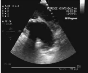
1 Department of Cardiology, Beytepe Military Hospital, Ankara-Turkey Video 1. A) Two-dimensional transthoracic echocardiography reve- aling dilated right atrium and ventricle,
©Copyright 2013 by AVES Yay›nc›l›k Ltd. A) Suprasternal window of TTE showing a linear structure at the descending aorta (yellow arrow) and patent ductus arteriosus under
Video 6: Midözefagial renkli Doppler görüntülemede mitral kapakta ileri düzeyde ekzantrik mitral yetersizliği izlenmektedir.. Murat Sünbül, Tarık Kıvrak,
Because of this type of connection is seen uncommonly at older ages and the patient was asymptomatic, selective aortic angiography was performed and the small PDA was seen between
In patients with patent ductus arteriosus (PDA) infective endarteritis is an important reason for hospital admission, with a higher incidence of 4,8 patients / 1000 hospital
Endarteritis associated with a clinically silent patent ductus arteriosus.. Silent patent ductus
Reopening was observed in only one case who received in- domethacine at 5th day of life. Although ductal closure was ob- tained at 8th day of life, a PDA recurrence was observed at
