The Effect of Thyroid Dysfunction and Treatment on Fat Tissue Adiponectin Levels in Rats
Page | 3790
The effect of thyroid dysfunction and treatment on fat tissue adiponectin levels in rats
Emine Atıci
1, Rasim Mogulkoc
2,*, Abdulkerim Kasim Baltaci
2, Esma Menevse
31Okan University, Health Sciences Faculty, Istanbul-Turkey
2Selcuk University, Medical Faculty, Department of Physiology, 3-Biochemistry Konya-Turkey
*corresponding author e-mail address: [email protected] ABSTRACT
Adipose tissue is a hormonal active system that produces and releases leptin, adiponectin and resistin. Seen from this aspect, it may be thought that adiponectin may interact with the thyroid axis. Thyroid function disturbance led to changes on the body weight, muscle volume and adipose tissue. Experimental findings show that adiponectin and thyroid hormones have biological interactin with each other. However, various results have been reported a relationship between thyroid hormones and adiponection levels. The aim of the present study was to determine adipose tissue adiponectin levels in rats with thyroid dysfunction and after treatment. Study was performed on 40 Sprague-Dawley male rats. Groups were designed as 1-control, 2-hypothyroidism (Hypo), 3- hypothyroidism + Thyroxine, 4-hyperthyroidism (Hyper), 5-hyperthyroidism + PTU. At the end of 3 weeks experimental period, adipose tissues of rats were analyed for adiponection by Elisa. Experimental hypothyroidism reduced adipose tissue adiponectin levels, but hyperthyrodism increased. Treatment of hypothyroidism and hyperthyroidism corrected disrupted adiponectin levels to control. The results of the present study show that experimental thyroid dysfunction has affected adipose tissue adiponection levels in rats.
Keywords: hypothyroidism, hyperthyroidism, adiponectin, rat.
1. INTRODUCTION
Until recently, adipose tissue was known just like an oil reservoir. However, it has been postulating as an endocrine and metabolic organ. Over the last two decade, it has been recognized that adipose tissue has endocrine, paracrine and autocrine effects and also fat tissue have important functions such as secretion of growth factors that affect cytokines and local fat tissue and different organs /tissues (CNS, liver, pancreas and skeletal muscles) [1]. Adipose tissue is a hormonal active system that produces and releases different bioactive substance such as leptin, adiponectin and resistin [2]. Leptin, adiponectin (ADP), vaspin, visfatin, IL-6 and resistin have been releasing from white adipose tissue [3]. Adiponectin has anti-atherogenic, antidiabetic and antiinflamator effects. Obese humans have lower plasma adiponectin levels compared to thin peoples [4]. However, a strong relationship is present ADP levels and insulin resistance [5,6]. Thyroid hormones have regulation of body metabolism [7,8]. Thyroid function disturbance led to changes on the body weight, muscle volume and adipose tissue [9]. If we consider that thyroid hormones participate in some physiological actions with ADP (eg increased thermogenesis and reduction of body fat by lipid oxidation), it may be thought that ADP may interact with the thyroid axis [10,11]. In the experimental studies on hypothyroid and hyperthyroid animals, controversial results regarding the association between ADP levels and thyroid hormones have been reported. Although serum ADP levels were found to be unchanged or increased in hyperthyroid rats, serum ADP levels in
hypothyroid rats did not change either [12,13]. ADP mRNA levels in the adipose tissue are reduced in hypothyroid rats when compared to controls, and this reduction is parallel to the reduction in triiodothyronine (T3), T4, fT3 and fT4 concentrations. ADP
expression in adipose tissue in hyperthyroid rats increased in parallel with the increase in thyroid hormones, whereas in hypothyroid rats it was reversed [14]. On the other hand, Kokkinos et al. [12] increased ADP levels observed in hypothyroid rats did not significantly change ADP levels after thyroid hormone administration [12]. Cabanelas et al. [15] note that T3 administration in rats does not have a significant effect on
ADP release in visceral (epididymal) and subcutaneous (adipose) adipose tissues, although downregulation of T3 and ADP mRNA
expression in the subcutaneous adipose tissue this downregulated did not determine in visceral adipose tissue (VAT).
In humans, the results of interaction studies between thyroid hormones and ADP are contradictory. Sieminska et al. [16] have reported that a positive correlation between adiponectin and free T3 and free T4 levels. However, Santini et al. [17] have reported
that there was no significant difference between serum adiponectin levels between euthyroid and hyperthyroid patients.
It has been understanding that there now is any consensus about thyroid dysfunction and adiponectin levels in human and experimental studies. The aim of the present study was to determine adipose tissue adiponectin levels in rats with experimental thyroid dysfunction and after treatment.
2. MATERIALS AND METHODS
The study included male Sprague-Dawley rats weighing between 250 and 300 g and supplied by the Baskent University’s Experimental Animal Breeding and Research Center. All procedures were planned in consideration of the “Guide for the
Care and Use of Laboratory Animals”. Study protocol was approved by Ethical Committee for Experimental Research on Animals of Baskent University’s School of Medicine (DA/14-27). After the subjects were let to adapt to the laboratory conditions
Volume 9, Issue 1, 2019, 3790 - 3793
ISSN 2069-5837
Open Access Journal
Received: 25.12.2018 / Revised: 28.01.2019 / Accepted: 11.02.2019 / Published on-line: 15.02.2019
Original Research Article
Biointerface Research in Applied Chemistry
www.BiointerfaceResearch.com
The effect of thyroid dysfunction and treatment on fat tissue adiponectin levels in rats
Page | 3791 (22oC±2oC, 12 hours light and 12 hours dark) for 2 weeks, they
were divided into groups according to the experimental protocol. Hormone analyses in the study were conducted at the Physiology Laboratory of Selcuk University’s School of Medicine.
Experiment Groups.
The rats were fed on a standard diet in a light- and heat-controlled environment, and all four groups except the control group were supplemented with thyroid hormones for 3 weeks.
Group 1 (n=8): Control: The rats in this group were sacrificed without being subjected to any procedure and adipose tissue was obtained.
Group 2 (n=8): 6-n-propyl-2-thiouracil (PTU): To induce hypothyroidism, the rats in this group were administered PTU (10 mg/kg/day) for 3 weeks [10].
Group 3 (n=8) PTU + L-thyroxin: After hypothyroidism was induced by 2-week PTU administration, the animals were administered high-dose L-thyroxin (1,5 mg/kg/day) for 1 week. Group 4 (n=8) L-thyroxin: To induce hyperthyroidism, the rats were injected with 0.3 mg/kg/day of L-thyroxin by intraperitoneally for 3 weeks.
Group 5 (n=8) L-thyroxin + PTU: After hyperthyroidism was induced by 2-week thyroxin injection, the animals were supplemented with 10 mg/kg/day PTU for one week.
At the end of three weeks rats were sacrificed. Adipose tissue samples were taken and kept at -80oC until the time of analysis. The samples were used to measure adipose tissue adiponectin levels utilizing relevant kits.
Adiponectin Analysis.
The fatty tissue of the rats was weighed and added to tubes. Then all the samples were homogenizated at 4°C by using a Misonix's Microscan ultrasonic tissue disrupter to produce 10% homogenate in 150 mM KCl. The obtained homogenates were centrifuged at 3000 rpm for 15 minutes. Supernatants were taken and adiponectin levels were analyzed according to the kit procedure. Elisa rat test kits (Phoenix Pharmaceuticals Catalog NO: EK-ADI-02) and RAYTO trade mark Elisa instrument (Indian) and washer (Indian) were used to analyze Adiponectin levels. Tissue values were calculated as ng/gram tissue.
Statistical Analysis.
SPSS statistics software was used for the statistical analyses. The results were described as mean ± standard deviation. Kruskal-Wallis variance analysis was used in the comparisons between groups and Mann Whitney U test was employed for the value of p<0.05, which was accepted as statistically significant.
3. RESULTS
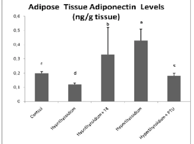
Adiponectin levels of experimental groups were 0.20±0.01; 0.12±0.01; 0.33±0.19; 0.43±0.08; 0.18±0.02 ng/gr for control, hypothyroidism, hypothyroidism +thyroxin, hyperthyroidism and hyperthyroidism plus PTU groups. Hyperthyroidism group (group 4) has higher adipose tissue adiponectin levels compared all other groups (P<0.001). However, hypothyroidism group which induced by 3 weeks PTU supplementation has the lowest adiponectin levels (P<0.001). Group 3 (PTU+ thyroxin treatment) has higher adiponectin levels compared to groups 1,2 and 5) (P<0.001).
Figure 1. a,b,c, and d show statistic differences among the groups (a>b>c>d). Adiponectin levels in experimental grups. Hyperthyroidsm
groups has the highest adiponectin levels compared to all other groups (a) (P<0.001). Hypothyroidism group has lower adiponectin levels compared
the all other groups (d) (P<0.001). T4 supplemnttaion increased adiponectin levels after hypothyroidism (b) (P<0.001). PTU reduces
tissue adiponectin levels in hyperthyroidism (c) (P<0.001). In the current study, the levels of adiponectin in the adipose tissue after thyroid dysfunction and treatment were examined in rats. When the results of the study were evaluated in general, it
was determined that while adiponectin levels decreased in hypothyroidism, hyperthyroidism increased. However, adiponectin levels, which vary with thyroid dysfunction, showed definite improvement after treatment of thyroid dysfunction. Thyroid hormones play a role in the regulation of body metabolism. The effects are stimulation of relaxation metabolic rate, increased energy expenditure, modulation of response to catecholamines and thermogenesis in fat tissue [7]. Disorders in thyroid function cause changes in body weight, muscle mass and fat tissue. Thyroid stimulating hormone (TSH) receptors were found in the fat area; suggesting that TSH plays a role in the regulation of adipocytokines, which play a role in the regulation of energy balance [17]. However, weight loss or insulin sensitizers cause an increase in ADP levels associated with the recovery of insulin resistance [4]. Recent studies on the relationship between adiponectin and hypothyroidism have produced conflicting results.
Most authors have reported that adiponectin levels remain unchanged in patients with thyroid hypofunction compared to euthyroid cases [6,18,19]. However, in the other study it has been reported that hypothyroidism led to lower adiponectin levels [20]. In the studies which performed on the human different findings have been reported related to adiponectin and hyperthyroidism. Some of them mentioned that hyperthyroidism led to higher adiponectin levels [21,22] but others reported that there were no differences between hyperthyroidism and euthyroidism in term of adiponectin [2,3,19]. It has been observed that ADP is interaction thyroid hormone synthesis especially free T4 and thereafter
persons who have high adiponectin and also higher T4 levels [23].
Emine Atıci, Rasim Mogulkoc, Abdulkerim Kasim Baltaci, Esma Menevse
Page | 3792 such as increased thermogenesis and lipid peroxidation in which
body fat is reduced and thus affect insuline resistance [10,11]. Controversial results have been reported on the association between ADP levels and thyroid hormones for experimental studies on hypo / hyperthyroid animals. In this manner, Aragao et al. [13] have reported that serum adiponectin levels of hyperthyroid rats 3.2 fold higher than that of euthyroid, whereas in experimental hypothyroidism did not change serum adiponetcin levels. Chang et al. [24] have reported increased adiponectin levels in Graves’ disease. As contrast, Seifi et al. [14] determined that hyperthyroidism which induced by 12 m/l of levothyroxine for 42 days increased adiponectin levels but hypothyrodism that induced by 250 mg/l methimazole for 6 weeks led to reducing adiponectin levels in rats. Similary, Lin et al. [25] reported that a positive correlation is a present adiponectin and freeT4 levels in thyroid cancer patients. Seifi et al. [26] and also reported that methimazole mRNA levels of AdipoR1 and AdipoR2 decreased in the white tissue in rats. However, in the same study experimental hyperthyroidism increased mRNA levels of AdipoR1 and AdipoR2. Iglesias et al. [5] and Yildiz et al. [27] determined that no differences in adiponectin levels between hyperthyroid and control groups and also no changes did not determine after normalization of circulating thyroid hormones. However,
Ahagarpour et al. [28] reported that hypotyhyroidism led to hyperadiponectinemia in a male mouse. In anoter study in which 65 hyperthyroid and 85 hypothyroid patients were evaluated for adiponectin, it has been reported that did not observe changes in adiponectin levels in any of the studied groups. In the sum of studies, it has been reported that increased adiponectin levels returned basal levels after the correction of hyperthyroidism [6,20,29]. In our study, increased tissue adiponectin levels were reduced to control levels that are similar to previous findings. However, in a study which performed on the dogs reduced serum adiponectin levels were reported [30]. They mentioned that this finding may relate to hyperlipidemia, insulin resistance and
adiponectine resistance. However, in another different study it has been reported that hypothyroidism led to increased higher adiponectin levels. But our finding showed reduced adiponectin levels in adipose tissue in rats. We think that different results could be experimental and animals species differences. ADP mRNA expression affected by thyroid [31]. Luvizotto et al. [32] experimental hyperthyroidism decrease gene expression and serum levels of adipokines in obesity. In this reduced adiponectin levels are related to weight loss, decreases all fat deposits and reduced adiponectin gene expression. Cabanelas et al. [15] reported that T3 administration in rats did not have a significant
effect on ADP release in visceral (epididymal) and subcutaneous (adipose) adipose tissues, although ADP mRNA expression with T3 was down regulated in the subcutaneous adipose tissue, such
effect did not occur in visceral adipose tissue (VAT). The authors suggest that ADP mRNA response to T3 changes depending on the type of WAT and its anatomical location. In humans, the results of interaction studies between thyroid hormones and ADP are contradictory. There may be differences in the nature of the patient, the nature and duration of thyroid hormone dysfunction, the metabolic effects of other hormones, and the possible effects of intermediate metabolism. Variations in the duration of studies and the small sample size involved in studies may be other possible reasons for this discrepancy. In addition, the presence of autoimmunity in thyroid diseases may play a role in this debate, because studies have shown a relationship between thyroid antibodies and ADP. Findings that we have obtained in experiments show differences and differences with the findings of the above-mentioned. The variability of the subjects of these differences was that in our study male rats were used, while other studies were performed on humans and dogs and mice in different ways. In addition, it is possible that the duration of the experiment (3 weeks in the current study) and the application is derived from the dose and the form. In some of them 6 weeks experimental period was applied.
4. CONCLUSIONS
The results of the study show that adiponectin levels in fat tissue altered in rats due to experimental hypothyroidism and hyperthyroidism, but deteriorated adiponectin levels were restored
after treatment. In future studies, determine the mRNA levels of adiponectin mentioned tissue or other tissues will be useful to determine molecular mechanisms in much more clearly.
5. REFERENCES
1. Warakomski, J.; Romuk, E.; Jarząb, B.; Krajewska, J.; Siemińska, L. Concentrations of Selected Adipokines, Interleukin-6, and Vitamin D in Patients with Papillary Thyroid Carcinoma in Respect to Thyroid
Cancer Stages. Int J Endocrinol 2018, 3,
https://doi.org/10.1155/2018/4921803.
2. Iglesias, P.; Alvarez, F.P.; Codoceo, R.; Diez, J.J.; Serum concentrations of adipocytokines in patients with hyperthyroidism and hypothyroidism before and after control of thyroid function. Clinical Endocrinology 2003, 59, 621-629, https://doi.org/10.1046/j.1365-2265.2003.01897.x.
3. Pontikides N., Krassas G.E., Basic endocrine products of adipose tissue instates of thyroid dysfunction. Thyroid 2007, 17, 421-431,
https://doi.org/10.1089/thy.2007.0016.
4. Gasbjerg, L.S.; Gabe, M.B.N.; Hartmann, B.; Christensen, M.B.; Knop, F.K.; Holst, J.J.; Rosenkilde, M.M. Glucose-dependent insulinotropic polypeptide (GIP) receptor antagonists as anti-diabetic
agents. Peptides 2018, 173-181,
https://doi.org/10.1016/j.peptides.2017.11.021.
5. Grigoraş, A.; Amalinei, C.; Balan, R.A.; Giuşcă, S.E.; Avădănei, E.R.; Lozneanu, L.; Căruntu, I.D. Adipocytes spectrum - From homeostasia to obesity and its associated pathology. Ann Anat 2018, 219, 102-120, https://doi.org/10.1016/j.aanat.2018.06.004.
6. Wen, F.; An, C.; Wu, X.; Yang, Y.; Xu, J.; Liu, Y.; Wang, C.; Nie, L.; Fang, H.; Yang, Z. MiR-34a regulates mitochondrial content and fat ectopic deposition induced by resistin through the AMPK/PPARα pathway in HepG2 cells. Int J Biochem Cell Biol 2018, 94, 133-145, https://doi.org/10.1016/j.biocel.2017.11.008. 7. Miljić, D.; Popovic, V. Metabolic Syndrome in Hypopituitarism. Front Horm Res 2018, 49, 1-19, https://doi.org/10.1159/000485997. 8. Wang, B.; Cheng, K.K. Hypothalamic AMPK as a Mediator of Hormonal Regulation of Energy Balance. Int J Mol Sci 2018, 11,
https://doi.org/10.3390/ijms19113552.
9. Pontikides, N.; Loustis, K.; Koliakos, G.; Constantinidis, T.H.; Kaltsas, T.H.; Krassas, G.E. Serum cytokines levels in hypothyroidism before and after treatment: relationship with body weight and body composition. Proceedings of the 31st European Thyroid Association Meeting, 2006.
The effect of thyroid dysfunction and treatment on fat tissue adiponectin levels in rats
Page | 3793 10. Bloemer, J.; Pinky, P.D.; Govindarajulu, M.; Hong, H.; Judd, R.;
Amin, R.H.; Moore, T.; Dhanasekaran, M.; Reed, M.N.; Suppiramaniam, V. Role of Adiponectin in Central Nervous System
Disorders. Neural Plast 2018, 4593530,
https://doi.org/10.1155/2018/4593530.
11. Sabir, S.; Akhtar, M.F.; Saleem, A. Endocrine disruption as an adverse effect of non-endocrine targeting pharmaceuticals, Environ Sci Pollut Res Int 2019, 26, 1277-1286, https://doi.org/10.1007/s11356-018-3774-4.
12. Delitala A.P., Steri M., Fiorillo E., Marongiu M., Lakatta E.G., Schlessinger D., Cucca F., Adipocytokine correlations with thyroid function and autoimmunity in euthyroid sardinians. Cytokine
2018, 111, 189-193, https://doi.org/10.1016/j.cyto.2018.08.027. 13. Cruz-Mejía, S.; Durán López, H.H.; Navarro, M.M., Xochihua, R.I.; De la Peña S.; Arroyo H.O.E. Body mass index is associated with interleukin-1, adiponectin, oxidative stress and ioduria levels in healthy adults. Nutr Hosp 2018, 841-846, https://doi.org/10.20960/nh.1614.
14. Seifi, S.; Tabandeh, M.R.; Nazifi, S.; Saeb, M.; Shirian, S.; Sarkoohi, P. Regulation of adiponectin gene expression in adipose tissue by thyroid hormones. Journal of Physiolology and Biochemistry 2012, 68, 193-203, https://doi.org/10.1007/s13105-011-0131-1.
15. Cabanelas, A.; Cordeiro, A.; Santos Almeida, N.A.; Monteiro de Paula, G.S.; Coelho, V.M.; Ortiga-Carvalho, T.M. Effect of triiodothyronine on adiponectin expression and leptin release by White adipose tissue of normal rats. Hormone and Metabolism Research
2010, 42, 254-260, https://doi.org/10.1055/s-0029-1246118.
16. Siemińska, L.; Foltyn, W.; Głogowska-Szeląg, J.; Kajdaniuk, D.; Marek, B.; Nowak, M.; Walczak, K.; Kos-Kudła, B. Relationships between adiponectin, sex hormone binding globulin and insulin resistance in hyperthyroid Graves' disease women. Endokrynology Polska 2013, 64, 26-29.
17. Santini, F.; Marsili, A.; Mammoli, C.; Valeriano, R.; Scartabelli, G.; Pelosini, C. Serum concentrations of adiponectin and leptin in patientswith thyroid dysfunctions. Journal of
Endocrinology and Investigation 2004, 5-7,
https://doi.org/10.1007/BF03346252.
18. Morishita, M.; Endo, T.; Baba, T.; Kuno, Y.; Ikeda, K.; Kiya, T.; Honnma, H.; Saito, T. Pioglitazone is effective for multiple phenotyepes of the Zucker fa/fa rat with polycystc ovary morphology and insulin resistance. J Ovarian Res 2018, 11, 24,
https://doi.org/10.1186/s13048-018-0395-y.
19. Ahmed, R.G.; El-Gareib, A.W.; Shaker, H.M. Gestational 3,3',4,4',5-pentachlorobiphenyl (PCB 126) exposure disrupts fetoplacental unit: Fetal thyroid-cytokines dysfunction. Life Sci 2018, 192, 213-220, https://doi.org/10.1016/j.lfs.2017.11.033.
20. Yaturu, S.; Prado, S.; Grimes, S.R. Changes in adipocyte hormones leptin, resistin, and adiponectin in thyroid dysfunction. Journal of Cell Biochemistry 2004, 491-496, https://doi.org/10.1002/jcb.20188. 21. Saito, T.; Kawano, T.; Saito, T.; Ikoma, A.; Namai, K.; Tamemoto, H. Elevation of serum adiponectin levels in Basedow disease.
Metabolism 2005, 54, 1461-1466,
https://doi.org/10.1016/j.metabol.2005.05.011.
22. Sieminska, L.; Niedziolka, D.; Pillich, A.; Kos-Kudla, B. Marek B., Nowak M., Borgiel-Marek H., Serum concentrationsof adiponectin and resistin in hyperthyroid Graves’disease patients. Journal of Endocrinology and Investigation 2008, 31, 745-749,
https://doi.org/10.1007/BF03349251.
23. Castelblanco, E.; Hernández, M.; Castelblanco, A.; Gratacòs, M.; Esquerda, A.; Molló, À.; Ramírez-Morros, A.; Real, J.; Franch-Nadal, J.; Fernández-Real, J.M.; Mauricio, D. Low-grade Inflammatory Marker Profile May Help to Differentiate Patients With LADA, Classic Adult-Onset Type 1 Diabetes, and Type 2 Diabetes. Diabetes Care
2018, 41, 862-868, https://doi.org/10.2337/dc17-1662.
24. Chang, C.L.; Lim, A.Y.; Tan, H.C.; Kovalik, J.P.; Tham, K.W.; Bee, Y.M. Physiological and Metabolic Changes During the Transition from Hyperthyroidism to Euthyroidism in Graves' Disease. Thyroid 2016, 26, 1422-1430, https://doi.org/10.1089/thy.2015.0602. 25. Lin, S.Y.; Hsu, W.H.; Lin, C.L.; Lin, C.C.; Lin, J.M.; Chang, Y.L.; Hsu, C.Y.; Kao, C.H. Evidence for an Association between Macular Degeneration and Thyroid Cancer in the Aged Population. Int J Environ Res Public Health 2018, 15, https://doi.org/10.3390/ijerph15050902. 26. Seifi, S.; Nazifi, S.; Tabandeh, M.R.; Saeb, M. AdipoR1 and AdipoR2 gene expression are regulated by thyroid hormones in adipose tissue Molecular and Cell Biochemistry 2013, 377, 55-63,
https://doi.org/10.1007/s11010-013-1570-5.
27. Yildiz, B.O.; Aksoy, D.Y.; Harmanci, A.; Unluturk, U.; Cinar, N.; Isildak, M. Effects of L-thyroxine therapy on circulating leptin and adiponectin levels in subclinical hypothyroidism: a prospective study. Archives of Medical Research 2013, 44, 317-320,
https://doi.org/10.1016/j.arcmed.2013.04.010.
28. Ahangarpour, A.; Alboghobeish, S.; Oroojan, A.A.; Zeidooni, L.; Samimi, A.; Afshari, G. Effects of Combined Exposure to Chronic High-Fat Diet and Arsenic on Thyroid Function and Lipid Profile in Male Mouse. Biological Trace Element and Research 2018, 182, 37-48, https://doi.org/10.1007/s12011-017-1068-1. 29. Atıci, E.; Mogulkoc, R.; Baltaci, A.K.; Menevse, E. The effect of thyroid dysfunction on nesfatin-1 and adiponectin levels in rats. Horm Mol Biol Clin Investig 2017, 32, https://doi.org/10.1515/hmbci-2017-0033.
30. Mazaki-Tovi, M.; Abood, S.K.; Kol, A.; Farkas, A.; Schenck, P.A. Increased serum concentrations of adiponectin in canine hypothyroidism. Veterinary Journal 2015, 203, 253-255,
https://doi.org/10.1016/j.tvjl.2014.12.007.
31. Gong, N.; Gao, C.; Chen, X.; Wang, Y.; Tian, L. Adipokine expression and endothelial function in subclinical hypothyroidism rats. Endocr Connect 2018, 295-304, https://doi.org/10.1530/EC-18-0007. 32. Luvizotto, R.A.; do Nascimento, A.F.; de Síbio, M.T.; Olímpio, R.M.; Conde, S.J.; Lima-Leopoldo, A.P. Experimental hyperthyroidism decreases gene expression and serum levels of adipokines in obesity. Scientific World Journal 2012, http://dx.doi.org/10.1100/2012/780890.
6. ACKNOWLEDGEMENTS
This study was supported by a grant Selcuk University, Scientific Research Council (Grant number is 14202034).
© 2019 by the authors. This article is an open access article distributed under the terms and conditions of the Creative Commons Attribution (CC BY) license (http://creativecommons.org/licenses/by/4.0/).