Arch Clin Exp Med 2017;2(1):26-28. e-ISSN: 2564-6567
DOI: 10.25000/acem.303499 Case Report / Olgu Sunumu
Atıf yazım şekli:
How to cite: Karabay Ö, Erdem MG, Hasbahçeci M. Jejunal fecaloma as a rare cause of intestinal obstruction: A case report. Arch Clin Exp Med. 2017;2(1):26-28.
Abstract
Fecalomas are usually found in the colon or the rectum. In adult ages, jejunal giant fecaloma is a very rare condition. It has been thought that there should be any kind of chronic diseases leading to the occurrence of such fecalomas at the unexpected localizations. In this case, it was aimed to present a 50-year-old male patient with small bowel obstruction caused by jejunal giant fecalomas. There was previous gastrojejunostomy with vagotomy as the peptic ulcer surgery 25 years ago. Jejunal resection was performed due to the presence of small necrotic areas on the affected segment caused by impacted jejunal fecaloma. Fecalomas may be considered as a differential diagnosis of small intestinal obstruction in a patient with previous peptic ulcer surgery.
Keywords: Fecaloma, Intestinal obstruction, Jejunum
Öz
Fekalomlar genellikle kolon ve rektumda bulunurlar. Yetişkinlerde jejunal dev fekalom bulunması oldukça nadir bir durumdur. Bazı kronik hastalıkların fekalomların alışılmadık lokalizasyonlarda gelişmesinde rolü olduğu düşünülmektedir. Bu olguda, dev jejunal fekaloma bağlı gelişen intestinal obstrüksiyonlu 50 yaşında erkek hastanın sunulması amaçlanmıştır. 25 yıl önce peptik ülser cerrahisi için yapılmış vagotomi ve gastrojejunostomi öyküsü mevcuttu. Jejunal fekalomun impakte olduğu segmentte küçük nekrotik alanların olmasından dolayı jejunal rezeksiyon ile tedavi edildi. Geçirilmiş peptik ülser cerrahisi olan hastalarda gelişen ince barsak obstrüksiyonunda fekalomlar ayırıcı tanıda düşünülmelidir.
Anahtar Kelimeler: Fekalom, İntestinal obstrüksiyon, Jejunum
Introduction
Jejunal giant fecaloma is a very rare condition in adults. Fecalomas are usually found in the colon or the rectum. It has been thought that presence of chronic constipation, psychiatric diseases, Chagas or Hirschsprung's disease may ease the development of such condition in these bowel segments. However, there has been limited number of cases in the literature reporting the occurrence of fecalomas beside the colon and the rectum [1,2].
Chronic constipation or intestinal obstruction is the main symptoms of the patients for their admission. Most fecalomas are treated successfully by conservative methods. If conservative methods are failed, surgical procedures can be used. Surgical techniques including removal of fecaloma via enterotomy or segmental resection of the bowel can be used based on intraoperative findings [1,2].
In this case, a 50-year-old male patient with small intestinal obstruction was treated via segmental resection of the jejunum due to the development of necrosis over the impacted jejunal fecaloma.
Case Report
A 50-year-old male patient was admitted to emergency service for nausea and vomiting for the last five days. It has been learned that there was severe diarrhea with colicky abdominal pain followed by obstipation. The patient had undergone bilateral truncal vagotomy and gastrojejunostomy for complicated peptic ulcer disease almost 25 years ago. On physical examination, there was distention over the whole abdomen and tenderness on the right upper quadrant. Laboratory findings revealed leukocytosis (18000 /uL), increase in CRP (151 mg/L) and creatinine (4 mg/dL) levels. After intravenous hydration and nasogastric drainage, computed tomography without intravenous contrast agent was taken. At tomography, small intestinal obstruction at the proximal jejunal segments was detected. There was an increased wall thickness with inflammatory changes on the small intestinal segments at the right upper quadrant leading to fecalization of the small bowel loops (Figure 1).
1 Fatih Medical Park Hospital, General
Surgery, Istanbul, Turkey.
2 Fatih Medical Park Hospital, Internal
Medicine, Istanbul, Turkey.
Conflict of Interest: No conflict of interest was declared by the authors.
Çıkar Çatışması: Yazarlar çıkar çatışması bildirmemişlerdir.
Financial Disclosure: The authors declared that this case has received no financial support.
Finansal Destek: Yazarlar bu olgu için finansal destek almadıklarını beyan etmişlerdir.
Received / Geliş Tarihi 02.04.2017
Accepted / Kabul Tarihi 11.04.2017
Published / Yayın Tarihi 18.04.2017
Corresponding author / Sorumlu yazar Önder Karabay
Adressess: Fatih Medikal Park Hastanesi, Genel Cerrahi, İstanbul, Türkiye
Tel: +90505 7330343
E-mail: [email protected] © Copyright 2017 ACEM
Figure 1: CT image. Fecalization of small bowel loops and increased wall thickness of a segment of the small intestine with mesenteric inflammation (white arrow)
Jejunal fecaloma as a rare cause of intestinal obstruction: A case
report
İntestinal obstrüksiyonun nadir bir nedeni jejunal fekalom: Olgu sunumu
Önder Karabay
1Arch Clin Exp Med 2017;2(1):26-28. Jejunum and fecaloma
P a g e | 27 The maximum diameter of the jejunum was measured as
7cm. Due to the findings gathered by physical examination, biochemical and imaging techniques, laparotomy was planned. After adhesiolysis, dilated jejunal segments were seen. Extensive exploration revealed that proximal jejunal segments were obstructed by an intraluminal impacted hard mass leading to necrosis on that point (Figure 2).
Due to the development of necrosis and inability to move this impacted lesion, segmental resection of the jejunum with side-to side anasside-tomosis was performed. Opening of the diseased segment revealed that there was a giant ulcer cavity with necrotic areas (Figure 3) and one giant fecaloma measuring up to 7 cm in its largest dimension impacted on the jejunum. It was also detected one more fecaloma intraluminally (Figure 4). Both fecalomas were hard in consistency by manual examination.
The patient was discharged on 5th day without a complication. Pathological examination was unremarkable except the development of a benign chronic ulcer with reactive lymph nodes within the mesentery.
Written consent was taken from the patient.
Figure 2: Intraoperative image of the small intestines. Necrotic area on the jejunum (black arrow)
Figure 3: Fecalomas
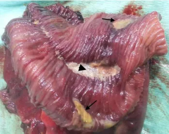
Figure 4: Internal view of the small bowel segment. Ulcer cavity at the base of the mesentery (black arrowhead) and ischemic areas (black arrows)
Discussion
Fecalomas are extreme hardening of the feces due to malfunction of the digestive system. It is more likely to be seen on the left side of the colon because the content is stiffer. Although they are most commonly found in the colon and rectum, there are limited numbers of publications in which fecalomas are seen in other bowel segments. Chronic constipation, Hirschsprung's disease, Chagas disease and psychiatric disorders are among the leading causes. Colonic obstruction, urinary retention or intraabdominal mass due to fecalomas have been reported rarely as case reports. Additionally, there are two case reports in relation with intestinal obstruction due to jejunal and ileal fecalomas in the literature [1,2].
After peptic ulcer surgery and vagotomy, it has been known that the risk of bezoar formation increases. In the literature, there are case reports related to bezoar development, especially after gastric bypass surgery [3,4]. Acceleration of gastric emptying after gastric surgery and decreased gastric acid secretion due to vagotomy facilitate the passage of undigested foods to the intestines. This situation is thought to contribute to the formation of bezoar [5]. In this case, it has been believed that bilateral truncal vagotomy and gastrojejunostomy for benign gastric ulcer leads to fecaloma formation. However, the pathophysiological explanation for this case remains still obscure.
Although microscopic examination of the fecalomas after extraction wasn’t performed, they have been evaluated as fecalomas due to external appearance and hard structure in the present case.
Conservative methods have been reported as the main treatment modality for the conditions caused by fecalomas, surgery may be needed in selected cases due to their delayed or untreated presentations.
In the literature, it has been reported that the authors are able to extract the fecalomas located in the jejunum and the ileum via enterotomy in previous case reports [1, 2]. However, the fecaloma in the present case could not be extracted with enterotomy, because of small necrotic areas over the jejunum caused by fecal impaction. It has been thought that delayed presentation causes prolonged intestinal obstruction and increased intraluminal pressure, consequently small intestinal ischemia.
As a result, intestinal obstruction due to fecalomas in the small bowel is a very rare situation. In those cases with previous peptic ulcer surgery, it is better to be kept in mind
Arch Clin Exp Med 2017;2(1):26-28. Jejunum and fecaloma
P a g e | 28 that fecalomas may be the etiology of intestinal obstruction besides
the bezoar development.
References
1. Mushtaq M, Shah MA, Malik AA, Wani KA, Thakur N, Parray FQ. Giant Fecaloma Causing Small Bowel Obstruction: Case Report and Review of the Literature. Bull Emerg Trauma 2015; 3: 70-2. 2. Yoo H, Park HW, Chang SH, Bae SH. Ileal Fecaloma Presenting with
Small Bowel Obstruction. Pediatr Gastroenterol Hepatol Nutr 2015; 18: 193-6.
3. Wapnick S, Lazarovitch I, Solowiejczyk M. Obstruction by bezoar after vagotomy and pyloroplasty. Lancet 1973; 23: 1454.
4. Grafskaia ND, Kotovskiĭ AE. Bezoar of the stomach after vagotomy. Khirurgiia 1978; (10): 127-8.
5. Sarhan M, Shyamali B, Fakulujo A, Ahmed L. Jejunal Bezoar Causing Obstruction After Laparoscopic Roux-en-Y Gastric Bypass. JSLS 2010; 14: 592–5.