https://doi.org/10.1177/1179547619852621
Creative Commons Non Commercial CC BY-NC: This article is distributed under the terms of the Creative Commons Attribution-NonCommercial 4.0 License (http://www.creativecommons.org/licenses/by-nc/4.0/) which permits non-commercial use, reproduction and distribution of the work without further permission provided the original work is attributed as specified on the SAGE and Open Access pages (https://us.sagepub.com/en-us/nam/open-access-at-sage).
Clinical Medicine Insights: Case Reports Volume 12: 1–3
© The Author(s) 2019 Article reuse guidelines: sagepub.com/journals-permissions DOI: 10.1177/1179547619852621
Introduction
Patency of the left main coronary artery (LMCA) may be nec-essary in patients with patent coronary bypass grafts to the left coronary arterial system for the perfusion of branches that could not be grafted. In patients that have complete ostial occlusion of the LMCA, so that it cannot be visualized in an antegrade fashion from the aortic root, left main coronary arte-rial recanalization cannot be achieved antegradely. In these patients, the presence of a patent saphenous vein graft may be an opportunity for recanalization of the LMCA.
A 70-year-old male patient has represented with chest pain upon mild exertion. Biochemical examination revealed his cre-atinine level as 1.04 mg/dL, high-sensitive Troponin I as 2.4 pg/mL (normal range: 0-26 pg/mL), hemoglobin 10.4 g/dL (normal range: 12-16 g/dL) with a hematocrit count of 35.3% (normal range: 33%-45 %), and serum sodium as 141 mmol/L (normal range: 135-145 mmol/L). He had a history of hyper-tension for 20 years and Parkinsonism for 5 years. He had coro-nary artery bypass surgery 3 years ago and had a successful percutaneous intervention to his circumflex and right coronary artery grafts a year ago. His LMCA and LIMA (left internal mammary artery) graft have been found as patent at that time.
His medical treatment consists daily doses of 125 mg levo-dopa, 75 mg clopidogrel, 180 mg diltiazem, 15 mg rivaroxa-ban, 750 mg ranolazine, 40 mg pantoprazole, and 20 mg rosuvastatin.
Upon echocardiographic assessment, it was found that the left ventricular ejection fraction was normal; there was mild mitral and tricuspidal regurgitation; and pulmonary arterial pressure was estimated to be 30 mm Hg. There were not any significant wall motion abnormalities. Electrocardiography (ECG) showed an atrial fibrillation rhythm with an average ventricular rate of 65 bpm.
Method
As the LIMA graft was found to be patent in previous angio-graphic assessment, for the operators’ ease of access, right radial artery was used rather than the left.
After cannulation of the right radial artery with a 5F Terumo Glidesheath radial artery sheath, the left coronary sinus was scanned with left Expo 3.5 and 4.0 Judkins of Boston Scientific (Tijuana, Baja California, Mexico) diagnostic cathe-ters, and the non-coronary and right coronary sinuses were scanned with right coronary diagnostic catheter. There were no signs of an LMCA. We thought that the patient’s symptoms were related to the main coronary occlusion. Namely, anasto-mosis-free large OM Obtuse Marginal and intermediate artery are only supplied through the graft connected to Obtuse Marginal 2 (OM2). Due to the critical stenosis in the proximal part of the anastomosis site of obtuse marginal artery, we thought that retrograde blood flow was not sufficient for inter-mediate artery and other great obtuse marginal vessels without
Successful Retrograde Visualization and Recanalization
of Chronic Ostial Occlusion of the Left Main Coronary
Artery via a Saphenous Vein Graft With the Drilled
Balloon Technique
Yakup Balaban
1and Murat Güçlü Elevli
21Department of Cardiology, Istinye Universty, VM Medicalpark Kocaeli Hospital, Bas¸iskele, Turkey. 2Sakarya Özel Adatıp Hastanesi, Kalp-Damar Cerrahisi Klinig˘i, Sakarya, Turkey.
ABSTRACT: We are representing a case of successful retrograde recanalization of a chronic ostial occlusive lesion of the left main coronary artery (LMCA) via a saphenous vein graft. A 70-year-old male patient, with a history of previous (3 years ago) coronary artery bypass surgery, was evaluated with coronary angiography because of his recent anginal symptoms. Left main coronary artery could not be visualized from the aortic root. It could be visualized with drilled balloon technique which was introduced retrogradely from a saphenous vein graft, and the chronic ostial lesion of the LMCA could be treated with balloon dilatation and stenting. Coronary occlusions can be treated retrogradely via the native collaterals and grafts. The coronaries that are not visualized with an antegrade fashion can be visualized with a retrograde way with the drilled balloon introduced through a patent graft. We could not find any publication about this technique on the web-based research. When encountered with a totally occluded ostial lesion of the LMCA, that cannot be visualized even from the aortic root, it may be tried to be visualized through a patent graft retrogradely. This is a safe and reliable method in patients with a total occlusion of the LMCA who also have a patent saphenous vein graft.
KeyWORDS: coronary artery bypass grafting, interventional cardiology, percutaneous coronary intervention, chronic total occlusion, retro-grade intervention
ReCeIVeD: April 3, 2019. ACCePTeD: April 9, 2019. TyPe: Case Report
FunDInG: The author(s) received no financial support for the research, authorship, and/or
publication of this article.
DeCLARATIOn OF COnFLICTInG InTeReSTS: The author(s) declared no potential
conflicts of interest with respect to the research, authorship, and/or publication of this article.
CORReSPOnDInG AuTHOR: Yakup Balaban, Department of Cardiology, Istinye
Universty, VM Medicalpark Kocaeli Hospital, Başiskele, 41140- Kocaeli, Turkey.
Email: yakupbalaban@gmail.com
852621ICR0010.1177/1179547619852621Clinical Medicine Insights: Case ReportsBalaban and Elevli
2 Clinical Medicine Insights: Case Reports
graft. For this reason, we decided that the LMCA should be revascularized.
Aorta-right coronary artery (Ao-RCA) and aorta-obtuse marginal artery (Ao-OM) grafts were visualized with a Medtronic, Launcher AL1 guiding catheter (Minneapolis, MN, USA). During the visualization of the OM graft, neither the state nor the location of the LMCA could be understood. For more clear visualization of the LMCA that was thought to be occluded from its ostium, Launcher 5F Amplatz 2 guiding catheter (Medtronic) was placed selectively into the Ao-RCA graft, and a 0.014 pilot 150 percutaneous transluminal coro-nary angioplasty (PTCA) guidewire (Abbott Vascular, Santa Clara, CA, USA) was placed through it to the left main root in a retrograde way so as to perform an intervention. After that,
2.0 mm × 12 mm Monorail Emerge balloon (Boston
Scientific–Galway, Galway, Ireland) was placed into the LMCA with the aid of this guidewire; 15 s of 10 atm predilata-tion was applied. Balloon was taken out and dilated with 4 atm pressure to punch with a needle to create 5 holes on it. It was
reintroduced in the LMCA for use as a multi-hole catheter and the lesion was visualized with it.1
Then, a 3.0 mm × 12 mm Monorail Emerge balloon (Boston Scientific–Galway) was placed into the lesion to dilate it suffi-ciently enough for stent insertion. A 3.0 mm × 15 mm drug eluting stent (Orsiro, Biotronik, Bülach, Switzerland) was intro-duced, but it could not advance beyond the stenotic lesion in the obtuse marginal coronary artery. For this reason, dilatation with a 2.0 mm × 20 mm Monorail Emerge balloon (Boston Scientific–Galway) was performed to this 90% stenotic critical lesion, located between the saphenous vein graft and the cir-cumflex artery. To have catheter backup, Medtronic Launcher 5F Amplatz 2 guiding catheter was advanced gently, during the balloon in the obtuse marginal lesion was still inflated to anchor it. The 3.0 mm × 15 mm drug eluting stent (Orsiro, Biotronik, Bülach, Switzerland) was reintroduced to the lesion, this time passing through the lesion in the OM lesion, and was applied to the ostial LMCA lesion successfully with 22 atm dilatation (Figure 1).
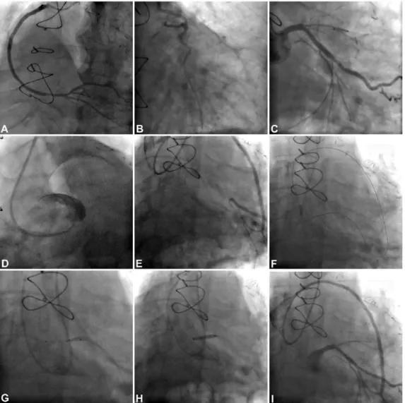
Figure 1. ABC. Same Patient’s Angiography in one year ago, (A) Picture of “LIMA-LAD graft” imaging. (B) The imaging of LMCA in one year ago. (C) Ao-RCA
Graft visualization. (D) Aortic root angiography: no silhouette of the LMCA appears. (E) Obtuse Marginal (OM) saphenous graft imaging: LMCA cannot be displayed. (F) The trace of a 0.014 guidewire was advanced to aorta through OM saphenous graft retrogradely. (G) The imaging of LMCA was obtained by the use of drilled balloon after the predilatation by 2.0 mm × 20 mm balloon; this image shows that the critical LMCA lesion still exists despite the balloon dilatation. (H) The imaging of 3.0 mm × 15 mm drug eluting stent application. (I) The final view of LMCA. LMCA indicates left main coronary artery.
Balaban and Elevli 3 No complications were encountered. A total of 75 mL
radi-opaque media, Omnipaque GE Healthcare (Cork, Ireland), was used. ECG evaluations performed preprocedure and immediate postprocedure; 24 h after, the procedure showed no significant changes. Serum creatinine and troponin levels did not show significant elevations at 12, 24, and 36 h after the procedure.
The patient is regularly followed up at 6-month intervals. He was last examined in January 2019. There are no active ischemic complaints.
Discussion
Web-based research shows there are publications about retro-grade coronary interventions via the saphenous vein graft, but there is only 1 case report about successful retrograde recanaliza-tion of a chronic ostial occlusive lesion of the LMCA via a saphenous vein graft.2-6 The drilled balloon technique was not
used in these studies. We think that the drilled balloon technique facilitates this process.
Left main coronary artery occlusions are frequently left untouched when the grafts are patent. We consider that, even if the left-sided grafts are patent, patency of the LMCA is benefi-cial for the patient as this will improve the perfusion of proximal left anterior descending, diagonal, septal, intermediary, and other circumflex branches that have not been grafted. We believe, if the grafts do not have competition with the native artery or if there are 2 or more sequential stenoses proximal to the grafted segment, recanalization of the left main or proximal lesion will be beneficial for the patient. If antegrade way is not available, retrograde approach through grafts may be used. In patients with previous coronary artery bypass grafting (CABG) surgery, native coronary lesions can be treated via the grafts. All these interven-tions may not only alleviate the symptoms but also decrease mortality in these patients.7-12
Conclusions
It seems it is essential to recanalize the occlusions and sten-otic lesions of the LMCA for improving the perfusion of non-grafted vessels of the coronary arterial system even if the left-sided grafts are patent. It is mostly probable in these patients that the retrograde approaches are going to be successful.
If the LMCA is occluded at its orifice and cannot be visual-ized, lesion may be recanalized through a graft in a retrograde fashion.
Author Contributions
YB; Conception or design of the work; acquisition, analysis, or; drafting the study or revising it critically for important
intellectual content. MGE; Conception or design of the work; interpretation of data for the study.
Ethical Approval
All procedures performed in studies involving human partici-pants were in accordance with the ethical standards of the institutional and/or national research committee and with the 1964 Helsinki Declaration and its later amendments or com-parable ethical standards.
Informed Consent
Informed consent was obtained from the participant patient included in the study.
ORCID iD
Yakup Balaban https://orcid.org/0000-0001-7553-5075
REfEREnCEs
1. Balaban Y, Bektas O, Bayramoglu A, Gumrukcuoglu HA, Kayisoglu AH. Imag-ing behind occluded areas with an iatrogenic perforated balloon: a safe, practical, and simple new method of visualizing the distal lumen in total occlusion. J Interv
Cardiol. 2017;30:544–549.
2. Dautov R, Manh Nguyen C, Altisent O, Gibrat C, Rinfret S. Recanalization of chronic total occlusions in patients with previous coronary bypass surgery and consideration of retrograde access via saphenous vein grafts. Circ Cardiovasc
Interv. 2016;9:30–35.
3. Hari P, Kirtane AJ, Bangalore S. Retrograde approach to an ostial left anterior descending chronic total occlusion through a left internal mammary artery graft.
Catheter Cardiovasc Interv. 2016;87:E224–E228.
4. Lin MM, Wang JH. Successful revascularization of an LCx CTO lesion by ret-rograde approach from an acute thrombotic SVG without protection device in an ACS patient. Int Heart J. 2016;57:372–375.
5. Burzotta F, Trani C, Coroleu S. Retrograde recanalization of left main from saphenous vein graft supported by percutaneous Impella Recover LP 2.5 assist device. J Invasive Cardiol. 2009;21:E147–E150.
6. Sekiguchi M, Taguchi T, Miyajima A, Hasegawa S, Yamazaki M, Kurabayashi MA. Novel wiring technique to insert a retrograde guidewire directly into the antegrade guiding catheter at the ascending aorta for retrograde percutaneous recanalization of an ostial coronary total occlusion. Int Heart J. 2016;57:503–506.
7. Biondi-Zoccai GG, Bollati M, Moretti C, et al. Retrograde percutaneous recan-alization of coronary chronic total occlusions: outcomes from 17 patients. Int J
Cardiol. 2008;130:118–120.
8. Rinfret S, Joyal D, Nguyen CM, Bagur R, Hui W. Retrograde recanalization of chronic total occlusions from the transradial approach; early Canadian experi-ence. Catheter Cardiovasc Interv. 2011;78:366–374.
9. Karmpaliotis D, Michael TT, Brilakis ES, et al. Retrograde coronary chronic total occlusion revascularization: procedural and in-hospital outcomes from a multicenter registry in the United States. JACC Cardiovasc Interv. 2012;5: 1273–1279.
10. Lee CK, Chen YH, Lin MS, et al. Retrograde approach is as effective and safe as antegrade approach in contemporary percutaneous coronary intervention for chronic total occlusion: a Taiwan single-center registry study. Acta Cardiol Sin. 2017;33:20–27.
11. Tanaka A, Ishii H, Oshima H, et al. Progression from stenosis to occlusion in the proximal native coronary artery after coronary artery bypass grafting. Heart
Vessels. 2016;31:1056–1060.
12. Vlasenko SV, Agarkov MV, Khilchuk AA, et al. Successful retrograde recanali-zation of an acute iatrogenic venous graft occlusion through the previously stented coronary anastomosis in a patient with non-ST elevation myocardial infarction. Radiol Case Rep. 2018;13:825–828.