he oculomotor nerve (the third cranial nerve) innervates the medial, inferior, superior recti muscles, the inferior oblique muscle, the lev-ator palpebrae superioris muscle, the iris sphincter muscle and the ciliary body.1
The third nerve nucleus is located in the midbrain near the cerebral aqueduct at the level of the superior colliculus. Each extraocular muscle
Isolated Unilateral Oculomotor Nerve Palsy
Due to Hematoma in Mesencephalone
AABBSS TTRRAACCTT Oculomotor nerve palsy causes reduced adduction, supraduction, infraduction, and ptosis with or without mydriasis. We report a case of isolated unilateral oculomotor nerve palsy due to hematoma in mesencephalone. A 30-years- old male patient presented with sudden onset of left ptosis, vertical diplopia, and difficulty in reading with his left eye that had persisted for one week. Direct and indirect light reflexes were negative on his left eye. There was incomplete ptosis on his left eyelid. In primary position, the left eye was deviated outward and downward. Supraduction of the left eye was absent, infraduction and adduction were reduced. MRI demonstrated hematoma in mesencephalone. Three months later, patient’s symptoms decreased, MRI demonstrated resolution of the hematoma and cavernoma in pons and gamma knife therapy was recommended. Isolated unilateral oculomotor nerve palsy due to hematoma related to cavernoma in mesencephalone is seen rarely and complete or incomplete recovery may be observed.
KKeeyywwoorrddss:: Hematoma; mesencephalon; oculomotor nerve diseases
Ö
ÖZZEETT Okülomotor sinir felci azalmış addüksiyon, supradüksiyon, infradüksiyon ve kapakta dü-şüklüğe neden olur, midriasis de olaya eşlik edebilir. Biz, mezensefalondaki hematomdan kaynak-lanan izole tek taraflı okülomotor sinir felci olan bir vakayı takdim ediyoruz. Bir hafta devam eden, ani başlangıçlı sol gözde kapakta düşüklük, dikey çift görme ve sol gözle yakını görememe şikaye-tiyle polikliniğimize başvuran 30 yaşındaki erkek hastada, yapılan muayenede sol gözde direkt ve indirekt ışık reflekslerinin negatif olduğu gözlendi. Sol göz üst kapakta kısmi düşüklük mevcuttu. Primer pozisyonda sol göz dışa ve aşağı doğru kaymış durumdaydı. Sol gözün supradüksiyonu yoktu, addüksiyon ve infradüksiyonu ise azalmıştı. MR görüntülerinde mezensefalonda hematom izlen-mekteydi. Üç ay sonra hastanın belirti ve bulguları azaldı, MR görüntülerinde hematomda çözülme ve ponsta kavernom izlendi ve hastaya gamma knife terapisi önerildi. Mezensefalondaki hema-tomdan kaynaklanan kavernom ile ilişkili izole tek taraflı okülomotor sinir felci nadiren görülür ve tam veya kısmi iyileşme gerçekleşir.
AAnnaahh ttaarr KKee llii mmee lleerr:: Hematom; mezensefalon; okülomotor sinir hastalıkları Servet ÇETİNKAYA,a
Emine MESTANb aClinic of Ophthalmology, Turkish Red Crescent Hospital, Konya
bDepartment of Neurology,
Dumlupınar University Faculty of Medicine, Kütahya
Re ce i ved: 09.03.2016
Received in revised form: 16.06.2016 Ac cep ted: 17.06.2016
Available online: 22.02.2018 Cor res pon den ce:
Servet ÇETİNKAYA
Turkish Red Crescent Hospital, Clinic of Ophthalmology, Konya, TÜRKİYE/TURKEY
Cop yright © 2018 by Tür ki ye Kli nik le ri
that receives innervation from the third nerve has a corresponding subnucleus. A single central nu-cleus (central caudal nunu-cleus) innervates both lev-ator palpebrae muscles. Distinct, bilateral subnuclei exist for the extraocular muscles. An additional bi-lateral subnucleus, the Edinger-Westphal nucleus, provides parasympathetic input to the ciliary body and pupillary sphincter. Each subnucleus inner-vates the ipsilateral corresponding extraocular muscle, with two exceptions, the single central caudal nucleus sends projections to both levator muscles and the superior rectus subnucleus inner-vates contralateral superior rectus muscle.2
Oculomotor fascicles travel ventrally through the midbrain, each passing through the red nu-cleus, substantia nigra and crus cerebri before they exit into subarachnoid space of the interpeduncu-lar fossa. Then the third nerve enters the cavernous sinus, wherein it is separated into superior and in-ferior divisions. The superior division innervates the superior rectus and levator muscles, while the inferior division supplies the inferior rectus, infe-rior oblique, medial rectus, ciliary body and pupil-lary sphincter muscles. After entering the superior orbital fissure, the oculomotor nerve lies within the annulus of Zinn and muscle cone and reaches the respective muscles.3
Damage to the third cranial nerve causes re-duced adduction, supraduction, infraduction, and ptosis with or without mydriasis.4 The aim of this case report is to represent the course of this rare case.
CASE REPORT
A 30-years-old male patient presented with sudden onset of left ptosis, vertical diplopia and difficulty in reading with his left eye that had persisted for one week. He had no systemic diseases. He never used tobacco or alcohol. On examination, he was conscious and well oriented, his vital signs were normal.
His uncorrected visual acuities were 20/20 on both eyes, his intraocular pressures were 16 mmHg in right eye and 15 mmHg in left eye respectively, and fundus examination was normal bilaterally.
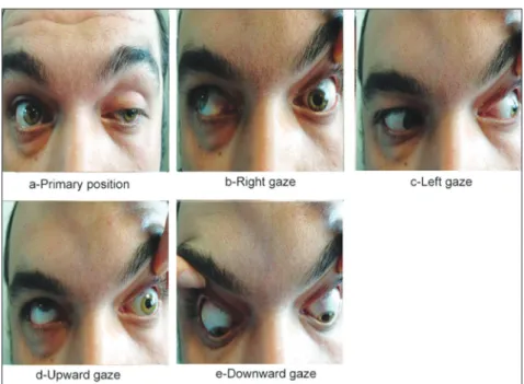
Di-rect and indiDi-rect light reflexes were positive on his right eye, and negative on his left eye. Left pupil was mydriatic and unresponsive to light. There was incomplete ptosis on his left eyelid. In primary po-sition, the left eye was deviated outward and downward. Supraduction of the left eye was absent, infraduction and adduction were reduced. All movements of the right eye were normal, as shown in Figure 1. Forced duction test was negative. Pa-tient’s diplopia complaint was getting worse, when he was forced to look upwards. His visual field analysis revealed no abnormalities. He had no other neurological signs or symptoms.
Complete blood count and biochemical analy-sis of the blood were normal. MRI demonstrated hematoma in mesencephalone on T1 and T2 weighted images, as shown in Figure 2. Patient also has arachnoid cyst seen in anterior part of the left temporal lobe on the MRI images, but that was not related to the patient’s clinical findings. Patient was consulted to Neurology and Neurosurgery Clinics and he was followed-up by neurologists and neu-rosurgeons, too. Since central caudal nucleus (the levator palpebralis nucleus) projects bilaterally and the superior rectus subnucleus projects contralat-erally, in our patient the pupillary involvement, unilateral ptosis and unilateral reduction in the movements of inferior, superior,medial recti and inferior oblique muscles, indicate that the fascicles within the midbrain were affected.
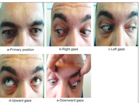
A gradual improvement of oculomotor nerve functions was observed during the subsequent fol-low-ups. Meanwhile he did not take any treat-ments. Three months later, patient’s symptoms decreased but didn’t resolve completely. Left pupil was responsive to light but lazy, ptosis was recov-ered, eye movements were better, but still they were reduced, as shown in Figure 3. MRI at that time demonstrated resolution of the hematoma and cavernoma or cavernous angioma (arterio-ve-nous malformation) in pons on T1 and T2 weighted images, shown in Figure 4. Neurosur-geons recommended gamma knife therapy (radia-tion therapy) for the patient to eradicate or minimize the lesion.
FIGURE 1: The primary position and the movements of right and left eyes of the patient.
DISCUSSION
Oculomotor nerve palsy may be congenital or ac-quired. Most acquired causes in adults are vascular diseases such as ischemia or infarction due to
dia-betes, hypertension and atherosclerosis, and hemor-rhage from arterio-venous malformation and hy-pertension, demyelinization and compression by intracranial aneurysms or other mass lesions like pri-mary or metastatic tumors. Infectious, inflammatory
FIGURE 3: The primary position and the movements of right and left eyes of the patient after three months.
and traumatic causes are less likely.5-14In our patient the cause was hematoma due to hemorrhage from cavernoma (arterio-venous malformation).
Identification of a total third nerve palsy is typically straightforward on examination, when there is ptosis, an eye that is deviated ‘down and out’ and a dilated pupil.[1,5]Our patient had total third nerve palsy, since he had ptosis, ‘down and out’ deviation and a dilated unreactive pupil. But it was incomplete third nerve palsy, because in-fraduction and adduction of the left eye were not absent but reduced and the ptosis was not com-plete.
Systemic hypertension and arterio-venous malformations represent the most common etiolo-gies of primary hematomas of the mesen-cephalone.15 De Mendonca et al.16 reported a 70-years-old hypertensive woman suffering from sudden onset of bilateral blepharoptosis, within few hours she developed focal signs attributable to a lesion of the mesencephalone and a stuporous state, from which she did not recover, neu-ropathological examination showed a hematoma in mesencephalone. Ramirez-Moreno et al. presented a case of hypertensive woman with an oculomotor nerve syndrome associated with limitation of horizantal gaze of the contralateral eye and reac-tive head inclination due to mesencephalic hematoma.17Durward et al. reported two patients presenting with a mesencephalic hematoma, the etiology was a proven arteriovenous malformation in one case and the other one was suspected to be due to hypertensive arteriolar rupture.18In our pa-tient, the etiology was also arteriovenous malfor-mation (cavernoma in pons).
The crossed projection of the superior rectus subnucleus indicates that unilateral third nerve le-sions with contralateral superior rectus involve-ment obligate a nuclear lesion, whereas a third nerve palsy with no contralateral superior rectus abnormality can not be caused by a nuclear lesion, furthermore, unilateral ptosis also shows that the lesion is not nuclear, because a single central nu-cleus (central caudal nunu-cleus), innervates both le-vator palpebrae muscles. Nuclear and fascicular nerve palsies often are associated with other
neu-rological signs such as pyramidal tract dysfunction, abnormalities of coordination and impaired con-sciousness, because of the large number of struc-tures located nearby, isolated palsies are very rare.2,15Due to unilateral ptosis and unilateral ex-traocular muscular involvement in our patient, the lesion was thought to be fascicular in origin. And other cranial nerves, motor, sensory, coor-dination and reflex systems were spared in our patient.
Edinger-Westphal nucleus provides para-sympathetic input to the pupillary sphincter and ciliary body. This nucleus and its fascicles are lo-cated at the top of the nuclear complex. Bilateral pupillary involvement with nuclear oculomotor palsy, therefore, is only expected in lesions with extension to the rostral midbrain.3,15 Pupil-spar-ing oculomotor palsy is most likely caused by mi-crovascular ischemia resulting from diabetes, hypertension and atherosclerosis.5Unilateral dif-ficulty in reading and unreactive pupil in our pa-tient were due to the destruction of the pupillomotor fibers located medially among the oculomotor fascicles.
Neurosurgical intervention (surgical evacua-tion) may be required in severe cases of mesen-cephalic hematoma.18 Careful observation for at least 6 months is essential before planning a stra-bismus and/or ptosis surgery, as some patients will show a partial or full spontaneous recovery. Most oculomotor nerve palsy due to microangiopathy re-solves completely in the first few months. When strabismus surgery is needed, the extent and sever-ity of extraocular muscle involvement and the presence of aberrant regeneration play a crucial role in surgical planning.5For our patient, neuro-surgeons recommended gamma knife therapy, which is a radiation therapy.
CONCLUSION
In conclusion, isolated unilateral oculomotor nerve palsy due to hematoma related to cavernoma in mesencephalone is seen very rarely and complete or incomplete recovery may be observed in such cases.
S
Soouurrccee ooff FFiinnaannccee
During this study, no financial or spiritual support was re-ceived neither from any pharmaceutical company that has a direct connection with the research subject, nor from a com-pany that provides or produces medical instruments and ma-terials which may negatively affect the evaluation process of this study.
C
Coonnfflliicctt ooff IInntteerreesstt
No conflicts of interest between the authors and / or family members of the scientific and medical committee members or
members of the potential conflicts of interest, counseling, ex-pertise, working conditions, share holding and similar situa-tions in any firm.
A
Auutthhoorrsshhiipp CCoonnttrriibbuuttiioonnss I
Iddeeaa//CCoonncceepptt:: Servet Çetinkaya; DDeessiiggnn:: Servet Çetinkaya; C
Coonnttrrooll//SSuuppeerrvviissiioonn:: Emine Mestan; DDaattaa CCoolllleeccttiioonn aanndd//oorr P
Prroocceessssiinngg:: Servet Çetinkaya; AAnnaallyyssiiss aanndd//oorr IInntteerrpprreettaattiioonn:: Servet Çetinkaya; LLiitteerraattuurree RReevviieeww:: Servet Çetinkaya; W Wrriitt--i
inngg tthhee AArrttiiccllee:: Servet Çetinkaya; CCrriittiiccaall RReevviieeww:: Emine Mes-tan.
1. Rucker JC. Acquired ocular motility disorders and nystagmus. In: Kidd DP, Newman NJ, Bi-ousse V, eds. Neuro-Ophthalmology. 4thed. Philadelphia: Elsevier Inc; 2008. p.312-31. 2. Donahue SP. Nuclear and fascicular disorders
of eye movement. In: Yanoff M, Duker JS, eds. Ophthalmology. 4th ed. Philadelphia, Pennsylvania: Elsevier Saunders; 2014. p.922-6.
3. Liu GT, Volpe NJ, Galetta SL. Eye movement disorders. In: Liu GT, Volpe NJ, Galetta S, eds. Neuro-Ophthalmology: Diagnosis and Management. 2nd ed. Philadelphia: Saunders Elsevier; 2010. p.491-550.
4. Trobe JD. Single ocular motor cranial nerve disorders. Rapid Diagnosis in Ophthalmology: Neuro-Ophthalmology. 1st ed. Philadelphia: Mosby/Elsevier; 2008. p.109-15.
5. Sadagopan KA, Wasserman BN. Managing the patient with oculomotor nerve palsy. Curr Opin Ophthalmol 2013;24(5):438-47. 6. Tokunaga M, Fukunaga K, Nakanishi R,
Watanabe S, Yamanaga H. Midbrain infarc-tion causing oculomotor nerve palsy and ipsi-lateral cerebellar ataxia. Intern Med 2014;53(18):2143-7.
7. Chang SI, Tsai MD, Wei CP. Posterior
com-municating aneurysm with oculomotor nerve palsy: clinical outcome after aneurysm clip-ping. Turk Neurosurg 2014;24(2):170-3. 8. Rajadhyaksha A, Phatak S, Nolkha N, Pathan
Y, Sonawale A. Rickettsial fever presenting with isolated third nerve palsy. J Assoc Physi-cians India 2013;61(2):144-5.
9. Chhabra MS, Golnik KC. Recovery of ocular motor cranial nerve palsy after herpes zoster ophthalmicus. J Neuroophthalmol 2014;34(1): 20-2.
10. Arrico L, Giannotti R, Ganino C, Lenzi T, Malagola R. Intracranial aneurysm and diplopia due to oculomotor nerve palsy: pre-and post-operative study. Clin Ter 2014; 165(4):258-62.
11. Sharma B, Gupta R, Anand R, Ingle R. Ocu-lar manifestations of head injury and incidence of post-traumatic ocular motor nerve involve-ment in cases of head injury: a clinical review. Int Ophthalmol 2014;34(4):893-900. 12. Adulkar NG, Arunkumar MJ, Kumar SM, Kim
U. Unusual case of temporal dermoid cyst pre-senting as oculomotor nerve palsy. Indian J Ophthalmol 2014;62(10):1032-4.
13. Mohebbi A, Jahandideh H, Harandi AA. Sphe-noid sinus mucocele as a cause of isolated
pupil-sparing oculomotor nerve palsy mimick-ing diabetic ophthalmoplegia. Ear Nose Throat J 2013;92(12):563-5.
14. Grabau O, Leonhardi J, Reimers CD. Recur-rent isolated oculomotor nerve palsy after ra-diation of a mesencephalic metastasis. Case report and mini review. Front Neurol 2014; 5:123.
15. Işikay CT, Yücesan C, Yücemen N, Culcuoglu A, Mutluer N. Isolated nuclear oculomotor nerve syndrome due to mesencephalic hematoma. Acta Neurol Belg 2000;100(4): 248-51.
16. de Mendonça A, Pimentel J, Morgado F, Ferro JM. Mesencephalic haematoma: case report with autopsy study. J Neurol 1990;237(1):55-8.
17. Ramírez-Moreno JM, Aguirre-Sánchez JJ, Durán-Herrera C, Parilla-Ramírez JL, Vialas-Simón J, González-Dorrego F. [Paralysis of the nucleus of the third cranial nerve second-ary to a mesencephalic hematoma]. Rev Neu-rol 1999;29(9):808-10.
18. Durward QJ, Barnett HJ, Barr HW. Presenta-tion and management of mesencephalic hematoma. Report of two cases. J Neurosurg 1982;56(1):123-7.