Full Terms & Conditions of access and use can be found at
https://www.tandfonline.com/action/journalInformation?journalCode=yhem20
Hematology
ISSN: (Print) 1607-8454 (Online) Journal homepage: https://www.tandfonline.com/loi/yhem20
The effects of iron treatment on viscosity in
children with cyanotic congenital heart disease
Semiha Terlemez, Fatma Sedef Tunaoğlu, Tayfun Göktaş, Bülent Çelik, Deniz
Erbaş, Ülker Koçak, Serdar Kula & Ayşe Deniz Oğuz
To cite this article: Semiha Terlemez, Fatma Sedef Tunaoğlu, Tayfun Göktaş, Bülent Çelik, Deniz Erbaş, Ülker Koçak, Serdar Kula & Ayşe Deniz Oğuz (2017) The effects of iron treatment on viscosity in children with cyanotic congenital heart disease, Hematology, 22:1, 30-35, DOI: 10.1080/10245332.2016.1207362
To link to this article: https://doi.org/10.1080/10245332.2016.1207362
Published online: 19 Jul 2016.
Submit your article to this journal
Article views: 832
View related articles
View Crossmark data
The effects of iron treatment on viscosity in
children with cyanotic congenital heart
disease
Semiha Terlemez
1, Fatma Sedef Tunaog
̆ lu
1, Tayfun Göktas
̧
2, Bülent Çelik
3,
Deniz Erbas
̧
2, Ülker Koçak
4, Serdar Kula
1, Ays
̧ e Deniz Oğuz
11
Medicine Faculty Pediatric Cardiology Department, Gazi University, Ankara, Turkey,2Medicine Faculty Physiology, Gazi University, Ankara, Turkey,3Chemistry Faculty Statistics, Gazi University, Ankara, Turkey,
4
Medicine Faculty Pediatric Hematology Department, Gazi University, Ankara, Turkey
Objective: This study was planned to determine the effects of iron treatment in children with cyanotic congenital heart disease.
Method and Materials: A total of 39 patients with cyanotic congenital heart disease including 20 (51%) females, 19 (49%) males and whose mean age was 9.9± 6.2 years, average weight was 33 ± 18.4 kg were evaluated. Patients were categorized into two groups as having iron deficiency and no iron deficiency with respect to their ferritin levels. 4 mg/kg/day iron treatment with two valences was applied to the groups with iron deficiency for 3 months. Clinical and laboratory findings of both groups were assessed at the outset and 3 months later and viscosity measurements were carried out.
Results:Iron deficiency was identified in 21 (53.8%) out of 39 patients. Average Hb and Hct values following 3-month iron treatment increased from 14.8± 2.4 g/dl to 16.0 ± 2.0 (P = 0.003) and from %45.8 ± 7.5 to %47.6± 7.2 (P = 0.052), respectively. Average viscosity value, however, was 5.6 ± 1.0 cP, it reduced to 5.5± 1.0 cP value by demonstrating very little reduction (P = 0.741). Nevertheless, O2sat value increased
from 71.7 to 75% and complaints such as headache, visual blurriness, having frequent sinusitis decreased. Conclusions: It was observed that iron treatment increased Hb and Hct levels in patients with cyanotic congenital heart disease without raising viscosity and it ensured improvement in clinical symptoms.
Keywords: Cyanotic congenital heart disease, Iron deficiency, Viscosity
Introduction
Secondary erythrocytosis is seen in cyanotic congeni-tal heart diseases due to low arterial oxygen saturation. This situation allows blood viscoelasticity to increase. Iron deficiency was observed in more than one-third of patients with cyanotic congenital heart disease.1,2 Microcytosis caused by iron deficiency, though, increases viscoelasticity and thrombosis.3 Replacement of iron deficiency is frequently rec-ommended in medical monitorings of patients with cyanotic congenital heart diseases. Nonetheless, while iron treatment restores microcytosis, it also increases hemoglobin level. Escalated hemoglobin and hematocrit values cause viscoelasticity to heighten. Given vicious cycle developed, advantage– disadvantage relation of iron treatment applied to cya-notic patients is unclear. How iron treatment affects
viscoelasticity in cyanotic congenital heart diseases has been unidentified.
This study was planned to determine the effects of iron treatment on viscoelasticity in children with cya-notic congenital heart disease.
Methods
Patients’ characteristics
Pediatric patients younger than 18 followed by Gazi University Medicine Faculty, diagnosed with cyanotic congenital heart disease were involved in the study. During evaluation, patients with bleeding findings that could lead to anemia, patients having blood trans-fusion in the last week and patients having iron treat-ment in the last 3 months were not included in the study.
Age, gender information, weight, height, and blood pressure values of patients were obtained. Detailed physical and neurological examinations were carried out in terms of neurological findings of increased vis-coelasticity in particular. Patients whose history
Correspondence to: Semiha Terlemez, Medicine Faculty Pediatric Cardiology Department, Gazi University, Ankara 06100, Turkey. Email:[email protected]
could be acquired (aged >5) were questioned with respect to hyperviscosity symptoms.
Definitions
During the first evaluation, complete blood count, serum iron, iron binding capacity (IBC), ferritin level, viscoelasticity, and transferrin saturation (TS) were examined in patients. Transferrin receptor level was also studied in the blood sample obtained to display iron deficiency in tissue level. Ferritin level was used for iron deficiency anemia in patients and patients with ferritin level lower than 12 ng/ml were diagnosed with iron deficiency anemia.4,5 Patients were divided into two groups according to their ferritin levels as with iron deficiency and non-iron deficiency. Iron deficiency treatment with two valences was orally initiated for patients with iron deficiency anemia as two doses, 4 mg/kg/day. Iron treatment was not applied to patients with no iron deficiency. Laboratory and clinical evaluations were repeated 3 months later in the groups that iron treatment was initiated and the treatment in question was not commenced.
Hemorheological measurements
Complete blood counts of patients were carried out by utilising Beckman Coulter LH 780 Analyzer and impedance method. Serum ferritin level was measured in Abbot Architect automatic analysis device. IBC was measured in Beckman Coulter AU 2700 plus autoana-lyzer. TSs were calculated as serum iron/total IBC × 100. Serum transferrin receptor determination was measured with ELISA method by using Makrosel human sTfR ELISA KIT. Viscoelasticity was measured with Vilastic Bioprofiler (Vilastic Scientific, Inc., Austin, TX, USA) by using oscillatory flow in a firm, cylindrical tube.
Statistics
Statistical Package for Social Sciences version 15 was utilized to evaluate results obtained from the study and form tables. Categorical variables (qualitative variables) were presented with frequency and percen-tage values, whereas quantitative variables were sub-mitted with mean, standard deviation, median, and minimum–maximum values. In the evaluation of cat-egorical variables, Chi-square test and when needed, Fisher exact test were used. Relations between vari-ables were analyzed with Pearson’s or Spearman’s cor-relation coefficients according to the situation of data. Receiver operating characteristic analysis was per-formed to determine cutoff points concerning some parameters. Materiality level was accepted as P < 0.05 values in all statistical analyses.
Results
A total of 39 patients were evaluated in the study. Of the patients, 20 (51%) were female, 19 (49%) were male, and it was established that their average age was 9.9± 6.2 years, average weight was 33 ± 18.4 kg. Average oxygen saturation was identified as 73.5± 8.2%.
The diagnoses of patients according to their rates, 12 (31%) Fallot tetralogy, 7 (17.9%) double outlet right ventricle, 7 (17.9%) ventricular septal defect (VSD)+ Eisenmenger syndrome, 4 (10%) transposi-tion of the great arteries+ pulmonary stenosis + VSD, 3 (7.7%) truncus arteriosus, 3 (7.7%) tricuspid atresia+ atrial septal defect (ASD) + VSD, 1 (2.6%) aorta–pulmonary window, 1 (2.6%) mitral atresia + ASD+ VSD, 1 (2.6%) pulmonary hypoplasia + VSD. Most frequently seen hyperviscosity symptom in patients was determined as headache. Of the hyper-viscosity symptoms, headache was seen in 25 (64.1%) patients, complaint of having sinusitis frequently in 23 (59%), visual blurriness in 21 (53.8%), nose bleed-ing in 9 (23.1%), tinnitus in 5 (12.8%).
Laboratory results
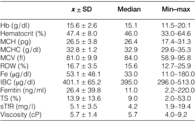
In the initial laboratory evaluations of patients, average hemoglobin levels were 15.6± 2.6 g/dl, hem-atocrit values were 47.4± 8.0%, MCH 26.5 ± 3.8 pg, MCHC 32.8± 1.2 g/dl, MCV 81.0 ± 9.9 fl, RDW 16.7± 3.5%, average iron levels 53.1 ± 48.1 μg/dl, IBC 401.1± 65.2 μg/dl, ferritin 26.4 ± 39.8 ng/ml, TS 13.9± 13.6%, sTfR 5.1 ± 5 mg/l, and viscosity was measured as 5.7± 1.4 cP (Table1).
Iron deficiency was determined in 21 (53.8%) out of 39 patients. No statistically significant difference was identified in terms of incidence of hyperviscosity symptoms between groups. No difference was estab-lished with respect to demographical features
Table 1 Initial laboratory data of patients
x ± SD Median Min–max Hb (g/dl) 15.6± 2.6 15.1 11.5–20.1 Hematocrit (%) 47.4± 8.0 46.0 33.0–64.6 MCH (pg) 26.5± 3.8 26.4 17.4–31.3 MCHC (g/dl) 32.8± 1.2 32.9 29.6–35.3 MCV (fl) 81.0± 9.9 84.0 58.9–95.8 RDW (%) 16.7± 3.5 15.6 12.7–25.9 Fe (μg/dl) 53.1± 48.1 33.0 11.0–180.0 IBC (μg/dl) 401.1± 65.2 395.0 296.0–513.0 Ferritin (ng/ml) 26.4± 39.8 11.0 2.2–220.0 TS (%) 13.9± 13.6 9.0 2.0–53.0 sTfR (mg/l) 5.1± 3.5 4.2 1.9–19.4 Viscosity (cP) 5.7± 1.4 5.7 4.0–9.2 Hb, Hemoglobin; Hematocrit, hematocrit; MCV, mean corpuscular volume; MCH, mean corpuscular hemoglobin; MCHC, mean corpuscular hemoglobin concentration; RDW, red blood cell distrubution width; Fe, iron; IBC, iron binding capacity; TS, transferrin saturation; sTfR, soluble transferrin receptor.
Terlemez et al. The effects of iron treatment on viscosity in children
between groups with iron deficiency and non-iron deficiency.
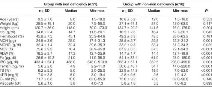
Average oxygen saturation value was 70% in the group diagnosed with iron deficiency and 75% in the group with non-iron deficiency. However, no statisti-cally significant difference was identified (P = 0.140). Average Hb, Hct values were measured, respect-ively, as: 14.8± 2.4 g/dl and 45.8 ± 7.5% in the group with iron deficiency, 16.5± 2.5 g/dl and 49.2 ± 8.3% in the group with non-iron deficiency. Hb and Hct values were found lower in the group with iron deficiency (Table 2). Other hematological par-ameters between groups with iron deficiency and non-iron deficiency were as shown in Table 2. Transferrin receptor levels were established as 7.0± 3.8 mg/l in patients diagnosed with iron deficiency and 2.8± 0.6 mg/l (P < 0.001) in patients with no iron deficiency (Table2).
Hematologic parameters displaying correlation that ferritin level in patients had been lower than 12 ng/ml value were evaluated; a correlation between MCH, MCHC, MCV lowness, and RDW highess was deter-mined; however, the highest correlation was estab-lished as (0.565) between MCH (0.565) and MCV (0.533) values. It was seen that values below 28 pg for MCH formed 83% susceptibility, 86% specificity, and 85% accurateness ( predictive value) and cutoff values below 82.8 fl values for MCV accounted for 89% susceptibility, 81% specificity, and 85% accurate-ness and cutoff value.
4 mg/kg/day divalent iron therapy was given to 21 patients diagnosed with iron deficiency orally by divid-ing two doses. It was identified in the group with iron treatment after 3 months that visual blurriness reduced in one patient; tinnitus recovered in two patients and decreased in one patient; headache complaint
disappeared in five patients and diminished in one; sinusitis rate reduced in four patients and nose bleed-ing frequency decreased in one patient.
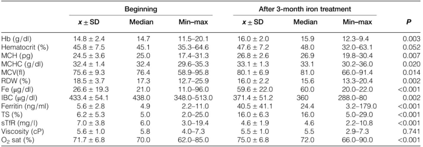
Initial hematologic values and those after 3 months were compared in patients who had been applied iron treatment. It was established that Hb value increased approximately from 14.8± 2.4g/dl to 16.0 ± 2.0 (P = 0.003) 3 months later. Although there was an increase in hemoglobin and hematocrit values, vis-cosity value was approximately 5.6± 1.0 cP initially, however, it was determined as 5.5± 1.0 cP after 3-month iron treatment (P = 0.741). In addition, initial average oxygen saturation increased from 71.7± 6.8 to 75.0± 6.8% in the group that iron treatment had been applied; this increase was found statistically sig-nificant (P < 0.001) (Table 3).
Hematologic evaluations of 18 patients who had not received iron treatment were repeated 3 months later. It was observed that there was a statistically significant decrease in Hb, Hct and MCHC levels of patients and a statistically considerable increase in RDW value. Though no iron deficiency developed in patients, there was a statistically significant decrease in serum iron, ferritin, TS, sTfR levels, on the other hand, no change was seen in viscosity values (Table4).
Pulmonary arterial hypertension (PAH) regarding congenital heart disease was present in 17 out of 39 patients in the study. Patients with pulmonary hyper-tension were compared to other patients in terms of clinical and laboratory findings. Iron treatment was applied to 9 out of 17 patients with pulmonary hyper-tension whose ferritin level was determined below 12 ng/ml. Iron deficiency was identified in 12 out of 22 patients with no pulmonary hypertension. Initial viscosity average of patients with pulmonary hyperten-sion was 5.4± 1.3 cP, viscosity value after 3-month
Table 2 Comparison of two groups with and without iron deficiency in terms of their demographical and laboratory values Group with iron deficiency (n:21) Group with non-iron deficiency (n:18)
P x ± SD Median Min–max x ± SD Median Min–max
Age (years) 9.2± 7.0 8.0 1.5–19.0 10.8± 5.2 12.0 1.5–18.0 0.553 Weight (kg) 29.5± 19.1 20.0 7.5–58.0 37.1± 17.1 37.0 13.0–63.0 0.117 Height (cm) 120.7± 36.9 118.0 70.0–173.0 134.7± 29.2 141.5 76.0–175.0 0.204 Hb (g/dl) 14.8± 2.4 14.7 11.5–20.1 16.5± 2.5 16.4 12.7–20.1 0.042 Hematocrit (%) 45.8± 7.5 45.1 35.3–64.6 49.2± 8.3 48.5 33.0–63.5 0.181 MCH (pg) 24.5± 3.6 25.0 17.4–31.3 28.8± 2.7 29.5 22.3–31.2 0.001 MCHC (g/dl) 32.4± 1.4 32.4 29.6–35.3 33.2± 0.8 33.4 31.2–34.3 0.029 MCV (fl) 75.6± 9.3 76.4 58.9–95.8 87.2± 6.5 87.5 72.1–94.3 <0.001 RDW (%) 18.5± 3.7 17.3 12.7–25.9 14.7± 1.7 14.4 12.7–19.9 <0.001 Fe (μg/dl) 26.6± 19.3 21.0 11.0–96.0 84.1± 53.4 61.0 28.0–180.0 <0.001 IBC (μg/dl) 433.4± 54.1 438.0 348.0–513.0 363.4± 57.1 352.5 296.0–495.0 0.001 Ferritin (ng/ml) 5.6± 2.8 4.9 2.2–11.0 50.6± 48.7 34.7 14.0–220.0 <0.001 TS (%) 6.2± 5.3 5.0 2.0–25.0 22.9± 14.8 19.5 7.0–53.0 <0.001 sTfR (mg/l) 7.0± 3.8 6.0 3.0–19.4 2.8± 0.6 2.6 1.9–4.2 <0.001 O2sat (%) 71.7± 6.8 70.0 62.0–85.0 75.6± 9.2 75.0 62.0–90.0 0.140 Viscosity (cP) 5.6± 1.0 5.8 4.0–7.3 5.8± 1.8 5.3 4.0–9.2 0.899 Hb, Hemoglobin; Hematocrit, hematocrit; MCV, mean corpuscular volume; MCH, mean corpuscular hemoglobin; MCHC, mean corpuscular hemoglobin concentration; RDW, red blood cell distrubution width; Fe, iron; IBC, iron binding capacity; TS, transferrin saturation; sTfR, soluble transferrin receptor.
treatment was measured as 5.4± 1.2 cP. Hb, Hct, and viscosity values did not show any difference with iron treatment, however, average oxygen saturation raised from 77.2± 6.8% (before treatment) to 78.6 ± 6.6% (after treatment) (P = 0.021).
Discussion
Hyperviscosity symptoms were questioned for the first time in pediatric patients with cyanotic congenital heart disease in the present study. In studies that adult patients with cyanotic heart disease were ana-lyzed with symptom scoring by Perloff et al.,6,7 mostly findings (hemoptysis, easy bruising, etc.) regarding hemostasis impairments were seen. This study indicated that hyperviscosity symptoms are different in children compared to adults.
Iron deficiency was determined in 21 (53.8%) out of 39 patients. Iron deficiency was seen in more than half of the patients. This result shows a similarity with the study outcomes carried out previously in children with
cyanotic congenital heart disease.8,9Although second-ary erythrocytosis was present in all patients in our study, Hb and Hct values of the group with iron deficiency were found lower than those without iron deficiency. Broberg et al.1 had carried out a similar study in adult patients with cyanotic congenital heart disease previously. By categorizing patients as those with and without iron deficiency, they compared both viscosities and exercise capacities of them. The results of our study show difference in some points compared to their study outcomes. Broberg et al. determined Hb levels of patients with iron deficiency lower than those without iron deficiency, however, Hct levels similar. Compared to our study, despite higher oxygen saturation in adult patients, higher Hb and Hct levels were established.1We identified lower Hb and Hct levels in spite of lower oxygen saturation in children with cyanotic congenital heart disease. In addition, Hct levels were determined lower in the group with iron deficiency compared to those
Table 3 Evaluation of iron treatment effects on viscosity and other hematologic parameters
Beginning After 3-month iron treatment
P x ± SD Median Min–max x ± SD Median Min–max
Hb (g/dl) 14.8± 2.4 14.7 11.5–20.1 16.0± 2.0 15.9 12.3–9.4 0.003 Hematocrit (%) 45.8± 7.5 45.1 35.3–64.6 47.6± 7.2 48.0 32.0–63.1 0.052 MCH (pg) 24.5± 3.6 25.0 17.4–31.3 26.8± 2.6 26.9 19.8–30.4 0.007 MCHC (g/dl) 32.4± 1.4 32.4 29.6–35.3 33.1± 1.3 33.1 30.2–36.0 0.020 MCV(fl) 75.6± 9.3 76.4 58.9–95.8 80.1± 6.9 81.0 66.0–91.4 0.014 RDW (%) 18.5± 3.7 17.3 12.7–25.9 16.0± 2.2 15.6 13.3–20.4 0.002 Fe (μg/dl) 26.6± 19.3 21.0 11.0–96.0 59.6± 22.0 60.0 20.0–22.0 <0.001 IBC (μg/dl) 433.4± 54.1 438.0 348.0–513.0 371.4± 51.2 360 288.0–80 0.002 Ferritin (ng/ml) 5.6± 2.8 4.9 2.2–11.0 40.5± 41.1 24.4 3.2–179.0 <0.001 TS (%) 6.2± 5.3 5.0 2.0–25.0 16.0± 6.3 16.0 5.0–29.0 <0.001 sTfR (mg/l) 7.0± 3.8 6.0 3.0–19.4 4.6± 1.9 4.6 2.2–10.8 <0.001 Viscosity (cP) 5.6± 1.0 5.8 4.0–7.3 5.5± 1.0 5.5 2.9–7.3 0.741 O2sat (%) 71.7± 6.8 70.0 62.0–85.0 75.0± 6.8 72.0 66.0–90.0 <0.001
Hb, Hemoglobin; Hematocrit, hematocrit; MCV, mean corpuscular volume; MCH, mean corpuscular hemoglobin; MCHC, mean corpuscular hemoglobin concentration; RDW, red blood cell distrubution width; Fe, iron; IBC, iron binding capacity; TS, transferrin saturation; sTfR, soluble transferrin receptor.
Table 4 Comparison of control hematologic values in the non-iron treatment group
Beginning After 3-month treatment
P x ± SD Median Min–max x ± SD Median Min–max
Hb (g/dl) 16.5± 2.5 16.4 12.7–20.1 15.7± 2.3 15.7 11.8–19.3 0.001 Hematocrit (%) 49.2± 8.3 48.5 33.0–63.5 47.6± 9.0 46.0 32.0–63.0 0.001 MCH (pg) 28.8± 2.7 29.5 22.3–31.2 28.1± 2.8 29.0 22.0–32.0 0.162 MCHC (g/dl) 33.2± 0.8 33.4 31.2–34.3 32.3± 1.5 32.9 29.0–34.4 0.002 MCV(fl) 87.2± 6.5 87.5 72.1–94.3 86.6± 6.6 86.0 70.0–94 0.085 RDW (%) 14.7± 1.7 14.4 12.7–19.9 15.5± 1.6 15.3 13.0–20 <0.001 Fe (μg/dl) 84.1± 53.4 61.0 28.0–180 57.2± 31.0 48.5 14.0–120 0.001 IBC(μg/dl) 363.4± 57.1 352.5 296–495 366.2± 43.8 360.0 305–480 0.983 Ferritin (ng/ml) 50.6± 48.7 34.7 14.0–220 31.5± 22.7 23.0 12.0–90 <0.001 TS (%) 22.9± 14.8 19.5 7.0–53.0 16.5± 8.0 15.5 5.0–34. 0.004 sTfR (mg/l) 2.8± 0.6 2.6 1.9–4.2 3.2± 0.9 3.0 2.2–5.8 0.001 Viscosity (cP) 5.8± 1.8 5.3 4.0–9.2 5.8± 1.8 5.5 3.8–9.2 0.407 O2sat (%) 75.6± 9.2 75.0 62.0–90.0 75.1± 9.0 74.0 62.0–90.0 0.070
Hb, Hemoglobin; Hematocrit, hematocrit; MCV, mean corpuscular volume; MCH, mean corpuscular hemoglobin; MCHC, mean corpuscular hemoglobin concentration; RDW, red blood cell distrubution width; Fe, iron; IBC, iron binding capacity; TS, transferrin saturation; sTfR, soluble transferrin receptor.
Terlemez et al. The effects of iron treatment on viscosity in children
without. The difference between adult patient groups and pediatric patients can be related to that children have higher physiological10 iron need and their iron consumption is faster in their growth and development period. Increased iron need due to secondary erythro-cytosis in children with cyanotic congenital heart disease is added to these factors. The results obtained from the study made us think that iron deficiency can pressure erythropoiesis despite secondary erythrocyto-sis in children with cyanotic congenital heart disease. Clinical and laboratory evaluations of patients whose iron levels were normal were repeated 3 months later. Though no iron deficiency anemia devel-oped, patients’ Hb, Hct, MCH, MCV, iron, ferritin, TS levels decreased, however, RDW, sTfR levels increased. Hematologic parameters changing in favor of iron deficiency were thought to be associated with continuing iron consumption owing to ongoing sec-ondary erythrocytosis in children with cyanotic conge-nital heart disease. In a study conducted in 44 cyanotic congenital heart disease pediatric patients by Onur et al.,9
it was seen in the patient group followed up with no treatment that Hb, Hct, ferritin values were decreasing after 3-month observation. These findings can bring forward iron treatment application from maintenance dose even if there is no iron deficiency in children with cyanotic congenital heart disease.
Although Hb, Hct values elevated after 3-month iron treatment application to patients with iron defi-cieny, viscosity values did not raise. This finding is a significant result obtained from the study. There are many causes affecting blood viscoelasticity, most widely known is the number of shaped elements of blood. Nevertheless, the morphology of erythrocytes plays a profound role in blood viscoelasticity. In pro-portion with the severity of iron deficiency, erythro-cytes lose their shape shifting characteristic.11 This situation causes viscosity to increase by aggravating their movement in blood’s downstream.8,12,13No stat-istically significant difference was identified in our study in terms of their Hct levels between cyanotic congenital heart diseased children with iron deficiency and those without iron deficiency. Viscosity values of patients with and without iron deficiency were also found similar. This result is not surprising when vis-cosity is thought to be closely related to hematocrit level. On the other hand, although Hct levels increased after 3-month iron treatment in patients with iron deficiency, no elevation was established in viscosity levels and some decrease was observed even it was of no significance statistically. This situation shows the effects of Hct level on viscosity and also the impor-tance of impairments in erythrocyte morphology seen in iron deficiency.
In a study performed on 123 adult patients with cya-notic congenital heart disease by Tay et al.14, they
applied oral iron treatment for 3 months by determin-ing iron deficiency in 25 patients. Three months later, it was seen that Hb and Hct values increased with exer-cise capacity. It was also observed in our study that there was a decrease in hyperviscosity symptoms fol-lowing iron treatment. Iron deficiency is a condition that must be treated in patients with cyanotic congeni-tal heart disease.
Of all the patients, PAH depending on congenital heart disease was determined in 17. Patients with PAH were also grouped into two with and without iron deficiency in itself. In the controls of PAH patients who had been applied iron treatment, a sig-nificant increase was established in Hb and Hct levels. Viscosity levels, however, stayed at the same values. Nevertheless, a considerable increase was observed in oxygen saturations of patients after iron treatment (P = 0.021). Pulmonary vascular resistance is high in patients with pulmonary hypertension patients differently from other patients. Hematocrit elevation may lead to increase of pulmonary arterial resistance and severity of PAH symptoms in PAH patients with cyanotic congenital heart disease.15,16 Viscosity did not increase with iron treatment in group with PAH in our study. Oxygen saturation levels, though, increased with iron treatment. Iron treatment appears to be beneficial in pediatric PAH patients with cyanotic congenital heart disease. However, the number of patients is very limited for our claim.
Another result obtained from the study was that values below 28 pg for MCH and ferritin level being under 12 ng/ml value and values below 28 pg for MCH accounted for 83% susceptibility, 86% speci-ficity, and 85% accurateness ( predictive value) and value below 82.8 fl for MCV formed 89% suscepti-bility, 81% specificity and 85% accurrateness and cutoff value. These values could be used to predict iron deficiency in patients with secondary erythrocytosis.
The issues that our study was carried out in single center and there were limited number patients involved were important restrictive factors. In addition, the effects of iron treatment on viscosity in patients with cyanotic congenital heart disease were compared on patients with and without iron deficiency. Study results could become more worthwhile if both groups are also compared with the patients who have no con-genital heart disease but are applied iron treatment due to their iron deficiency situation.
Conclusions
Iron deficiency is a frequently seen condition in patients with cyanotic congenital heart disease. Results obtained from the study indicated that iron treatment increased Hb and Hct levels without
elevating viscosity in patients with cyanotic congenital heart disease and it caused improvement in clinical symptoms.
Acknowledgments
We thank the staff of Gazi University Pediatric Cardiology a lot for their help.
Disclaimer statement
Contributors Grant support: Gazi University research Project, Number: 01/2011-93.
Funding None.
Conflicts of interest None.
Ethics approval Gazi University Medicine Faculty ethics committee approval was obtained for the study. Families and patients were informed regarding the study and informed voluntary confirmation form approval was acquired from families and pediatric school-age patients.
References
1 Broberg CS, Bax BE, Okonko DO, Rampling MW, Bayne S, Harries C, et al. Blood viscosity and its relationship to iron deficiency, symptoms, and exercise capacity in adults with cyano-tic congenital heart disease. J Am Coll Cardiol. 2006;48(2): 356–65.
2 Kaemmerer H, Fratz S, Braun S, Koelling K, Eicken A, Brodherr-Heberlein S,et al. Erythrocyte indexes, iron metab-olism, and hyperhomocysteinemia in adults with cyanotic conge-nital cardiac disease. Am J Cardiol.2004;94(6):825–8. 3 Milligan DW, MacNamee R, Roberts BE, Davies JA. The
influ-ence of iron-deficient indices on whole blood viscosity in poly-cythaemia. Br J Haematol.1982;50(3):467–71.
4 Thomas DW, Hinchliffe RF, Briggs C, Macdougall IC, Littlewood T, Cavill I. British Committee for Standards in Haematology. Guideline for the laboratory diagnosis of func-tional iron deficiency. Br J Haematol.2013;161(5):639–48. 5 Turkish Hematology Association. Iron Deficiency Anemia in
Children, Diagnosis and Treatment Guidelines2011.
6 Perloff JK, Rosove MH, Child JS, Wright GB. Adults with cya-notic congenital heart disease: hematologic management. Ann Intern Med.1988;109:406–13.
7 Rosove MH, Perloff JK, Hocking WG, Child JS, Canobbio MM, Skorton DJ. Chronic hypoxaemia and decompensated ery-throcytosis in cyanotic congenital heart disease. Lancet.1986;2: 313–5.
8 Linderkamp O, Klose HJ, Betke K, Brodherr-Heberlein S, Bühlmeyer K, Kelson S, et al. Increased blood viscosity in patients with cyanotic congenital heart disease and iron deficiency. J Pediatr.1979;95(4):567–9.
9 Onur CB, Sipahi T, Tavil B, Karademir S, Yoney A. Diagnosing iron deficiency in cyanotic heart disease. Indian J Pediatr.
2003;70(1):29–31.
10 Berglund S, Domellöf M. Meeting iron needs for infants and children. Curr Opin Clin Nutr Metab Care. 2014;17(3): 267–72.
11 Brandao MM, Castro Mde L, Fontes A, Cesar CL, Costa FF, Saad ST, et al. Impaired red cell deformability in iron deficient subjects. Clin. Hemorheol. Microcirculation.2009;43 (3):217–21.
12 Schmid-Schönbein H, Wells R, Goldstone J. Influence of deformability of human red cells upon blood viscosity. Circ Res.1969;25(2):131–43.
13 Whitmore RL. Rheology of the circulation. 1st ed. London: Pergamon Press; 196.
14 Tay EL, Peset A, Papaphylactou M, Inuzuka R, Alonso-Gonzalez R, Giannakoulas G. Replacement therapy for iron deficiency improves exercise capacity and quality of life in patients with cyanotic congenital heart disease and/or the Eisenmenger syndrome. Int J Cardiol.2011;151(3): 307–12.
15 Barer GR, Bee D, Wach RA. Contribution of polycythaemia to pulmonary hypertension in simulated high altitude in rats. J Physiol.1983;336:27–38.
16 Benis AM, Usami S, Chien S. Effect of hematocrit and inertial losses on pressure-flow relations in the isolated hindpaw of the dog. Circ Res.1970;27:1047–68.
Terlemez et al. The effects of iron treatment on viscosity in children