©2017 by Quintessence Publishing Co Inc.
Reconstruction of Localized Alveolar Defects
Nur Altiparmak, DDS, PhD1/Sina Uckan, DDS, PhD2/Burak Bayram, DDS, PhD3/Sıdıka Soydan, DDS, PhD4 Purpose: The aim of this study was to compare the complication rates of recipient sites prepared using
two incision techniques: crestal and tunnel. Materials and Methods: In this prospective study, patients
underwent augmentation procedures (68 patients; 75 sites) by the same surgeon that were performed consecutively using the crestal incision technique (27 horizontal, 10 vertical; crestal group) or the tunnel incision technique (27 horizontal, 11 vertical; tunnel group). Autogenous bone block grafts were harvested with a piezoelectric surgical device, and the grafts were fixed at the recipient sites by two titanium screws in both groups. The authors evaluated minor exposure, transient paresthesia, major exposure, permanent paresthesia, gingival recession at adjacent teeth, surgery time, and visual analog scale pain scores. Results:
Soft tissue dehiscence and graft failure were significantly lower in patients undergoing the tunnel technique.
Conclusion: The tunnel incision technique significantly decreased soft tissue exposure, the most common
complication of augmentation procedures with autogenous onlay bone grafts. This technique should be considered an alternative to the crestal incision technique for preparation of the recipient site. Int J Oral
MaxIllOfac IMplants 2017;32:1103–1110. doi: 10.11607/jomi.5275
Keywords: alveolar ridge augmentation, autogenous bone, complication, failure, minimally invasive surgery,
subperiosteal tunnel technique
I
nsufficient bone height and width is one of the most common problems in dental rehabilitation of partially and totally edentulous patients. Such defects involve hard and soft tissues in a wet, mobile environment, and the alveolar ridge is one of the most difficult areas of the human body to reconstruct. A favorable interarch relationship must be restored before endosseous implants can be inserted. Alveolar augmentation procedures are frequently used in maxillofacial surgery. Several bone grafting techniques, such as augmenta-tion with autologous block bone grafts, distracaugmenta-tion osteogenesis, and guided tissue regeneration, as well as several bone substitute materials and synthetic materials, have been developed.1,2Bone grafting techniques for alveolar reconstruction are well documented in the literature. A crestal inci-sion from the top of the crest is the preferred inciinci-sion technique for preparation of the recipient site in onlay bone augmentation procedures with autogenous grafts. Despite excellent results reported in studies,1–5 failure of onlay bone grafts may occur from instability, infection, and exposure of the graft in clinical practice. However, the most common complication is exposure of the bone graft during the early healing period.6–8
Several surgical techniques have been developed to minimize the graft exposure rates and maintain the soft tissue. The tunnel incision technique is a less invasive method used in an effort to decrease wound dehiscence complications in augmentation procedures with autogenous or synthetic bone grafts. Ridge aug-mentation using subperiosteal tunneling dissection is a closed and partially blind procedure because it does not permit a direct view of the deficient ridge; however, this technique allows access to the recipient area with minimal tissue dissection and handling. The procedure requires patience and delicate surgical maneuvers to dissect and develop the subperiosteal flap that enables formation of the pocket.
The tunnel technique is used to insert particulated bone graft materials; however, the success rate associated with use of particulated materials is low.9 Although particulated graft material is easily inserted through the flap, augmentation 1Fellow, Department of Oral and Maxillofacial Surgery,
Faculty of Dentistry, Baskent University, Ankara, Turkey.
2Dean, Department of Oral and Maxillofacial Surgery,
Faculty of Dentistry, Medipol University, Istanbul, Turkey.
3Associate Professor, Department of Oral and Maxillofacial
Surgery, Faculty of Dentistry, Baskent University, Ankara, Turkey.
4Assistant Professor, Department of Oral and Maxillofacial
Surgery, Faculty of Dentistry, Baskent University, Ankara, Turkey.
Correspondence to: Dr Nur Altiparmak, Baskent Universitesi
Dishekimligi Fakultesi, 11.sok no. 26, 06490 Bahcelievler, Ankara, Turkey. Fax: +903122152962. Email: [email protected]
without fixation is controversial.9 Since the introduction of new membranes and bone graft materials, several studies have been published highlighting improvements to this technique.10–14 Particulate graft materials were used in these studies, and membrane fixation through tunneling dissection resulted in reasonable success; however, the technique is complex and requires a high level of surgical skill and extended time for the procedure.
To the best of the authors’ knowledge, no controlled studies have reported the success of augmentation pro-cedures with autogenous block bone grafts at recipient sites prepared via the tunnel technique. The purpose of this study was to compare minor and major complication rates of recipient sites prepared via the crestal incision and tunnel incision techniques. The investigators hypoth-esized that the tunnel technique prevents or diminishes postoperative graft exposure with fewer postoperative complications than those induced by the crestal technique. Accordingly, the aim of this study was to demonstrate a lower graft exposure rate by the tunnel technique than by the crestal incision technique during the healing period.
MATERIALS AND METHODS
Study Design
This controlled prospective study was approved by the Baskent University Medical and Health Sciences Research Council and Ethics Committee, Ankara, Turkey (project no. D-KA13/04) and supported by the Baskent University Research Fund.
The same surgeon performed all operations, which were done consecutively using the crestal incision technique (crestal group) or the tunnel incision tech-nique (tunnel group). Thus, an augmentation procedure performed using the tunnel technique was followed by an augmentation procedure performed using the crestal technique.
The ramus or symphysis was used as the donor site. Selection of the recipient site was based on the amount of required bone and anatomical variations. The mean size of the harvested graft was 4 mm in width, 10 to 12 mm in length, and 6 mm in height for both the symphysis and ramus grafts.
Fig 1 (Left) Incision at recipient site in the tunnel group.
Fig 2 (Right) Dissection at recipient site in the tunnel group.
Figs 3a to 3f Bone block graft adaptation and fixation through the tunnel.
a b c
Surgical Technique
A vertical incision was planned in the tunnel group. The incision was made as long as possible without harming the anatomical structures of the recipient site (5 mm in front of the defect) after infiltrative articaine anesthetic (Ultracain DS Forte, Aventis) was administered. After the incision was made, the tunnel in which the graft was to be placed was prepared at the recipient site by dissecting the periosteum from the alveolar bone in the tunnel, with care taken not to traumatize the soft tissue (Figs 1 and 2).
In the crestal incision group, the length of the incision was determined by the size of the defect in the recipient site. After administration of infiltrative anesthetic, the incision was made from the top of the crest, and relaxing vertical incisions were then made by removal of the mucoperiosteal flap. Preparation of the recipient site was completed by making horizontal periosteal incisions parallel to the crestal incision for tension-free closure.
After completion of the ramus or symphysis donor site, a platelet-rich fibrin membrane was prepared by centrifugation of 10-mL blood samples from patients for 12 minutes at 2,700 rpm (PC-02 centrifuge, Process). In both groups, immobilization of the block grafts to the recipient site was ensured by placement of two titanium screws with an 8- or 10-mm length and a 1.3- or 1.5-mm diameter (Synthes). In the tunnel group, titanium screws were placed by dissecting the tunnel. The screws could be placed through the tunnel because all recipient sites were reconstructed with the insertion of one or two implants in one or two edentulous areas. If a lengthy edentulous area had been augmented by means of the tunnel technique, it might have been difficult to reach the posterior side to place the bone screws. After the particulated bone graft material (0.25 to 1.00 mm, 0.5 mL, Bio-Oss, Geistlich Pharma AG) was placed in the gaps between the recipient site and the block graft, the block graft and particulate graft were covered with the platelet-rich fibrin15 membrane. The recipient site was primarily closed with 3-0 Vicryl suture (Figs 3 and 4).
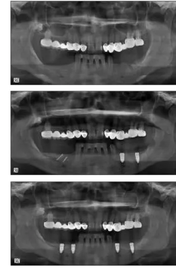
Fig 4 (a) Preoperative clinical image. (b) Postoperative clinical image. (c) Image of augmented crest at time of implant inser-tion. (d) Preoperative radiographic image. (e) Postoperative ra-diographic image. (f) Rara-diographic image after implant insertion.
a b c d e f
Study Variables
Incision type (tunnel or crestal) was the primary predic-tor variable. The other study variables were the recipi-ent site (maxilla or mandible) and the augmrecipi-entation procedure characteristic (vertical or horizontal). The primary outcomes of this study were minor and major graft exposure, and the secondary outcomes were skin or mucosal paresthesia and gingival recession at adjacent teeth.
Data Collection Methods
Follow-up examinations were performed at 3 days, 7 days, 15 days, 21 days, 1 month, and 3 months during the 6-month waiting period before implant placement. In both the tunnel and crestal groups, implants (Straumann or Nobel Biocare) were placed in the augmented sites if no changes in the treatment prognosis related to major complications had occurred.
Complications that precluded the proposed treatment procedure and prosthetic rehabilitation were classified as major. Complications that necessitated no management or those that could be managed easily were considered minor. In both groups, the following parameters were evaluated at the follow-up visits during the 6-month waiting period before placement of the implants. • Minor complications (transient paresthesia [yes/
no], minor exposure of the graft [yes/no])
• Major complications (major exposure of the graft [yes/no], permanent paresthesia [yes/no]) • Operation time (time from the first incision to
placement of the last suture)
• Patient-reported intraoperative (ie, immediately after surgery) and postoperative (ie, 1 month after surgery) visual analog scale (VAS) pain scores (0 cm = no pain, 10 cm = most severe pain)
• Gingival recession at adjacent teeth (comparison of preoperative and 6-month follow-up photographs) • Implant placement in augmented area (yes/no)
Data Analyses
Data analysis was performed using statistical software (SPSS version 17.0, IBM SPSS). Categorical variables were analyzed with Pearson’s chi-square or Fisher’s exact test, where appropriate. Multiple binary logistic regression analyses were performed to determine the effects of the incision techniques (crestal vs tunnel) on both primary (ie, minor and major graft exposure) and secondary (eg, temporary skin or mucosal paresthesia) outcomes after adjustment for the study variables (procedure and recipient site). The odds ratio (OR) and 95% confidence interval (CI) were also calculated for each variable. A P value of < .05 was considered statistically significant.
RESULTS
The study was composed of 68 volunteers (39 women and 29 men; mean age, 41.5 years) with alveolar crest atrophy who were admitted to the Oral and Maxillofacial Surgery Clinic of Baskent University, Faculty of Dentistry, for placement of dental implants from February 2013 to January 2014. In this study, patients require a maximum of three implants after block bone grafting. All participants had an American Society of Anesthesiologists physical status of I ( a normal, healthy patient), and all provided informed consent for participation.
Thirty-four ramus and 41 symphysis grafts were used for the alveolar crest reconstruction. A tunnel incision was used in 38 augmentation procedures (27 horizontal, 11 vertical), and a crestal incision was used in 37 aug-mentation procedures (27 horizontal, 10 vertical). Table 1 All Study Variables Versus the
Predictor Variable (Crestal or Tunnel Incision)
Crestal (n = 37)
No. (%) Tunnel (n = 38) No. (%) P value
Augmentation procedure .853 Horizontal 27 (73.0) 27 (71.1) Vertical 10 (27.0) 11 (28.9) Recipient site .298 Maxilla 21 (56.8) 17 (44.7) Mandible 16 (43.2) 21 (55.3)
Fig 5 Postoperative incision clinical image; major exposure ob-served on crestal line.
Table 1 presents the comparisons between the pre-dictor variable (experimental and control groups) and the study variables (vertical or horizontal, maxilla or mandible). No statistically significant difference in the horizontal or vertical location was present between the crestal and tunnel groups (P = .853). In addition, no statistically significant difference was observed in the position of the recipient site (maxilla or mandible) between the crestal and tunnel groups (P = .298).
At the 6-month follow-up examination, minor exposure was observed at 4 of 38 augmented recipient sites in the tunnel group and at 12 of 37 augmented recipient sites in the crestal group. The minor exposure did not affect the treatment prognosis; therefore, the implants were placed as planned.
Major exposure was observed at one recipient site in the tunnel group, and the graft was lost as a result of uncontrollable infection. Thus, the implant could not be placed at that recipient site. In the crestal group, total resection of the graft necessitated by major exposure
was performed in two patients and partial resection was performed in one patient (Fig 5).
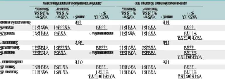
Comparisons between the primary outcome variable (minor or major complications) and the research variables (vertical or horizontal, maxilla or mandible) are shown in Table 2. The incidence of minor complications was 4.080 times higher in the crestal group than in the tunnel group (95% CI, 1.176–14.153; P = .021). The investigators did not observe any statistically significant effect of a crestal incision on the incidence of major complications (OR, 3.265; 95% CI, 0.324–32.909; P = .358) (Table 3).
In all patients, implants were inserted into the aug-mented sites 6 months after the augmentation surgery (59 implants in the tunnel group, 63 implants in the crestal group).
Table 4 presents the logistic regression models indicating whether the primary predictor variables (tunnel or crestal incision) had any effect on the primary outcome variables (minor or major complications) after adjusting for the other study variables (vertical or horizontal, maxilla or mandible). Table 3 Primary Predictor Variable Versus Primary Outcome Variable
Minor exposure Major exposure
Absent (n = 59) No. (%)
Present (n = 16)
No. (%) P value (95% CI)OR
Absent (n = 71) No. (%)
Present (n = 4)
No. (%) P value (95% CI)OR
Incision technique .021 .358 Tunnel 34 (57.6) 4 (25.0) 1.000 37 (52.1) 1 (25.0) 1.000 Crestal 25 (42.4) 12 (75.0) 4.080 (1.176–14.153) 34 (47.9) 3 (75.0) 3.265 (0.324–32.909) OR = odds ratio; CI = confidence interval.
Table 2 All Study Variables Versus Primary Outcome Variables
Minor complications Major complications
Absent (n = 59) No. (%)
Present (n = 16)
No. (%) P value (95% CI)OR
Absent (n = 71) No. (%)
Present (n = 4)
No. (%) P value (95% CI)OR
Procedure .010 .064 Horizontal 47 (79.7) 7 (43.7) 1.000 53 (74.6) 1 (25.0) 1.000 Vertical 12 (20.3) 9 (56.3) 5.036 (1.557–16.284) 18 (25.4) 3 (75.0) 8.833 (0.863–90.379) Recipient site .235 .054 Maxilla 32 (54.2) 6 (37.5) 1.000 38 (53.5) 0 (0.0) 1.000 Mandible 27 (45.8) 10 (62.5) 1.975 (0.635–6.141) 33 (46.5) 4 (100) Not calculated OR = odds ratio; CI = confidence interval.
Table 4 Multiple Logistic Regression Analyses
Minor complications Major complications
OR 95% CI P value OR 95% CI P value
Crestal 5.867 1.436–23.973 .014 3.709 0.344–40.007 .280
Vertical 5.589 1.491–20.947 .011 9.519 0.905–100.131 .061
Mandibular 1.807 0.484–6.750 .379
Permanent paresthesia was not observed in any of the recipient sites in either group. In addition, no statistically significant effects of the incision tech-nique, procedure, or recipient site on the occurrence of transient mucosal paresthesia were observed (P = .516, P = .266, and P = .736, respectively). Simi-larly, no statistically significant effects of the incision technique, procedure, or recipient site on the occur-rence of transient skin paresthesia were observed (P = .615, P = .834, and P = .615, respectively). Gingival recession at adjacent teeth secondary to a vertical incision was observed at four recipient sites in the tunnel group at the 6-month follow-up (Fig 6). No gingival recession occurred in the crestal group. No statistically significant effects of the incision technique, procedure, or recipient site on gingival
recession at adjacent teeth were observed (P = .115, P =.571, and P =.358, respectively) (Table 5).
Table 6 presents logistic regression models indicating whether the primary predictor variables (tunnel or crestal incision) affected the secondary outcome variables (other complications) after adjustment for the other study variables (vertical or horizontal, maxilla or mandible).
Bonferroni correction revealed no statistically sig-nificant difference in the median intraoperative or postoperative VAS score for the symphysis donor site between the crestal and tunnel groups (P = .373 and P = .017, respectively). Bonferroni correction also revealed no statistically significant difference in the median intraoperative or postoperative VAS score for the ramus donor site between the crestal and tunnel groups (P = .317 and P = .257, respectively).
Table 5 All Study Variables Versus Primary Outcome Variables
Gingival recession at adjacent teeth Temporary mucosal paresthesia Temporary skin paresthesia
Absent (n = 71) No. (%)
Present (n = 4)
No. (%) P value (95% CI)OR
Absent (n = 65) No. (%)
Present (n = 10)
No. (%) P value (95% CI)OR
Absent (n = 72) No. (%)
Present (n = 3)
No. (%) P value (95% CI)OR
Incision technique .115 .516 .615
Tunnel 34 (47.9) 4 (100.0) 1.000 34 (52.3) 4 (40.0) 1.000 37 (51.4) 1 (33.3) 1.000
Crestal 37 (52.1) 0 (0.0) Not calculated 31 (47.7) 6 (60.0) 1.645
(0.424–6.381)
35 (48.6) 2 (66.7) 2.114
(0.183–24.368)
Procedure .571 .266 1.000
Horizontal 50 (70.4) 4 (100.0) 1.000 45 (69.2) 9 (90.0) 1.000 52 (72.2) 2 (66.7) 1.000
Vertical 21 (29.6) 0 (0.0) Not calculated 20 (30.8) 1 (10.0) 0.250 (0.030–2.108) 20 (27.8) 1 (33.3) 1.300 (0.112–15.144) Recipient site .358 .736 .615 Maxilla 37 (52.1) 1 (25.0) 1.000 32 (49.2) 6 (60.0) 1.000 37 (51.4) 1 (33.3) 1.000 Mandible 34 (47.9) 3 (75.0) 3.265 (0.324–32.909) 33 (50.8) 4 (40.0) 0.646 (0.167–2.507) 35 (48.6) 2 (66.7) 2.114 (0.183–24.368) OR = odds ratio; CI = confidence interval.
Fig 6 (a) Preoperative clinical image. (b) Postoperative clinical image at 6 months; gingival recession at the adjacent tooth due to tunnel incision.
a b
Table 6 Multiple Logistic Regression Analyses for Secondary Outcomes
Temporary mucosal paresthesia Temporary skin paresthesia
OR 95% CI P value OR 95% CI P value
Crestal 1.611 0.406–6.390 .497 2.346 0.199–27.709 .498
Vertical 3.817 0.434–33.540 .227 0.262 0.030–2.302 .959
Mandibular 1.158 0.284–4.722 .837 0.863 0.212–3.518 .518
When the donor site was in the symphysis region, the median operation times for the crestal and tunnel groups were statistically similar (96 and 95 minutes, respectively; P = .321). The same finding was obtained when the donor site was in the ramus region (110 and 105 minutes, respectively; P = .510). In both groups, however, the median operation time was significantly higher when the donor site was in the ramus region than in the symphysis region (P < .001).
DISCUSSION
A review of the literature reveals slow progress in mini-mally invasive bone augmentation during the past three decades. Direct comparisons are hampered by a lack of comparable or quantitative data.9 The purpose of the current clinical investigation was to compare the commonly used crestal incision technique with the tunnel technique. This report describes a tunneling inci-sion technique as an alternative to the crestal inciinci-sion technique during alveolar bone grafting using intraoral autogenous block bone grafts.
In a recent experimental animal (dog) study, Xuan et al16 claimed that when a tunneling procedure was used to place xenogenous bone blocks for vertical ridge augmentation, bone formation in the graft sites was significantly greater than when a standard flap procedure was used. However, the literature contains no articles comparing tunnel and crestal incision techniques for autogenous block bone grafting, and current knowledge regarding this incision technique is limited.
Khoury et al17 evaluated the complication rate associ-ated with the tunnel technique in a case series of 173 reconstruction procedures performed with autogenous block grafts. They reported one major complication
due to flap necrosis and two minor complications due to minor graft exposure. Moraes18 reported a 97.50% success rate and a 6.21% resorption rate associated with the tunnel technique for block grafting. The success rate in the tunnel group in the present controlled study was 97.4%, very close to that reported by Moraes.18
Tunnel dissection required a longer operating time in the present study than in the study by Moraes.18 The present authors found no statistically significant differ-ence in the median operating time for either the ramus or symphysis donor site between the crestal and tunnel groups. This lack of a statistically significant difference may be related to the advantages of dissection with direct vision and the shorter augmentation time in the crestal group and the shorter time required for soft tissue closure at the donor site prepared in the form of a tunnel. During recipient site preparation, paresthesia of the skin or mucosa may occur secondary to nerve damage during incision or dissection. This nerve damage may affect various mandibular and maxillary nerve branches.19 In the present study, statistically similar rates of transient paresthesia of the skin or mucosa were observed in the crestal and tunnel groups. No permanent paresthesia of the skin or mucous membranes occurred in any of the patients. Transient paresthesia was observed in the early postoperative period, but it resolved completely after day 21. This was likely due to the resolution of postoperative edema and inflammation.
This study also compared the postoperative VAS scores between the tunnel and crestal groups. The VAS scores for the ramus donor site and those for the symphysis donor site were compared separately between the two groups to eliminate any differences associated with the donor site. No statistically significant differences were found between the tunnel and crestal groups in the median intraoperative or postoperative VAS scores in the ramus or symphysis donor site.
Vertical incisions were performed for 33 teeth adjacent to the site to be augmented and at the recipient sites prepared using the tunnel incision technique; gingival recession was observed in 4 of these 33 teeth during the postoperative period. One of these four cases of gingival recession occurred in association with four teeth that had been treated with conjunctive graft material. No treatment was required for the minor gingival recession of the other three teeth, and no clinical problems were observed.
The limitation of the tunnel incision technique is the inability to perform open sinus elevation surgery simultaneously with the grafting process. Patients with indications for simultaneous open sinus elevation surgery were excluded from the present study because recipi-ent site preparation with a crestal incision would have been required. Also, the tunnel technique is intended for limited areas of augmentation; it can be performed Table 5 All Study Variables Versus Primary Outcome Variables
Gingival recession at adjacent teeth Temporary mucosal paresthesia Temporary skin paresthesia
Absent (n = 71) No. (%)
Present (n = 4)
No. (%) P value (95% CI)OR
Absent (n = 65) No. (%)
Present (n = 10)
No. (%) P value (95% CI)OR
Absent (n = 72)
No. (%)
Present (n = 3)
No. (%) P value (95% CI)OR
Incision technique .115 .516 .615
Tunnel 34 (47.9) 4 (100.0) 1.000 34 (52.3) 4 (40.0) 1.000 37 (51.4) 1 (33.3) 1.000
Crestal 37 (52.1) 0 (0.0) Not calculated 31 (47.7) 6 (60.0) 1.645
(0.424–6.381)
35 (48.6) 2 (66.7) 2.114
(0.183–24.368)
Procedure .571 .266 1.000
Horizontal 50 (70.4) 4 (100.0) 1.000 45 (69.2) 9 (90.0) 1.000 52 (72.2) 2 (66.7) 1.000
Vertical 21 (29.6) 0 (0.0) Not calculated 20 (30.8) 1 (10.0) 0.250 (0.030–2.108) 20 (27.8) 1 (33.3) 1.300 (0.112–15.144) Recipient site .358 .736 .615 Maxilla 37 (52.1) 1 (25.0) 1.000 32 (49.2) 6 (60.0) 1.000 37 (51.4) 1 (33.3) 1.000 Mandible 34 (47.9) 3 (75.0) 3.265 (0.324–32.909) 33 (50.8) 4 (40.0) 0.646 (0.167–2.507) 35 (48.6) 2 (66.7) 2.114 (0.183–24.368) OR = odds ratio; CI = confidence interval.
in recipient regions limited to one or two missing teeth. Otherwise, it might be impossible to reach the distal region of the graft to insert the fixation screw. Moreover, the soft tissue must be healthy to ensure successful dissection when the tunnel incision technique is used. Therefore, for patients who required tooth extraction at the recipient site, the grafting procedure was performed 6 weeks after the tooth was extracted.
The tunnel incision technique significantly reduced the rate of minor graft exposure. Despite the lack of a statistically significant decrease in the rate of major graft exposure, three times fewer major exposures were observed in the tunnel group than in the crestal group, and this technique decreased the rate of major graft exposure to 2.6%. These results indicate that the lesser known tunnel incision technique may become a challenging alternative to the more commonly used crestal incision technique. The most important advan-tage of the tunnel technique is its ability to significantly decrease the incidence of graft exposure at the incision line. The two disadvantages of the technique are limited vision and, although rare, moderate gingival recession at adjacent teeth.
The main limitation of the present study is the small sample size, which prevented comparison of the two treatment groups in terms of the characteristics of the procedures (vertical vs horizontal). However, this small sample size did not affect the results of the study. All four major complications occurred in the mandible, and three of the four were vertical augmentations. One major and three minor complications occurred after the vertical augmentation procedures in the tunnel group, and the success rate of horizontal onlay bone grafting was 100% in the tunnel group.
CONCLUSIONS
The tunnel incision technique significantly decreased soft tissue exposure, which is the most commonly reported complication of augmentation processes with autogenous onlay bone grafts. Therefore, this technique should be considered an alternative to the crestal incision technique for preparation of the recipient site. Further studies with larger sample sizes should be performed to compare the success of the tunnel technique with that of the routine crestal inci-sion technique.
ACKNOWLEDGMENTS
The authors declare no conflicts of interest.
REFERENCES
1. von Arx T, Hardt N, Wallkamm B. The TIME technique: A new meth-od for localized alveolar ridge augmentation prior to placement of dental implants. Int J Oral Maxillofac Implants 1996;11:387–394. 2. von Arx T, Buser D. Horizontal ridge augmentation using
autog-enous block grafts and the guided bone regeneration technique with collagen membranes: A clinical study with 42 patients. Clin Oral Implants Res 2006;17:359–366.
3. Roccuzzo M, Ramieri G, Bunino M, Berrone S. Autogenous bone graft alone or associated with titanium mesh for vertical alveolar ridge augmentation: A controlled clinical trial. Clin Oral Implants Res 2007;18:286–294.
4. Sacco AG, Chepeha DB. Current status of transport-disc-distrac-tion osteogenesis for mandibular reconstructransport-disc-distrac-tion. Lancet Oncol 2007;8:323–330.
5. Chiapasco M, Zaniboni M, Rimondini L. Autogenous onlay bone grafts vs. alveolar distraction osteogenesis for the correction of vertically deficient edentulous ridges: A 2-4-year prospective study on humans. Clin Oral Implants Res 2007;18:432–440.
6. Misch CM, Misch CE. The repair of localized several ridge defects for implant placement using mandibular bone grafts. Implant Dent 1995;4:261–267.
7. Schwartz-Arad D, Levin L, Sigal L. Surgical success of intraoral autogenous block onlay bone grafting for alveolar ridge augmenta-tion. Implant Dent 2005;14:131–138.
8. Chiapasco M, Gatti C, Gatti F. Immediate loading of dental implants placed in severely resorbed edentulous mandibles reconstructed with autogenous calvarial grafts. Clin Oral Implants Res 2007;18:13–20. 9. Tamimi F, Torres J, Lopez-Cabarcos E, et al. Minimally invasive
maxil-lofacial vertical bone augmentation using brushite based cements. Biomaterials 2009;30:208–216.
10. Block MS, Degen M. Horizontal ridge augmentation using human mineralized particulate bone: Preliminary results. J Oral Maxillofac Surg 2004;62(suppl):s67–s72.
11. Block MS. Horizontal ridge augmentation using particulate bone. Atlas Oral Maxillofac Surg Clin North Am 2006;14:27–38. 12. Kfir E, Kfir V, Eliav E, Kaluski E. Minimally invasive guided bone
regeneration. J Oral Implantol 2007;33:205–210.
13. Smiler D, Soltan M, Lee JW. A histomorphogenic analysis of bone grafts augmented with adult stem cells. Implant Dent 2007;16:42–53. 14. Mazzocco C, Buda S, De Paoli S. The tunnel technique: A different
approach to block grafting procedures. Int J Periodontics Restor-ative Dent 2008;28:45–53.
15. Dohan DM, Choukroun J, Diss A, et al. Platelet-rich fibrin (PRF): A second-generation platelet concentrate. Part I: Technological concepts and evolution. Oral Surg Oral Med Oral Pathol Oral Radiol Endod 2006;101:e37–e44.
16. Xuan F, Lee CU, Son JS, Fang Y, Jeong SM, Choi BH. Vertical ridge augmentation using xenogenous bone blocks: A comparison between the flap and tunneling procedures. J Oral Maxillofac Surg 2014;72:1660–1670.
17. Khoury F, Antoun H, Missika P. Bone Augmentation in Oral Implan-tology. London: Quintessence, 2007.
18. Moraes RP. A prospective clinical evaluation of a new technique of autologous bone graft: Subperiosteal tunneling. Int J Oral Maxil-lofac Surg 2011;40:e22.
19. Lamas Pelayo J, Peñarrocha Diago M, Martí Bowen E, Peñarrocha Diago M. Intraoperative complications during oral implantology. Med Oral Patol Oral Cir Bucal 2008;13:e239–e243.