Role of the Hereditary Thrombophilic Abnormalities in
Retinal Vein Occlusions
Retinal Ven Tıkanıklıklarında Herediter Trombofilinin Rolü
Handan Canan1, A. Nihal Demircan2
1
Baskent University School of Medicine, Department of Ophthalmology, Adana Clinic and Research Center, ADANA
2
Cukurova University School Of Medicine, Department of Ophthalmology, ADANA
Cukurova Medical Journal 2015;40(4):723-729.
ABSTRACT
Purpose: The aim of our study was to evaluate the relation between hereditary thrombophilic factors leading to
coagulation disorders and retinal vein occlusion (RVO).
Material and Methods: A total of 45 consecutive patients with RVO group and 42 healty subjects (Control group) were
enrolled. The mean follow-up period was 15.2±5.5 months. The following investigations were performed in both groups: Factor V Leiden (FVL), prothrombin G20210A and methylenetetrahydrofolate reductase (MTHFR) enzyme mutations, antithrombin III, protein C and S activities, fibrinogen, factor VII and VIII levels, D-dimer, activated partial thromboplastin time and prothrombin time/INR, complete blood count, ESR and blood biochemistry.
Results: Factor V leiden heterozygote mutation was found in four (9%) patients in RVO and one (2.4%) in Control
groups. Homozygote FVL mutation and PT G20210A mutation were not found in neither of the groups. In the RVO group, 26 patients (57.8%) had MTHFR C677T heterozygote mutation and four (8.9%) had homozygote mutation. In the Control group 14 (33.3%) patients had MTHFR C677T heterozygote mutation and four (9.5%) had homozygote mutation. There was a significant difference in MTHFR C677T genotype distribution between the 2 groups (p=0,032). The serum triglyceride, glucose, fibrinogen and ESR levels were significantly higher in patients compared to the controls
Conclusion: We believe that, in addition to all related systemic and ophthalmological investigations, hematological
screening tests to detect hypercoagulation should be performed while investigating the etiology in patients with RVO.
Key words: Hereditary Thrombophilia, Retinal Vein Occlusion
ÖZET
Amaç: Çalışmamızda retina ven tıkanıklıkları (RVT) ile koagülasyon bozukluklarına yol açan herediter trombofili
faktörleri arasındaki ilişkiyi incelemeyi amaçladık.
Materyal ve Metod: Çalışmamıza 45 hasta RVT grubu olarak, 42 sağlıklı olgu kontrol grubu olarak dahil edildi. Takip
süresi ortalama 15.2±5.5 aydı. Her 2 grupta Faktör V Leiden (FVL), protrombin G20210A ve metilentetrahidrofolat redüktaz (MTHFR) enzim mutasyonu, antitrombin III, protein C ve S aktivitesi, fibrinojen, faktör VII ve VIII seviyeleri, D-dimer, aktive parsiel tromboplastin zamanı, protrombin zamanı/INR, CBC, ESR ve tam kan biyokimyası incelemeleri yapıldı.
Bulgular: RVT grubunda 4 (%9), kontrol grubunda ise 1 (%2.4) olguda FVL heterozigot mutasyonu tespit edildi. Her 2
grupta da homozigot FVL mutasyonu ve PT G20210A mutasyonu tespit edilmedi. RVT grubunda 26 (%57.8) hastada heterozigot, 4 olguda (%8.9) homozigot MTHFR C677T mutasyonu tespit edildi. Kontrol grubunda ise 14 (%33.3) olguda heterozigot ve 4(%9.5) olguda ise homozigot MTHFR C677T mutasyonu vardı. Gruplar arasında MTHFR
C677T mutasyonu açısından istatistiksel olarak anlamlı fark vardı (p=0,032). RVT grubunda serum trigliserid, glukoz , fibrinojen ve ESR seviyeleri kontrol grubuna göre belirgin olarak yüksek bulundu.
Sonuç: RVT olan hastaların etyolojisini araştırırken tüm ilişkili sistemik ve oftalmolojik muayenenin yanısıra
hiperkoagülasyon değerlendirmesi için hematolojik testlerin yapılması gerektiğini düşünmekteyiz.
Anahtar kelimeler: Kalıtsal trombofili, Retina ven tıkanıklığı INTRODUCTION
Retinal vein occlusion (RVO) is the second most common type of retinal vascular disorder after diabetic retinal disease, and may have manifestations of hemorrhage, ischemia, neovascularization and edema in the retina1. RVO may be related to systemic diseases such as hypertension, genetic disorders and hematological diseases1. Thrombophilia is an abnormality of blood coagulation that increases the risk of thrombosis. The etiology of venous thrombosis is multifactorial. Abnormalities of the haemostatic system and hematological factors may cause thrombophilic states resulting in systemic vascular thrombosis. Most important risk factors include factor V Leiden (resistance to activated protein C [APC]), prothrombin 20210A, MTHFR C677T gene mutations and deficiencies in antithrombin, protein C, and protein S 2-4.
The aim of this study was to assess the association between hereditary thrombophilic factors and RVO.
MATERIAL and METHODS
In this prospective study, a total of 45 consecutive patients with RVO treated between 2001 and 2002 at Cukurova University Faculty of Medicine, Department of Ophthalmology formed the RVO group. Fourty-two healty subjects with normal ophthalmological examination and without systemic disease formed the Control group. All patients in both groups were informed about the design of the study and written informed concent was obtained.
In the RVO group, all patients underwent routine ophthalmological examination and fundus fluorescein angiography to determine the degree of retina perfusion. Detailed medical histories for systemic diseases were acquired from all patients.
Factor V Leiden (FVL), prothrombin G20210A and methylenetetrahydrofolate reductase (MTHFR) enzyme mutations, antithrombin III (AT-III), protein C and S activities, fibrinogen, factor VII and VIII levels, D-dimer, activated partial thromboplastin time (APTT) and prothrombin time (PT), International Normalization Ratio (INR), complete blood count, erythrocyte sedimentation rate (ESR), and blood biochemictry were investigated in both groups.
Blood samples of 4 ml were obtained for mutation studies at hematology laboratory. Of the 4 ml samples, 2 ml was stored at a maximum temperature of +4°C for one week until DNA isolations were performed. DNA isolation was performed in complete blood using MagNa Pure LC DNA Isolation Kit (Roche Applied Sciences) and MagNa Pure LC automatic DNA RNA isolation device (Roche Applied Sciences). Isolated DNA samples were stored at -20°C until the mutation analyses were performed. Factor V Leiden, Prothrombin G20210A and MTHFR C766T mutations were detected using Detection Kits (Roche Applied Sciences) and the LightCycler device.
Statistical Analysis: The data were installed
and analyzed using the SPSS pocket program version 10.0. The non-parametric Mann-Whitney U test was used for differences in group means or distributions. The Chi-square test was used for the differences in distributions or for relationships between groups and categorical factors. Risk factors for disease were predicted using the logistic regression analysis. The measure of the risk: odds ratio estimated relative risk was calculated. Relative risk (RR) <1 meant a decrease in risk, and RR >1 meant an increase in risk. Values of p<0.05 were considered statistically significant.
RESULTS
The RVO group included 45 eyes of 45 patients, 18 of eyes (40%) had central retinal vein occlusion and 27 eyes (60%) had branch retinal vein occlusion. The mean follow-up period was 15.2±5.5 months (7-24 months). The demographic
characteristic of the RVO and Control groups are shown in Table 1. Systemic medical conditions of patients were given in Table 2. No patient in any of the groups had a known history of blood disorders.
Table 1. Demografic characteristics in the RVO and Control groups.
RVO group (n=45) Control group (n=42)
Age 58.6 ± 9 54.3 ± 11.3
Gender (M/F) 23 / 22 22 / 20
Tobacco use 9 ( %20 ) 8 ( %19 )
Alcohol Consumption 4 ( %8.8 ) 4 ( %9.5 )
Table 2. Systemic diseases in the RVO group
Patient (n=45) Diabetes mellitus (type 2) 10 ( %22.2 )
Hypertansion 31 ( %69 )
Diabetes and hypertansion 3 ( %6.6 )
Cardic diseises 8 ( %17.8 )
Serum lipid level elevation 17 ( %37.8 ) Deep venous thrombosis 1 ( %2.2 )
Factor V leiden heterozygote mutation was found in four (9%) patients in the RVO group and one (2.4%) in the Control group. No homozygote FVL mutation was found in either of the groups. There was no significant difference in genotype distribution of patients and controls (p=0.362). No patient had the prothrombin G20210A variant. In the RVO group, 26 patients (57.8%) had MTHFR C677T heterozygote mutation and four (8.9%) had homozygote mutation. In the control group 14 (33.3%) patients had MTHFR C677T heterozygote mutation and four (9.5%) had homozygote mutation. There was a significant difference in genotype distribution between the 2 groups (p=0,032) (Table 3). Three patients (6.7%) had concurrent FVL and MTHFR C677T heterozygote mutation. APTT, PT, INR and complete blood
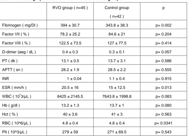
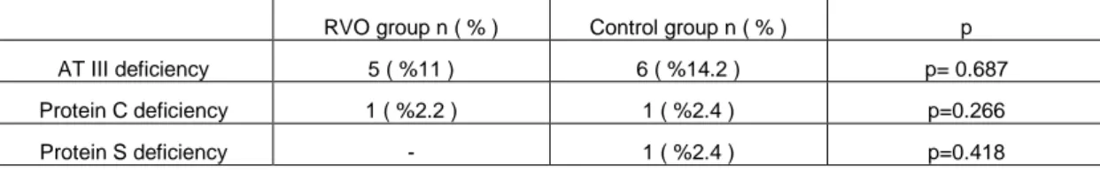
count plasma levels were normal in all patients in both groups (Table 4). ESR and fibrinogen levels were significantly higher in the RVO group (p=0.013, p=0.002, respectively). There was no significant difference between the two groups for antithrombin III, protein C and S levels (Table 5).
The mean serum triglyceride level (mg/dL) was 166.8±71 (73-398) in the RVO group, and 114.2±26.4 (73-150) in the Control group. The mean serum glucose level (mg/dL) was 123±57 (35-325) in the RVO group and 93.2±9.9 (72-110) in the Control group. The serum triglyceride and glucose levels were significantly higher in the RVO group (p=0.001, p=0.022, respectively). Other biochemical parameters included liver enzymes, blood urea nitrogen, creatinine, total cholesterol and low density lipoprotein were normal range in both groups .
Table 3. The frequencys of gene mutations in the RVO and Control groups.
RVO group ( n=45 ) Control group ( n=42 ) p
FVL Normal 41 ( %91 ) 41 ( %97.6 ) Heterozygote 4 ( %9 ) 1 ( %2.4 p=0.362= Homozygous - - MTHFR C677T Normal 15 ( %33.3 ) 24 ( %57.2 ) Heterozygote 26 ( %57.8 ) 14 ( %33.3 ) p=0.032 Homozygous 4 ( %8.9 ) 4 ( %9.5 ) PTG20210A Normal 45 ( %100 ) 42 ( %100 ) Heterozygote - - Homozygous - -
Table 4. Hematologic parameters in the RVO and Control groups.
RVO group ( n=45 ) Control group
( n=42 ) p Fibrinogen ( mg/Dl ) 394 ± 30.7 343.8 ± 38.3 p= 0.002 Factor VII ( % ) 78.2 ± 25.2 84.6 ± 21 p= 0.204 Factor VIII ( % ) 122.5 ± 73.5 127 ± 77.5 p= 0.414 D-dimer (aeg / dL ) 0.4 ± 0.3 0.3 ± 0.1 p= 0.057 PT ( dk ) 13.1 ± 0.5 13.7 ± 3.1 p= 0.586 APTT ( sn ) 28.2 ± 1.9 28.5 ± 2.2 p= 0.555 INR 1 ± 0.04 1.1 ± 0.4 p= 0.915 ESR ( mm/h ) 20.5 ± 16 15 ± 12.5 p= 0.013 WBC ( 10^3/µL ) 8425 ± 2145.5 7643.8 ± 1998.8 p= 0.083 Hb ( g/dl ) 13.2 ± 1.3 13.7 ± 1 p= 0.080 Hct ( % ) 40 ± 3.6 41 ± 3 p= 0.563 RBC ( 10^6/µL ) 4.8 ± 0.4 4.8 ± 0.4 p= 0.0341 Plt ( 10^3/µL ) 279 ± 59 271 ± 69.5 p= 0.543 726
Table 5. AT III, Protein C and S deficiency in the RVO and Control groups.
RVO group n ( % ) Control group n ( % ) p AT III deficiency 5 ( %11 ) 6 ( %14.2 ) p= 0.687 Protein C deficiency 1 ( %2.2 ) 1 ( %2.4 ) p=0.266
Protein S deficiency - 1 ( %2.4 ) p=0.418
DISCUSSION
Hypertension and primary open angle glaucoma have been identified as major risk factors for retinal vein occlusion5,6. Although it has been shown that genetic risk factors are important in the development of venous thrombosis, their role in RVO is not clear yet 2-4 . Factor V Leiden mutation is a relatively common heritable abnormality, and the prevalence of heterozygote traits vary between 1% to 8%, in different regions7,8. Salomon et al. found that there was no significant difference between the prevalence of this mutation in the patient and the control groups2. In present study, we found the prevalence of FVL heterozygote mutation as 8.9% in the RVO group and 2.4% in the Control group. The difference was not significant between the two groups. However, some studies reported that there was a positive correlation between FVL mutation and RVO, and this mutation was considered as a risk factor in patients with RVO3. Prothrombin G20210A mutation is another common cause of hereditary thrombophilia. This mutation has been defined as a risk factor in cases of familial venous thrombosis9. Similar to FVL mutation, Prothrombin G20210A mutation is very rare in Asian or African populations10. Many studies on prothrombin G20210A mutation and RVO were not able to demonstrate a significant relationship between mutation and RVO2,4. Salomon et al.2 found no significant relationship between this mutation and RVO. Similarly, we also could not find this mutation in neither of the groups. Mutation in the thermolabile variant of methylenetetrahydrofolate reductase enzyme causes a decrease of enzyme activity which render these individuals more prone to thrombotic events11,12. The prevalence of this
mutation shows a significant geographic variation (1.4% to 29.7%)13,14. In Turkey, the prevalence of MTHFR heterozygote mutation has been reported as 37.4% to 42% in the normal population, whereas homozygote mutation has been reported as 5.8% to 6.3% 15. Salomon et al. reported the risk factors in patients with RVO as hypertension, family history of stroke and MTHFR C677T homozygote mutation2. Loewenstein et al. defined in 1999 the significant association between RVO and a mutation in MTHFR C677T 16. They enrolled 59 consecutive patients with newly diagnosed RVO and reported that 26 patients (44%) were heterozygotes and 11 (18.6%) were homozygotes for MTHFR C677T mutation. They found that RVO may be associated with a mutation in MTHFR. The authors suggested screening for this mutation in patients with RVO and administration of folic acid to prevent the development of thrombosis and to downgrade retinal vascular changes in individuals with positive screening16 . We also found a positive correlation between RVO and MTHFR C677T mutation. The Eye Disease Case-Control Study Group concluded that the RVO risk increases with hypertension, diabetes mellitus type 2, elevated ESR (especially in women) and history of glaucoma; however, the risk decreases with physical activity, alcohol consumption, and exogenous estrogen administration in postmenopausal women5,6. In our study, ESR and fibrinogen level were significantly higher in patients with RVO compared to the control group; however there was no gender difference. Hayreh et al. found that the most common systemic disease associated with RVO was hypertension17. In our study, 69% of the patients with RVO had hypertension, 6.6% had
diabetes mellitus and hypertension, and 17.8% had a history of cardiovascular disease. The logistic regression model, revealed elevated blood glucose and triglyceride levels and MTHFR677C-T gene mutation as other important risk factors for RVO. Logistic regression analysis involving the parameters for which significant differences were detected an odds ratio of 2.67 for MTHFR C677T gene mutation (95% confidence interval 1.1-6.4), an odds ratio of 1.049 for serum glucose level (95% confidence interval 1.008-1.091) and an odds ratio of 1.034 for serum triglyceride level (95% confidence interval 1.014-1.055).
RVO is a multifactorial retinal vascular disorder that commonly leads to severe visual impairment. Our results favour performing hematological investigations for the thrombophilic disorders in patients with retinal vascular occlusion. If a hereditary defect is found, the patient should undergo consultation of the hematology department and prophylactic anticoagulant treatment should be administered to prevent other thrombotic events. This approach may potentially prevent a fatal disorder such as cerebral or cardiac infarct.
Acknowledgement: This study was presented as
a poster at the 38th National Congress of Turkish Ophthalmology Society.
REFERENCES
1. Recchia FM, Brown GC. Systemic disorders associated with retinal vascular occlusion. Curr Opin Ophthalmol. 2000;11:462-7.
2. Salomon O, Moissiev J, Rosenberg N, Vidne O, Yassur I, Zivelin A, et al. Analysis of genetic polymorphisms related to thrombosis and other risk factors in patients with retinal vascular occlusion. Blood Coagul Fibrinolysis. 1998;9:617-22.
3. Albisinni R, Coppola A, Loffredo M, Cerbone AM, Di Minno G, Greco GM. Retinal vein occlusion and inherited conditions predisposing to thrombophilia. Thromb Haemos. 1998;80:702-3.
4. Backhouse O, Parapia L, Mahomed I, Lee D. Familial thrombophilia and retinal vein occlusion. Eye. 2000;14:13-7.
5. The Eye Disease Case-Control Study Group. Risk factors for central retinal vein occlusion. Arch Ophthalmol. 1996;114:545-54.
6. The Eye Disease Case-Control Study Group. Risk factors for branch retinal vein occlusion. Am J Ophthalmol. 1993;116:286-96.
7. Bertina RM, Koeleman BP, Koster T, Rosendaal FR, Dirven RJ, de Ronde H, et al. Mutation in blood coagulation factor V associated with resistance to activated protein C. Nature. 1994;369:64-7.
8. Bowen DJ, Bowley S, John M, Collins PW. Factor V Leiden (G1691A), the prothrombin 3´-untranslated region variant (G20210A) and thermolabile methylenetetrahydrofolate reductase (C677T): a single genetic test genotypes all three loci. Determination of frequencies in the S. Wales population of the UK. Thromb Haemost. 1998;79:949-54.
9. Poort SR, Rosendaal FR, Reitsma PH, Bertina RM. A common genetic variation in the 3’-untranslated region of the prothrombin gene is associated with elevated plasma prothrombin levels and an increase in venous thrombosis. Blood. 1996;88:3698-703. 10. Rosendaal FR, Doggen CJ, Zivelin A, Arruda VR,
Aiach M, Siscovick DS, et al. Geographic distribution of the 20210 G to A prothrombin variant. Thromb Haemost. 1998;79:706-8.
11. Stein JH, McBride PE. Hyperhomocysteinaemia and atherosclerotic disease. Arch Intern Med. 1998;158:1301-6.
12. Cahill M, Karabatzaki M, Donoghue C, Meleady R, Mynett-Johnson LA, Mooney D, et al. Thermolabile MTHFR genotype and retinal vascular occlusive disease. Br J Ophthalmol. 2001;85:88-90.
13. Haghighatgoo A, Valles-Ayoub Y, Saechao C, Esfandiarifard S, Martinez SL, Pietruszka M, et al. MTHFR C677T genotype frequency in patients of Middle Eastern descent as determined by real-time PCR and melting curve analysis. Genet Test Mol Biomarkers. 2009;13:471-6.
14. Khandanpour N, Willis G, Meyer FJ, Armon MP, Loke YK, Wright AJ, et al. Peripheral arterial disease
and methylenetetrahydrofolate reductase (MTHFR) C677T mutations: A case-control study and meta-analysis. J Vasc Surg. 2009;49:711-8.
15. Balta G, Gürgey A. Methylenetetrahydrofolate reductase (MTHFR) C677T mutation in Turkısh patıents wıth thrombosis. Turk J Pediatr. 1999;41:197-9.
16. Lowenstein A, Goldstein M, Winder A, Lazar M, Eldor A. Retinal vein occlusion associated with methylenetetrahydrofolate reductase mutation. Ophthalmology. 1999;106:1817-20.
17. Hayreh SS, Zimmerman B, McCarthy MJ, Podhajsky P. Systemic disease associated with various types of retinal vein occlusion. Am J Ophthalmol. 2001;131:61-77.
Yazışma Adresi / Address for Correspondence: Dr. Handan Canan
Baskent University School of Medicine,
Department of Ophthalmology, Adana Clinic and Research Center Yuregir, ADANA
E-mail: [email protected] Geliş tarihi/Received on : 05.03.2015 Kabul tarihi/Accepted on: 13.04.2015