Treatment of saphenous vein graft lesions with paclitaxel- and
sirolimus-eluting stents: comparison of short- and long-term clinical outcomes
Safen ven greft lezyonlar›n›n paklitaksel ve sirolimus sal›n›ml› stentler ile tedavisi: K›sa ve uzun
dönem klinik sonuçlar›n karfl›laflt›r›lmas›
Selçuk Görmez, Refik Erdim, Onur Erdo¤mus, Murat Civan
1, Alp Burak Çatako¤lu, Murat Gülbaran
1,
Cemflid Demiro¤lu, Vedat Aytekin
1Department of Cardiology, Florence Nightingale Hospital, ‹stanbul
1
Department of Cardiology, Faculty of Medicine, ‹stanbul Bilim University, ‹stanbul, Turkey
A
BSTRACT
Objective: The purpose of this study was to compare treatment of saphenous vein graft (SVG) lesions with paclitaxel-eluting (PES) and
sirolimus-eluting stents (SES) in daily practice with regard to short- and long-term clinical outcomes.
Methods: Between August 2002 and September 2006, a total of 71 patients with SVG lesions who were implanted PES or SES with percutaneous
coronary intervention in our center were evaluated retrospectively. Forty-six patients with PES (PES group) were compared to twenty-five patients treated with SES (SES group) in terms of in-hospital, 30-day, six-months and 1-year clinical outcomes. Statistical analyses were performed using Chi-Square statistics or Fisher’s exact and independent sample t test. Survival analysis was done using Kaplan-Meier method and log-rank test.
Results: Baseline clinical characteristics were similar in both groups except for a tendency toward a lower age in the SES group. No statistically
significant difference was found between two groups by means of lesion and procedural characteristics. All clinical outcomes at 30-day, 6-month and 1-year after the interventions were similar in both groups. Early stent thrombosis was detected in one patient (2.2%) of PES group (p=0.65). Late stent thrombosis was not observed in both groups. The rate of major adverse cardiac events at 1-year was 8.7% in the PES group and 16% in the SES group (p=0.44).
Conclusion: Short- and long-term clinical outcomes of PES and SES in the treatment of SVG lesions are similar. The results of our study showed
that both drug-eluting stents are effective and safe in real-world patient with diseased SVGs. (Anadolu Kardiyol Derg 2008; 8: 431-6)
Key words: Drug-eluting stents, saphenous vein, angioplasty, outcomes, survival analysis
Ö
ZET
Amaç: Bu çal›flmada amac›m›z günlük pratikte paklitaksel sal›n›ml› (PSS) ve sirolimus sal›n›ml› (SSS) stentler ile tedavi edilen safen ven greft (SVG)
lezyonlu hastalar› k›sa ve uzun dönem klinik sonuçlar aç›s›ndan karfl›laflt›rmakt›r.
Yöntemler: Merkezimizde A¤ustos 2002 ile Eylül 2006 tarihleri aras›nda SVG lezyonu için perkütan koroner giriflim ile PSS veya SSS uygulanm›fl olan
toplam 71 hasta retrospektif olarak de¤erlendirildi. Hastane içi, 30. gün, 6. ay ve 1. y›l klinik sonuçlar› aç›s›ndan PSS uygulanan 46 hasta (PSS gru-bu), SSS uygulanan 25 hasta (SSS grubu) ile karfl›laflt›r›ld›. ‹statistiksel de¤erlendirmede Ki-Kare, “Fisher exact” ve ba¤›ms›z örneklem t testleri kul-lan›ld›. Sa¤kal›m analizi Kaplan-Meier yöntemi ve log-rank testi ile yap›ld›.
Bulgular: Temel klinik özellikler her iki grupta da SSS grubundaki hastalar›n yafl ortalamas›n›n daha düflük olmas› d›fl›nda benzerdi. Lezyon ve ifllem
özellikleri aç›s›ndan her iki grup aras›nda istatistiksel olarak anlaml› bir fark bulunmad›. Giriflim sonras› 30. gün, 6. ay ve 1. y›lda tüm klinik sonuçlar her iki grupta da benzerdi. Erken stent trombozu PSS grubundaki 1 hastada (%2.2) saptand› (p=0.65). Geç stent trombozu her iki grupta da görülme-di. Birinci y›lda majör istenmeyen kardiyak olay oran› PSS grubunda %8.7, SSS grubunda %16 idi (p=0.44).
Sonuç: Safen ven greft lezyonlar›n›n tedavisinde paklitaksel ve sirolimus sal›n›ml› stentlerin k›sa ve uzun dönem klinik sonuçlar› birbirlerine
benzer-dir. Çal›flmam›zda elde etti¤imiz bulgular, her iki ilaç sal›n›ml› stentin de SVG lezyonu bulunan hastalar›n tedavisinde etkin ve güvenli oldu¤unu gös-termektedir. (Anadolu Kardiyol Derg 2008; 8: 431-6)
Anahtar kelimeler: ‹laç sal›n›ml› stent, safen ven, anjiyoplasti, sonuçlar, sa¤kal›m analizi
Address for Correspondence/Yaz›flma Adresi: : Prof. Dr. Vedat Aytekin, Istanbul Bilim Üniversitesi, Florence Nightingale Hastanesi Abide-i Hürriyet Cd. No:290/1 34403
fiiflli, ‹stanbul, Türkiye Phone: +90 212 224 49 50-4099 Fax: +90 212 296 52 22 E-mail: [email protected]
Presented at the 23rd National Cardiology Congress of the Turkish Society of Cardiology , October 19-23, 2007, Belek, Antalya, Turkey
©Telif Hakk› 2008 AVES Yay›nc›l›k Ltd. fiti. - Makale metnine www.anakarder.com web sayfas›ndan ulafl›labilir. ©Copyright 2008 by AVES Yay›nc›l›k Ltd. - Available on-line at www.anakarder.com
Introduction
Treatment of stenotic lesions of saphenous vein grafts
(SVGs) remains one of the challenging issues of cardiovascular
medicine. Effective and satisfactory therapies are still lacking.
Almost half of all SVGs are totally occluded and up to 40% of
patent SVGs are severely diseased within a decade following
coronary bypass surgery due to a tendency of degenerative
process in vessel wall (1, 2). As repeat coronary artery bypass
grafting was shown to be associated with a high morbidity and
mortality, percutaneous coronary interventions (PCI) have
become the preferred revascularization procedure for SVG
lesions (3, 4). However, balloon angioplasty in degenerated soft
and friable SVG lesions is associated with a high complication
rate and a high incidence of restenosis (5-7). Bare-metal
stents (BMS) deployment in SVG lesions has been shown to
improve procedural and clinical outcomes when compared
with balloon angioplasty (8-9). However, the results of BMS in
diseased SVGs are less favorable than those in de novo
coronary lesions due to high restenosis rates (10, 11).
Drug-eluting stents (DES) have reduced the rate of restenosis
and repeat revascularization with respect to BMS in de novo
native coronary artery lesions in selected patient population
(12-14). Some studies have proposed superiority of DES in SVG
lesions when compared to BMS in short and mid-term
follow-up (15-23). A recent trial, however, reported unfavorable
clinical outcomes with DES compared to BMS in long-term
(>3 years) follow-up (24). Since the clinical trials comparing
different DES to treat diseased SVGs are currently limited, we
performed this retrospective study to evaluate short-and
long-term clinical outcomes with sirolimus-eluting stents (SES)
and paclitaxel-eluting stents (PES).
Methods
This was a retrospective study, which analyzes the PCI
database of our institute. We identified a total of 77
consecutive patients who underwent percutaneous
revascularization for SVG lesions which were treated with PES
(Taxus™, Boston Scientific Corp., Natick, Massachusetts) or
SES (Cypher™, Cordis Corp., Miami, Florida) between August
2002 and September 2006. Six patients were excluded from the
analysis due to lack of proper office-visit records and contact
information. The remaining 71 patients with 88 lesions in 80
SVGs were included in the study. The PES group included
forty-six patients with 59 SVG lesions and SES group included
twenty-five patients with 29 SVG lesions. Out of 101 stents
deployed totally, 68 were PES and 33 were SES. The data about
patients and procedural characteristics were collected from
hospital records. All patients were clinically followed-up by
hospital visits at 30-day, 6-month and 1-year. Both groups were
compared with regard to their baseline clinical, procedural and
lesion characteristics and in-hospital, 30-day, 6-month and
1-year clinical outcomes. The PCI procedures were performed
by standard percutaneous techniques using the femoral
approach and standard stent applications as described
previously (25). All patients underwent 12-lead
electrocardiog-raphy pre- and post interventions. Creatine kinase-MB (CKMB)
enzymes were also routinely collected before the PCI and at 6
and 24 hours after intervention. Platelet glycoprotein IIb/IIIa
receptor inhibitors and distal protection devices as
AngioGuard ™ (Cordis Corp., Miami, Florida) and Filterwire
EX™ (Boston Scientific Corp. Natick, Massachusetts) were
used at the discretion of each operator. Written informed
consent for a routine procedure was taken from all patients
before the procedure. Patients were treated with lifelong
aspirin and clopidogrel for at least six months after DES
implantation. Digital coronary angiograms were analyzed
offline by an expert operator blinded to the procedure using
Philips QCA-DCI software. Minimal lumen diameter (MLD),
reference vessel diameter (RVD) and percent diameter stenosis
at baseline and post-procedure were measured, respectively.
Definitions and clinical end-points. Target vessel
revascu-larization (TVR) was defined as all ischemia-driven PCI or
coronary bypass surgery. ST-elevation myocardial infarction
(MI) was defined as presence of new pathological Q waves in the
electrocardiogram associated with an elevation of CKMB ≥3 x
the upper limit of the normal value. A non-ST elevation MI was
defined as creatine kinase-MB enzyme elevation ≥3 x the upper
limit of the normal value. Major adverse cardiac events (MACE)
were defined as death, non-fatal MI (ST-elevation or non-ST
elevation) and TVR. All deaths were considered as cardiac
unless otherwise recorded. Clinical outcomes of all patients
were obtained from previous clinical visit records and telephone
contacts, which were conducted by trained cardiologists. Stent
thrombosis was classified as definite, probable, or possible in
accordance with criteria developed in 2006 by the Academic
Research Consortium. Early stent thrombosis included all events
occurring within 30 days of PCI, late stent thrombosis included
those occurring between 31 to 360 days after PCI (26). Procedural
success was defined as successful stent deployment and
residual stenosis <30% assessed by quantitative analysis,
with-out MACE during the hospital stay.
Statistical analysis
Statistical analysis was performed using SPSS statistical
software package (version 15.0 for Windows, SPSS Inc. Chicago,
Illinois). Categorical variables were expressed as frequencies
and continuous variables as mean±1 standard deviation.
Continuous variables were compared using independent sample
t test. Categorical variables were compared with Chi-Square
sta-tistics or Fisher’s exact test. Survival free of MACE was
estimat-ed using the Kaplan-Meier method and the differences between
the two survival curves were compared with the log-rank test. A
p value <0.05 was considered statistically significant, and all
reported p values are two-sided.
Results
Clinical characteristics at baseline were similar in both
groups except for a lower age in the SES group (67.02±8.26
versus 61.92±8.59, p=0.017 (Table 1). The frequency of male
gender was 89% and the percentage of patients with diabetes
was 46.5% in our study population. Mean SVG age in PES and
SES groups was 12.15±4.64 years and 10.92±4.67 years,
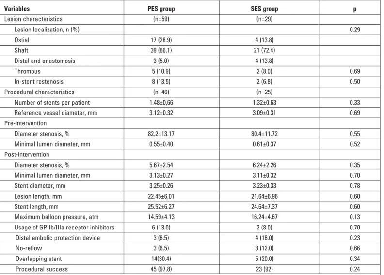
respec-tively (p=0.29). Procedural and lesion characteristics are
shown in Table 2. There was no statistically significant
difference between two groups by means of lesion and
procedure characteristics. Procedural success was 97.8% in
PES group and 92% in SES group (p=0.24). The frequency of
glycoprotein IIb/IIIa inhibitor and distal embolic protection
device usage in SES and PES groups was 13% vs. 8% (p=0.70)
and 6.5% vs. 16% (p=0.23), respectively.
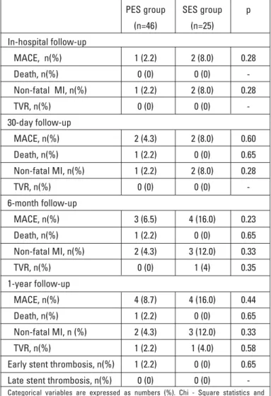
In-hospital results and clinical follow-up outcomes
During the in-hospital period non-fatal MI was observed in
1 (2.2%) patient of PES and in 2 (8%) patients of SES (p=0.28)
group while there was no death and TVR. One patient died in
PES group within the first month after discharge. There was no
statistically significant difference between two groups in terms
of clinical outcomes at 30-day, six month and 1-year (Table 3).
The rates of MACE at 30-days and six months were 4.3% and
6.5% for the PES group and, 8% and 16% for the SES group. At
the end of six months after index procedure, there was no
additional death recorded in both groups while only one TVR
was performed in SES group. In the PES and SES groups,
MACE rates at 1-year were 8.7% and 16%, respectively
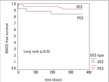
(p=0.44). The rate of MACE-free survival was 91.3% in the PES
group and 84% in the SES group (p=0.33) (Fig. 1). One patient
(2.2%) was regarded as having early stent thrombosis in the
PES group, whereas there was no early stent thrombosis in
SES group (p=0.65). Late stent thrombosis was not observed in
each group. Nine patients had undergone coronary angiography
due to ischemic symptoms and/or a positive functional
ischemia study at the end of one-year. As routine angiographic
follow-up was not the planned end-point; the results of the
angiographic examinations during the follow-up period were
not analyzed in the study.
Discussion
The results of our study showed that short- and long-term
clinical outcomes of PES versus SES in the treatment of SVG
lesions are similar and both drug-eluting stents seem to be
effective and safe in real world patients with diseased SVG’s.
In most centers, PCI for SVG lesions accounts for 10% to
15% of all coronary interventions (27). Intervention for SVG
lesions is associated with a higher risk of peri-procedural
com-plications and late cardiac events when compared with the
native coronary PCI (5-9). In general, patients with diseased
SVGs tend to be older and likely to have co morbidities, which
may reflect relatively high-risk population. Moreover, the
tendency for distal atheroembolization of friable and soft
plaque results in peri-procedural no-reflow and myocardial
infarction in SVG interventions (5). High late cardiac adverse
event risk is related to increased restenosis rate at the target
site and progression of disease at other sites of SVG’s (8).
Several devices and drugs have been tested in SVG lesions
to reduce these adverse cardiac outcomes. In two randomized
Figure 1. Kaplan-Meier event free survival curve of both study groups
DES - drug-eluting stent, MACE - major adverse composite events, PES - paclitaxel-eluting stent, SES - sirolimus-eluting stent
MACE-free survival 0 100 200 300 400 DES type SES SES PES PES time (days) Long rank p=0.33 1.0 .9 .8 .7 .6 .5 .4 .3 .2 .1 0.0
Parameters PES group (n=46) SES group (n=25) p*
Age, yrs 67.02±8.26 61.92± 8.59 0.017
Male, n (%) 43 (93.5) 20 (80) 0.12
Family history of CAD, n (%) 13(28.3) 11(44) 0.18
Hypercholesterolemia, n (%) 38 (82.6) 21 (84) 0.88 Smoking, n (%) 12 (26.1) 10 (40) 0.22 Hypertension, n (%) 35 (76.1) 19 (76) 0.99 Diabetes mellitus, n (%) 18 (39.1) 15 (60) 0.09 Prior PCI, n (%) 18 (39.1) 9 (36) 0.79 Prior MI, n (%) 24(52.2) 14 (56) 0.75 Unstable angina, n (%) 19 (41.3) 12 (48) 0.58
Mean SVG age, years 12.15±4.64 10.92±4.67 0.29
LVEF <40, % 6 (13) 4 (16) 0.73
Categorical variables are expressed as numbers (%) and continuous variables as mean±1 standard deviation. *- independent samples t test, Chi-Square statistics and Fisher’s exact tests. CAD - coronary artery disease, LVEF - left ventricular ejection fraction, MI - myocardial infarction, PCI - percutaneous coronary interventions,PES - paclitaxel-eluting stent, SES - sirolimus-eluting stent, SVG - saphenous vein graft
trials, a significant reduction in periprocedural complications
and 30-day adverse events has been demonstrated with distal
embolic protection devices in SVG interventions (28-29).
Thrombectomy catheters may be useful to prevent distal
thromboembolism if large luminal thrombi are present (30).
However, the use of membrane-covered stents or platelet
glycoprotein IIb/IIIa inhibitors has shown no benefit in this
lesion subset (31-36). Although, stenting of SVG lesions with
BMS reduces acute complications of PCI, the incidence of
restenosis and repeat revascularization is still high. There are
several studies suggesting favorable outcomes with DES
compared to BMS in the treatment of SVG lesions in short- and
mid-term follow-up (15-23). Ge et al. (17) reported significantly
lower cumulative MACE at six months in DES group compared
to BMS group (11.5% vs 28.1%, p=0.02). Lee et al. (18) also
demonstrated reduced composite MACE rates with DES in
9.1±2.1 months follow-up (10% in DES vs 37% in BMS, p=0.035).
In a randomized trial, Vermeersch et al. showed that SES
significantly reduced restenosis rate and repeat
revasculariza-tion procedures in de novo SVG lesions with respect to BMS at
six month (22). On the other hand, in the current literature number
of the studies comparing SES with PES in SVG interventions is
limited. In a prospective and non-randomized study, comparing
SES with PES to treat SVG lesions Chu et al. showed that PES and
SES have similar efficacy and clinical outcomes at the end of a
six-month follow-up (37). Their six-month MACE rate was 10.5%
in the PES group and 8.5% in the SES group (p=0.75). Consistent
with this finding, the six-month MACE rate in our study also has
not shown a significant difference between two groups (6.5 % in
PES group and 16 % in SES group, p=0.23). We have also found
non-significant relationship between both groups regarding
1-year clinical outcomes. In addition, when compared with
previous studies on SVG interventions with DES, our study
population have considerably more diabetic patients (39% in PES
group and 60% in SES group, p=0.09) which probably reflect a
higher risk profile (15-23, 37).
In the present study, there was only one case of stent
thrombosis during the 1-year follow-up. A patient from the PES
Variables PES group SES group p
Lesion characteristics (n=59) (n=29)
Lesion localization, n (%) 0.29
Ostial 17 (28.9) 4 (13.8)
Shaft 39 (66.1) 21 (72.4)
Distal and anastomosis 3 (5.0) 4 (13.8)
Thrombus 5 (10.9) 2 (8.0) 0.69
In-stent restenosis 8 (13.5) 2 (6.8) 0.50
Procedural characteristics (n=46) (n=25)
Number of stents per patient 1.48±0,66 1.32±0.63 0.33
Reference vessel diameter, mm 3.12±0.32 3.09±0.31 0.69
Pre-intervention
Diameter stenosis, % 82.2±13.17 80.4±11.72 0.55
Minimal lumen diameter, mm 0.55±0.40 0.61±0.37 0.52
Post-intervention
Diameter stenosis, % 5.67±2.54 6.24±2.26 0.35
Minimal lumen diameter, mm 3.13±0.27 3.11±0.32 0.70
Stent diameter, mm 3.25±0.26 3.23±0.33 0.78
Lesion length, mm 22.45±6.01 21.64±6.96 0.60
Stent length, mm 25.52±6.27 24.64±7.37 0.60
Maximum balloon pressure, atm 14.59±4.13 16.24±4.67 0.13
Usage of GPIIb/IIIa receptor inhibitors 6 (13.0) 2 (8.0) 0.70
Distal embolic protection device 3 (6.5) 4 (16.0) 0.23
No-reflow 3 (6.5) 3 (12.0) 0.66
Overlapping stent 14(30.4) 5 (20.0) 0.34
Procedural success 45 (97.8) 23 (92) 0.24
Categorical variables are expressed as numbers (%) and continuous variables as mean±1 standard deviation. * - independent samples t test, Chi-Square statistics and Fisher’s exact tests.
GP - glycoprotein, PES - paclitaxel-eluting stent, SES - sirolimus-eluting stent
group died suddenly within the first month after index PCI while
taking dual anti-platelet treatment. Thus, it was categorized as a
probable PES thrombosis. In recent years, some concerns were
raised about higher rates of late adverse cardiac events after
DES implantation because of several reports suggesting
increased very late stent thrombosis with DES at long-term
follow-up (38-40). In recently published randomized prospective
RRISC trial comparing SES to BMS in patients with diseased
SVGs, Vermeersch et al. reported a significant increase in total
mortality at the end of three-year clinical follow-up and the
reduced revascularization rates with SES proven at 6 months
was no longer obtained at 3 years (24). This may be related to
delayed endothelialization, increased late stent thrombosis risk
or delayed restenosis due to reduced drug effectiveness of DES
at late period. In our study, we have found no stent thrombosis in
SES group while only one case in PES group developed stent
thrombosis at the end of 1-year. As our study was limited to
1-year follow-up, no analyzes could be performed to evaluate
very late (>1 year) stent thrombosis. In addition, because of the
limited sample size, our study may have been underpowered to
detect the rate of stent thrombosis.
Study limitations
This study has several limitations. First, it is a single center,
retrospective and non-randomized observational study.
Second, like similar studies related to SVG interventions, the
sample size of our study was small too. Finally, the use of
glycoprotein inhibitors and distal embolic protection devices
were limited due to social insurance reimbursement problem
and there was no routine angiographic follow-up. Despite
limitations mentioned above, to the best of our knowledge, this
study represents, the longest clinical follow-up of consecutive
patients treated with SES versus PES for diseased SVGs.
Conclusions
Short and long-term clinical outcomes of paclitaxel- and
sirolimus-eluting stents in the treatment of SVG lesions are
similar. Findings of our study suggest that both drug-eluting
stents seem to be effective and safe in real world patients with
diseased SVGs. Further studies with larger patient groups are
needed to clarify the effectiveness of different DESs on SVG
lesions.
References
1. Fitzgibbon GM, Leach AJ, Kafka HP, Keon WJ. Coronary bypass graft fate: long-term angiographic study. J Am Coll Cardiol 1991; 17: 1975-80.
2. Fitzgibbon GM, Kafka HP, Leach AJ, Keon KW, Hooper GD, Burton JR. Coronary bypass graft fate and patients outcome: Angiographic follow-up of 5,065 grafts related to survival and reoperation in 1,388 patients during 25 years. J Am Coll Cardiol 1996; 28: 616-26.
3. Foster ED. Reoperation for coronary artery disease. Circulation 1985; 72 (6 Pt 2): V59-V64.
4. Weintraub WS, Jones EL, Morris DC, King SB 3rd, Guyton RA, Craver JM. Outcome of reoperative coronary bypass surgery versus coronary angioplasty after previous bypass surgery. Circulation 1997; 95: 868-77.
5. de Feyter PJ, van Suyken RJ, de Jaegere PP, Topol EJ, Serruys PW. Balloon angioplasty for the treatment of lesions in saphenous vein bypass grafts. J Am Coll Cardiol 1993; 21: 1539-49. 6. Morrison DA, Crowley ST, Veerakul G, Barbiere CC, Grover F, Sacks J. Percutaneous transluminal angioplasty of saphenous vein grafts for medically refractory unstable angina. J Am Coll Cardiol 1994; 23: 1066-70.
7. Webb JG, Carere RG, Virmani R, Baim D, Teirstein PS, Whitlow P, et al. Retrieval and analysis of particulate debris following saphe-nous vein graft intervention. J Am Coll Cardiol 1999; 34: 468-75. 8. Savage MP, Douglas JS Jr., Fischman DL, Pepine CJ, King SB 3d,
Werner JA, et al. SAVED Investigators. Stent placement compared with balloon angioplasty for obstructed coronary bypass grafts. Saphenous Vein De Novo Trial Investigators. N Engl J Med 1997; 337: 740-7.
9. Hanekamp CE, Koolen JJ, Den Heijer P, Schalij MJ, Piek JJ, Bär FWHM, et al. Randomized study to compare balloon angioplasty and elective stent implantation in venous bypass grafts: the Venestent study. Catheter Cardiovasc Interv 2003; 60: 452-7. 10. Keeley E, Velez C, O’Neill W. W, Safian RD. Long-term clinical
outcome and predictors of major adverse cardiac events after percutaneous interventions on saphenous vein grafts. J Am Coll Cardiol 2001; 38: 659-65.
PES group SES group p (n=46) (n=25) In-hospital follow-up MACE, n(%) 1 (2.2) 2 (8.0) 0.28 Death, n(%) 0 (0) 0 (0) -Non-fatal MI, n(%) 1 (2.2) 2 (8.0) 0.28 TVR, n(%) 0 (0) 0 (0) -30-day follow-up MACE, n(%) 2 (4.3) 2 (8.0) 0.60 Death, n(%) 1 (2.2) 0 (0) 0.65 Non-fatal MI, n(%) 1 (2.2) 2 (8.0) 0.28 TVR, n(%) 0 (0) 0 (0) -6-month follow-up MACE, n(%) 3 (6.5) 4 (16.0) 0.23 Death, n(%) 1 (2.2) 0 (0) 0.65 Non-fatal MI, n(%) 2 (4.3) 3 (12.0) 0.33 TVR, n(%) 0 (0) 1 (4) 0.35 1-year follow-up MACE, n(%) 4 (8.7) 4 (16.0) 0.44 Death, n(%) 1 (2.2) 0 (0) 0.65 Non-fatal MI, n (%) 2 (4.3) 3 (12.0) 0.33 TVR, n(%) 1 (2.2) 1 (4.0) 0.58
Early stent thrombosis, n(%) 1 (2.2) 0 (0) 0.65 Late stent thrombosis, n(%) 0 (0) 0 (0)
-Categorical variables are expressed as numbers (%). Chi - Square statistics and Fisher’s exact tests
MACE - major adverse cardiac event, MI - myocardial infarction, PES - paclitaxel-elut-ing stent, SES - sirolimus-elutpaclitaxel-elut-ing stent, TVR - target vessel revascularization
11. Silber S, Albertsson P, Aviles FF, Camici PG, Colombo A, Hamm C, et al. Task Force for Percutaneous Coronary Interventions of the European Society of Cardiology. Guidelines for percutaneous coronary interventions: the task force for percutaneous coronary interventions of the European Society of Cardiology. Eur Heart J 2005; 26: 804-47.
12. Moses J, Leon M, Popma J, Fitzgerald PJ, Holmes DR, O'Shaughnessy C, et al. SIRIUS Investigators. Sirolimus-eluting stents versus standard stents in patients with stenosis in a native coronary artery. N Engl J Med 2003; 349: 1315-23.
13. Morice MC, Serruys PW, Sousa JE, Fajadet J, Ban Hayashi E, Perin M, et al. RAVEL Study Group. Randomized study with the sirolimus-coated bx velocity balloon- expandable stent in the treatment of patients with de novo native coronary artery lesions. A randomized comparison of a sirolimus eluting stent with a standard stent for coronary revascularization. N Engl J Med 2002; 346: 1773-80. 14. Stone GW, Ellis SG, Cox DA, Hermiller J, O'Shaughnessy C, Mann
JT, et al. A polymer-based, paclitaxel-eluting stent in patients with coronary artery disease. N Engl J Med 2004; 350: 221-31. 15. Hoye A, Lemos PA, Arampatzis CA, Saia F, Tanabe K, De¤ertekin
M, et al. Effectiveness of the sirolimus-eluting stent in the treat-ment of sphenous vein graft disease. J Inv Cardiol 2004; 16: 230-3. 16. Price M, Sawhney N, Kao JA, Madrid A, Schatz RA, Teirstein PS.
Clinical outcomes after sirolimus-eluting stent implantation for de novo saphenous vein graft lesions. Catheter Cardiovasc Interv 2005; 65: 208-11.
17. Ge L, Iakovou I, Sangiorgi G, Chieffo A, Melzi G, Cosgrave J, et al. Treatment of saphenous vein graft lesions with drug eluting stents. J Am Coll Cardiol 2005; 45: 989-94.
18. Lee MS, Shah AP, Aragon J, Chieffo A, Melzi G, Cosgrave J, et al. Drug-eluting stenting is superior to bare metal stenting in saphenous vein grafts. Catheter Cardiovasc Interv 2005; 66: 507-11.
19. Sharma A, Minutello RM, Yang F, Wing E, Bhagan S, Chou ET. One-year clinical follow up of sirolimus-eluting stents in the treatment of saphenous vein graft disease. J Am Coll Cardiol 2005; 45 (3 Suppl 1): 25A.
20. Tsuchida K, Ong ATL, Aoki J, van Mieghem CA, Rodriguez-Granillo GA, Valgimigli M, et al. Immediate and one-year outcome of percutaneous intervention of saphenous vein graft disease with paclitaxel-eluting stents. Am J Cardiol 2005; 96: 395-8. 21. Chu WW, Rha SW, Kuchulakanti PK, Cheneau E, Torguson R,
Pinnow E, et al. Efficacy of sirolimus-eluting stents compared with bare metal stents for saphenous vein graft intervention. Am J Cardiol 2006; 97: 34-7.
22. Vermeersch P, Agostoni P, Verheye S, Van den Heuvel P, Convens C, Bruining N, et. al. Randomized double-blind comparison of sirolimus-eluting stent versus bare-metal stent implantation in diseased saphenous vein grafts: six-month angiographic, intravascular ultrasound, and clinical follow-up of the RRISC Trial. J Am Coll Cardiol 2006; 19; 48: 2423-31.
23. Hoffmann R, Pohl T, Köster R, Blindt R, Boeckstegers P, Heitzer T. Implantation of paclitaxel-eluting stents in saphenous vein grafts: clinical and angiographic follow-up results from a multicentre study. Heart 2007; 93: 331-4.
24. Vermeersch P, Agostoni P, Verheye S, Van den Heuvel P, Convens C, Van den Branden F, et al. Increased late mortality after sirolimus-eluting stents versus bare-metal stents in dis-eased saphenous vein grafts results. From the randomized DELAYED RRISC Trial. J Am Coll Cardiol 2007; 50: 261-7.
25. Colombo A, Tobis J. Techniques in Coronary Artery Stenting. London: Martin Dunitz Publishers; 2000.
26. Mauri L, Hsieh WH, Massaro JM, Ho KK, D'Agostino R, Cutlip DE. Stent thrombosis in randomized clinical trials of drug-eluting stents. N Engl J Med 2007; 356: 1020-9.
27. Baim D. Percutaneous treatment of saphenous vein graft dis-ease: the ongoing challenge. J Am Coll Cardiol 2003; 42: 1370-2. 28. Baim DS, Wahr D, George B, Leon MB, Greenberg J, Cutlip DE, et
al. On behalf of the Saphenous vein graft Angioplasty Free of Emboli Randomized (SAFER) Trial Investigators. Randomized trial of a distal embolic protection device during percutaneous inter-vention of saphenous vein aorto-coronary bypass grafts. Circulation 2002; 105: 1285-90.
29. Stone GW, Rogers C, Hermiller J, Feldman R, Hall P, Haber R, et al. Randomized comparison of distal protection with a filter-based catheter and a balloon occlusion and aspiration system during percutaneous intervention of diseased saphenous vein aorto-coronary bypass grafts. Circulation 2003; 108: 548-53. 30. Stone GW, Cox DA, Babb J, Nukta D, Bilodeau L, Cannon L, et al.
Prospective, randomized evaluation of thrombectomy prior to percutaneous intervention in diseased saphenous vein grafts and thrombus-containing coronary arteries. J Am Coll Cardiol 2003; 42: 2007-13.
31. Schachinger V, Hamm CW, Munzel T, Haude M, Baldus S, Grube E, et al. A randomized trial of polytetrafluoroethylene-membrane covered stents compared with conventional stents in aortocoro-nary saphenous vein grafts. J Am Coll Cardiol 2003; 42: 1360-9. 32. Stankovic G, Colombo A, Presbitero P, van den Branden F,
Inglese L, Cernigliaro C, et al. Randomized evaluation of polyte-trafluoroethylene-covered stent in saphenous vein grafts: the Randomized Evaluation of polytetrafluoroethylene COVERed stent in Saphenous vein grafts (RECOVERS) trial. Circulation 2003; 108: 37-42.
33. Schachinger V, Hamm CW, Munzel T, Haude M, Baldus S, Grube E, et al. STENTS (STents IN Grafts) Investigators. A randomized trial of polytetrafluoroethylene membrane-covered stents com-pared with conventional stents in aortocoronary saphenous vein grafts. J Am Coll Cardiol 2003; 42: 1360-9.
34. Ellis S, Linkoff A, Miller D, Tcheng JE, Kleiman NS, Kereiakes D, et al. Reduction in complications of angioplasty with abciximab occurs largely independently of baseline lesion morphology. J Am Coll Cardiol 1998; 32: 1619-23.
35. Roffi M, Mukherjee D, Chew DP, Bhatt DL, Cho L, Robbins MA, et al. Lack of benefit from intravenous platelet glycoprotein IIb/IIIa receptor inhibition as adjunctive treatment for percutaneous interventions of aortocoronary bypass grafts. A pooled analysis of five randomized clinical trials. Circulation 2002; 106: 3063-7. 36. Karha J, Gurm H, Rajagopal V, Fathi R, Bavry AA, Brener SJ, et al.
Use of platelet glycoprotein IIb/IIIa inhibitors in saphenous vein graft percutaneous coronary intervention and clinical outcomes. Am J Cardiol 2006; 98: 906-10.
37. Chu WW, Kuchulakanti PK, Wang B, Clavijo LC, Suddath WO, Pichard AD, et al. Efficacy of sirolimus-eluting stents as compared to paclitaxel-eluting stents for saphenous vein graft intervention. J Interven Cardiol 2006; 19: 121-5.
38. McFadden EP, Stabile E, Regar E, Cheneau E, Ong AT, Kinnaird T, et al. Late thrombosis in drug-eluting coronary stents after discontinuation of antiplatelet therapy. Lancet 2004; 364: 1519-21. 39. Lakovou I, Schmidt T, Bonizzoni E, Ge L, Sangiorgi GM, Stankovic G, et al. Incidence, predictors, and outcome of thrombosis after successful implantation of drug-eluting stents. JAMA 2005; 293: 2126-30.
40. Pfisterer M, Brunner-La Rocca HP, Buser PT, Rickenbacher P, Hunziker P, Mueller C, et al. Late clinical events after clopidogrel discontinuation may limit the benefit of drug-eluting stents an observational study of drug-eluting versus bare-metal stents. J Am Coll Cardiol 2006; 48: 2584-91.