RESEARCH ARTICLE
Gene-environment interaction in
molar-incisor hypomineralization
Mariana BezamatID1, Juliana F. Souza2, Fernanda M. F. SilvaID3, Emilly G. Corrêa4, Aluhe
L. Fatturi2, João A. BrancherID5, Fla´via M. Carvalho6, Tayla Cavallari4, Laı´s BertolazoID4, Cleber Machado-SouzaID7, Mine KoruyucuID8, Merve Bayram9, Andrea Racic1, Benjamin
M. HarrisonID1, Yan Y. SweatID10, Ariadne LetraID11, Deborah Studen-PavlovichID12, Figen Seymen8, Brad Amendt10, Renata I. Werneck4, Marcelo C. Costa3,
Adriana Modesto12, Alexandre R. VieiraID1*
1 Department of Oral Biology, University of Pittsburgh, Pittsburgh, Pennsylvania, United States of America, 2 Department of Stomatology, Federal University of Parana´, Curitiba, State of Parana´ , Brazil, 3 Department of Pediatric Dentistry and Orthodontics, Federal University of Rio de Janeiro, Rio de Janeiro, Rio de Janeiro, Brazil, 4 Graduate Program of Dentistry, Pontifical Catholic University of Parana´ , Curitiba, State of Parana´, Brazil, 5 Graduate Program of Dentistry, Positivo University, Curitiba, State of Para´, Brazil, 6 Department of Genetics, Oswaldo Cruz Foundation (FIOCRUZ), Rio de Janeiro, Rio de Janeiro, Brazil, 7 Graduate Program of Applied Biotechnology to Child and Adolescent Health, Pequeno Prı´ncipe College, Curitiba, State of Para´ , Brazil, 8 Department of Pedodontics, Istanbul University, Istanbul, Turkey, 9 Department of Pedodontics, Medipol Istanbul University, Istanbul, Turkey, 10 Craniofacial Anomalies Research Center and Department of Orthodontics, College of Dentistry, The University of Iowa, Iowa City, Iowa, United States of America, 11 Department of Diagnostic and Biomedical Sciences, and Center for Craniofacial Research, UTHealth School of Dentistry at Houston, Houston, Texas, United States of America, 12 Department of Pediatric Dentistry, University of Pittsburgh, Pittsburgh, Pennsylvania, United States of America
Abstract
Molar incisor hypomineralization (MIH) is an enamel condition characterized by lesions ranging in color from white to brown which present rapid caries progression, and mainly affects permanent first molars and incisors. These enamel defects usually occur when there are disturbances during the mineralization or maturation stage of amelogenesis. Both genetic and environmental factors have been suggested to play roles in MIH’s development, but no conclusive risk factors have shown the source of the disease. During head and neck development, the interferon regulatory factor 6 (IRF6) gene is involved in the structure for-mation of the oral and maxillofacial regions, and the transforming growth factor alpha (TGFA) is an essential cell regulator, acting during proliferation, differentiation, migration and apoptosis. In this present study, it was hypothesized that these genes interact and con-tribute to predisposition of MIH. Environmental factors affecting children that were 3 years of age or older were also hypothesized to play a role in the disease etiology. Those factors included respiratory issues, malnutrition, food intolerance, infection of any sort and medica-tion intake. A total of 1,065 salivary samples from four different cohorts were obtained, and DNA was extracted from each sample and genotyped for nine different single nucleotide polymorphisms. Association tests and logistic regression implemented in PLINK were used for analyses. A potential interaction between TGFA rs930655 with all markers tested in the cohort from Turkey was identified. These interactions were not identified in the remaining cohorts. Associations (p<0.05) between the use of medication after three years of age and
a1111111111 a1111111111 a1111111111 a1111111111 a1111111111 OPEN ACCESS
Citation: Bezamat M, Souza JF, Silva FMF, Corrêa EG, Fatturi AL, Brancher JA, et al. (2021) Gene-environment interaction in molar-incisor hypomineralization. PLoS ONE 16(1): e0241898. https://doi.org/10.1371/journal.pone.0241898
Editor: JJ Cray, Jr., Ohio State University, UNITED
STATES
Received: August 6, 2020 Accepted: October 22, 2020 Published: January 6, 2021
Peer Review History: PLOS recognizes the
benefits of transparency in the peer review process; therefore, we enable the publication of all of the content of peer review and author responses alongside final, published articles. The editorial history of this article is available here: https://doi.org/10.1371/journal.pone.0241898
Copyright:© 2021 Bezamat et al. This is an open access article distributed under the terms of the Creative Commons Attribution License, which permits unrestricted use, distribution, and reproduction in any medium, provided the original author and source are credited.
Data Availability Statement: All relevant data are
within the paper and itsSupporting information files.
Funding: F.M.F.S. was supported by Fundac¸ão de Amparoà Pesquisa do Estado do Rio de Janeiro–
MIH were also found, suggesting that conditions acquired at the age children start to social-ize might contribute to the development of MIH.
Introduction
The hardest biological material in the human body is tooth enamel, composed of both mineral and organic phases [1]. Disturbances during amelogenesis impact maturation or mineraliza-tion stages, and can lead to defects in enamel translucence, known as enamel hypomineraliza-tion [2]. Molar-incisor hypomineralization (MIH) is an example of an enamel condition, and is characterized by the appearance of lesions ranging from white to brownish coloration which present rapid caries progression and hypersensitivity [3]. These asymmetrical lesions affect the first permanent molars, usually with the permanent incisors [4], and more recently MIH has been reported to affect canines too [5]. Prevalence of MIH varies but has been consistently reported to range between 1% and 35% in all parts of the world, with most reported frequen-cies around 12% [6–22].
Regarding the timing of these lesions’ appearance, reports have shown a variation in the chronology of crown completion and enamel formation of incisors and molars. Gleiser and Hunt documented crown completion of first permanent molars to be between 2.5 and 4.4 years [23]. Boyde reported the chronological age of enamel completion in the upper central incisor as 4.64 years and Reid documented complete crown formation in lower central incisors to be at 3.8 years [24].
The enamel maturation stage is particularly important for tissue development, and a very sensitive stage in terms of hypomineralized enamel dysplasia [25].
Different systematic reviews concluded that most of the previously studied environmental factors that were believed to be risk factors for MIH did not fully explain the disease etiology [5,26,27]. In contrast, etiological factors associated with enamel forming genes [2,28] or in immune response-related genes [28,29] have been reported to play a role in the condition’s onset.
Over the past few years, our group has focused on studying different phenotypes that may be present in the same individuals with the hypothesis that they may be influenced by the same underlying genetic variation [30]. Conditions such as cleft lip and palate, have shown an increased frequency of abnormal tooth sizes and morphology [31]. Furthermore, this work has shown that the frequency of dental anomalies as a consequence of disturbances in dental devel-opment was much higher in individuals born with clefts, indicating that dental phenotypes should be considered an extended phenotype of oral clefts [32]. Later, it was also shown that
IRF6 and TGFA interaction might contribute to oral clefts [33], and that genetic associations for MIH can be identified when studied in combination with cleft lip and palate [34].IRF6 is a
gene involved in the structure formation of the oral and maxillofacial regions and has been documented as responsible for not only influence amelogenesis, but also play a role in root and crown anomalies of first molars in mice [35]. This same study showed thatirf6 conditional
knockout mouse presented reduced enamel density preeruption and delayed enamel forma-tion [35].TGFA on the other hand, is an essential cell regulator, acting during proliferation,
differentiation, migration and apoptosis.TGFA has also been associated with orofacial clefts
[36], and a previous study using the case-parent trio design raised the possibility thatIRF6 and TGFA interact and lead to orofacial clefts [37], supporting the finding from our group [33].
FAPERJ process E-26/201.745/2019 (https://www. google.com/search?source=univ&tbm=isch&q= Funda%C3%A7%C3%A3o+de+Amparo+%C3%A0 +Pesquisa+do+Estado+do+Rio+de+Janeiro%E2% 80%93+FAPERJ+process+E-26/201.745/ 2019&sa=X&ved=2ahUKEwiyt97Vw_XqAhW-hXIEHbfJCoEQsAR6BAgBEAE&biw=956&bih= 665). E.G.C. was supported by CAPES. A.R. was supported by the University of Pittsburgh School of Dental Medicine Dean’s Summer Research program. The work was supported in part by a grant from Arauca´ria Foundation awarded to R.I.W (www.fappr.pr.gov.br). This paper is based in part on a thesis submitted to the graduate faculty, Federal University of Rio de Janeiro, in partial fulfillment of the requirements for the PhD degree (for F.M.F.S.) and on a thesis submitted to the graduate faculty, Pontifical Catholic University of Parana´, in partial fulfillment of the requirements for the MS degree (for E.G.C.). The funders had no role in study design, data collection and analysis, decision to publish, or preparation of the manuscript.
Competing interests: The authors have declared
In light of the aforementioned evidence, it was hypothesized thatIRF6 and TGFA interact
and contribute to predisposition of MIH. Additionally, it was hypothesized that environmental factors affecting children that were 3 years of age or older could play a role in the etiology of the disease. Those factors included the presence of any respiratory issues, malnutrition, any type of food intolerance, infection of any sort, and the use of any type of medication.
Results
One thousand and sixty-five individuals from both Brazilian and Turkish cohorts were ana-lyzed. All four cohorts are described in detail in the (S1–S4Files) and a breakdown of the pop-ulations included in the study is described inTable 1. All markers were in Hardy-Weinberg equilibrium (PLINK threshold p-value bellow 0.001), with the exception of rs930655 in the sample from Curitiba (Federal University of Parana´), that was excluded from further analysis (Table 2). It was also excluded rs2073487, rs642961 and rs2902345 in the sample from Rio de Janeiro, and rs1523305 and rs2902345 in the sample from Curitiba (Pontifical Catholic Uni-versity of Parana´). The numbers in bold inTable 2highlight the distribution of the genotypes deviating from Hardy-Weinberg equilibrium.
Single marker analysis
No significant associations between the selected SNPs and MIH were detected in the study populations.
Gene-gene interaction analyses
The cohort from Rio de Janeiro showed a trend toward statistical evidence of interaction betweenTGFA rs1523305 and IRF6 rs642961 (p = 0.03, OR = 1.77) and between IRF6
rs2073487 andTGFA rs2902345 (p = 0.04, OR = 1.60). Significant results (p<0.0004) were
found for the cohort from Istanbul betweenTGFA rs930655 and all IRF6 markers (Table 3).
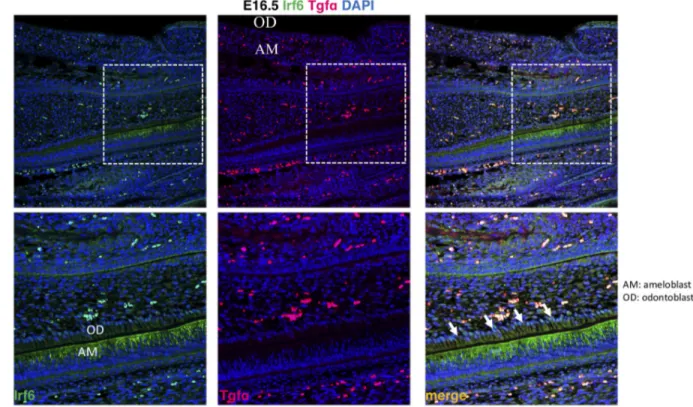
Gene expression analysis
It was investigated the localization of Tgfa and Irf6 in wild type mice at critical stages of dental development. It was observed expression of Irf6 in ameloblasts, apparently more membrane-bound than cytoplasmic or nuclear, whereas expression of Tgfa was not observed (Fig 1).
Environmental factors
Associations were identified between MIH and any type of medications taken at three years of age (Table 4). This variable was designed to capture medication used early in life. In this analy-sis MIH was used as the dependent variable and medication intake as the independent vari-able. In the Curitiba cohort (Federal University of Parana´), out of 191 individuals who
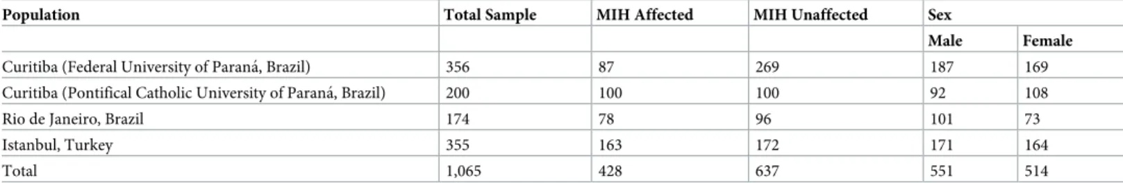
Table 1. Breakdown of populations used in the study.
Population Total Sample MIH Affected MIH Unaffected Sex
Male Female
Curitiba (Federal University of Parana´, Brazil) 356 87 269 187 169
Curitiba (Pontifical Catholic University of Parana´, Brazil) 200 100 100 92 108
Rio de Janeiro, Brazil 174 78 96 101 73
Istanbul, Turkey 355 163 172 171 164
Total 1,065 428 637 551 514
reported having taken medications, 52 were affected by MIH and 139 were not. In contrast, out of 166 individuals who reported not taking any medications, 36 were affected with MIH and 130 were not. In the cohort from Rio de Janeiro, 35 individuals reported having taken medications and were affected by MIH, 15 reported having taken medications and were not affected by MIH, 43 did not take any medications and were affected with MIH and 81 did not take medications and were not affected by MIH. In both groups it was found statistical
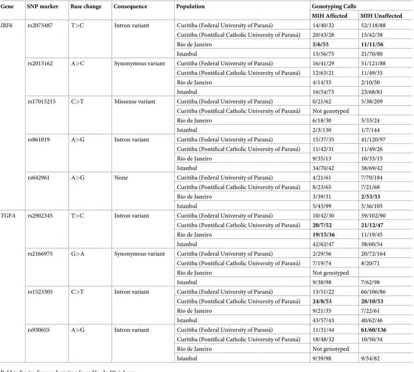
Table 2. Characteristics of the selected variants and genotyping frequencies.
Gene SNP marker Base change Consequence Population Genotyping Calls
MIH Affected MIH Unaffected
IRF6 rs2073487 T>C Intron variant Curitiba (Federal University of Parana´) 14/40/32 52/118/88 Curitiba (Pontifical Catholic University of Parana´) 20/43/28 15/42/38
Rio de Janeiro 5/6/55 11/11/56
Istanbul 15/56/75 21/70/80
rs2013162 A>C Synonymous variant Curitiba (Federal University of Parana´) 16/41/29 51/121/88 Curitiba (Pontifical Catholic University of Parana´) 12/63/21 11/49/35
Rio de Janeiro 4/14/33 2/10/50
Istanbul 16/54/73 23/68/81
rs17015215 C>T Missense variant Curitiba (Federal University of Parana´) 0/21/62 5/38/209 Curitiba (Pontifical Catholic University of Parana´) Not genotyped
Rio de Janeiro 6/18/30 5/33/24
Istanbul 2/3/130 1/7/144
rs861019 A>G Intron variant Curitiba (Federal University of Parana´) 15/37/35 41/120/97 Curitiba (Pontifical Catholic University of Parana´) 11/42/31 11/49/26
Rio de Janeiro 9/35/13 10/33/15
Istanbul 34/70/42 38/69/42
rs642961 A>G None Curitiba (Federal University of Parana´) 4/21/61 7/70/184 Curitiba (Pontifical Catholic University of Parana´) 8/23/65 7/21/68
Rio de Janeiro 3/39/31 2/53/33
Istanbul 5/43/99 5/36/105
TGFA rs2902345 T>C Intron variant Curitiba (Federal University of Parana´) 10/42/30 59/102/90 Curitiba (Pontifical Catholic University of Parana´) 20/7/52 21/12/47
Rio de Janeiro 19/15/36 11/19/45
Istanbul 42/62/47 38/60/54
rs2166975 G>A Synonymous variant Curitiba (Federal University of Parana´) 2/29/56 20/72/164 Curitiba (Pontifical Catholic University of Parana´) 7/19/74 8/20/71
Rio de Janeiro Not genotyped
Istanbul 9/38/98 7/62/98
rs1523305 C>T Intron variant Curitiba (Federal University of Parana´) 13/51/22 66/106/86 Curitiba (Pontifical Catholic University of Parana´) 24/8/53 28/10/53
Rio de Janeiro 9/21/35 7/22/61
Istanbul 43/57/43 40/62/46
rs930655 A>G Intron variant Curitiba (Federal University of Parana´) 11/31/44 61/60/136
Curitiba (Pontifical Catholic University of Parana´) 18/48/32 10/50/34
Rio de Janeiro Not genotyped
Istanbul 9/39/98 9/54/82
Bold indicates figures deviating from Hardy-Weinberg. https://doi.org/10.1371/journal.pone.0241898.t002
evidence for an interaction ofIRF6 and TGFA genotypes and medication intake at 3 years of
age (Table 4).
Discussion
MIH was originally defined as hypomineralization of systemic origin of one to four permanent first molars frequently associated with affected incisors [38]. This definition influenced the work done for more than a decade that focused on the identification of an environmental etio-logical factor [27]. Our group brought to light the possibility that MIH, in fact, has a multifac-torial mode of inheritance [2,5], and therefore has a genetic component, which was estimated in a proportion explaining the variation in the population of at least 20% based on data from twins [39]. Further, our group has proposed that MIH is possibly a localized and multifactorial expression of amelogenesis imperfecta [40].
Table 3. Summary results of gene-gene interaction analysis betweenTGFA rs930655 and the other IRF6 markers in the samples from Istanbul. The odds of the less common allele ofTGFA rs930655 in addition to the IRF6 marker
allele to increase the chance of MIH to occur.
SNP Allele Prevalence Ratio (95% confidence interval) p-value
rs17015215 T 1.33 (1.25–1.42) 2.603e-016 rs2013162 A 1.25 (1.16–1.34) 2.992e-019 rs861019 A 1.34 (1.26–1.42) 1.379e-016 rs2073487 C 1.24 (1.16–1.34) 8.509e-019 rs642961 A 1.39 (1.31–1.47) 1.842e-015 https://doi.org/10.1371/journal.pone.0241898.t003
Fig 1. Expression of Irf6 and Tgfa in sagittal sections of E16.5 wild type murine embryos. Irf6 is expressed in ameloblasts (AM) but not
odontoblasts (OD), whereas Tgfa expression was not detected. https://doi.org/10.1371/journal.pone.0241898.g001
Some argue that the genetic condition affecting enamel is called amelogenesis imperfecta, and that affect all teeth and have a Mendelian mode of inheritance. On the other hand, enamel hypomineralization (enamel hypoplasia or enamel hypocalcification) would be simply caused by impaired ameloblast function and not by altered genes. Therefore, enamel hypomineraliza-tion would be a chronological disturbance that would encompass MIH. This line of thought has led to the hypothesis that the cause of the impaired ameloblast function could be identified because that would be the result of systemic or local causes and this hypothesis has hindered progress of the field for the last 20 years. MIH, like dental caries or periodontitis, fits well in the complex or multifactorial inheritance framework, and similar to cardiovascular diseases as an example, is determined by more than one gene and can be influenced by the environment.
The evidence that MIH frequency varies across the world [6–22] is supported by the results found in the present study. The association found in the Turkish population was different from the results obtained in the cohorts from Brazil, which indeed indicates that genetic fac-tors contributing to the MIH phenotype may vary depending on geographic origin. Both MIH and clefts apparently have higher frequencies in the north of Europe and this frequency declines as one travels toward the Mediterranean [36,41]. Interestingly enough, these differ-ences are not clearly seen for the figures from the Americas, with Brazil and the United States with reported frequencies of 10% to 13% [16,19–21]. Additional evidence that interferon regu-latory factor 6(IRF6) and transforming growth factor alpha (TGFA) may interact, as previously
suggested in the formation of cleft lip and palate [33], is also supported here as for the forma-tion of MIH in the Turkish populaforma-tion. It was not possible to determine the exact mechanism underlying this population-specific association. The interpretation of these results should be analyzed cautiously. When one looks at the frequency of the less common allele of rs17015215 in the Turkish sample, there are two thirds of the individuals affected by MIH in comparison to 1/7 of the individuals that served as comparison. The frequency of the less common allele of rs930655 is also low: 9/39 in the affected individuals and 9/54 in the unaffected. When using these small numbers, one obtains very high odds ratios between 25 and 40. The issue of overestimation of odds ratios is well documented in the literature [42–52]. To avoid the
Table 4. Summary of gene-environment interaction results considering medications (taken after three years of age) and its association with MIH phenotype in the cohort from Curitiba (Federal University of Parana´) and Rio de Janeiro.
Sample from Curitiba (Federal University of Parana´, Brazil)
Gene SNP Allele Odds Ratio (95% confidence interval) p-value with the genetic influence p-value of medications and MIH only
IRF6 rs17015215 T 2.49 (1.31–4.69) 0.028 0.22 rs2013162 A 2.33 (1.29–4.38) 0.04 rs861019 G 2.5 (1.29–4.72) 0.026 rs2073487 C 2.34 (1.29–4.39) 0.04 rs642961 A 2.36 (1.28–4.44) 0.037 TGFA rs2166975 A 2.43 (1.3–4.57) 0.032 rs1523305 C 2.3 (1.3–4.32) 0.044 rs2902345 T 2.4 (1.33–4.47) 0.038
Sample from Rio de Janeiro, Brazil
Gene SNP A1 Odds Ratio (95% confidence interval) p-value with the genetic influence p-value of medications and MIH only
IRF6 rs17015215 T 4.11 (1.81–7.42) 0.0009 0.0002
rs2013162 A 5.16 (2.43–8.87) 0.0002
rs861019 G 6.86 (3.33–10.4) 0.0004
rs2073487 T 5.74 (2.47–10.02) 1.9e-005
overestimation of the strength of the association, when one wishes odds ratios to be approxi-mations of relative risk, prevalence ratios were calculated, which are the ones presented in
Table 3.
Our group has previously reported an interaction ofTGFBR1 and childhood pneumonia
as a possible gene-environment mechanism that increases the chance of MIH occurrence. Additionally, it was reported statistical evidence of potential interaction between genes related to tooth development and immune response [29]. This present work reports evidence for both gene-gene (IRF6-TGFA) and gene-environment (IRF6-medications taken and TGFA-medications taken) interactions, which support the hypothesis that MIH is a complex
genetic condition [5]. In the gene expression analysis, it was observed expression of Irf6 in ameloblasts, but expression of Tgfa was not observed. These results can possibly be explained by the timing of the analysis or simply meant thatTGFA and IRF6 do not colocalize, and
hence, have no direct joint effect of enamel disturbances. The variable medication intake at 3 years of age was created to capture children with history of medication intake from birth and past 3 years and to serve as a surrogate for children more prone to infections and/or illnesses. The 52 children that were included in these analyses had a perfect correlation of medication used before 1 year of age and medication used at 3 years of age. Three years of age was the cutoff chosen with the assumption that mothers would more likely remember that phase since their toddler was able to speak approximately 200 words and being mostly understood. Knowing that enamel development of first permanent molars is completed around 4 years of age [23], a possible mechanism underlying MIH is an interference in enamel formation due to an organic stress in response to illness or infection. This organic stress could happen at any time between birth and past 4 years of age in children with a particular genetic back-ground that includes hypomorphic alleles ofIRF6, TGFA, and other genes such as TGFBR1
[29], andAQP5 [28]. It is likely that disturbances of first permanent molar development may happen after the first year of life, since mineralization of these teeth is not complete until later.
Previous work suggested that MIH is associated with medication intake although this find-ing was not confirmed by others [26]. The use of medications, which is a surrogate for illness, apparently can lead to MIH depending on genetic variation inIRF6 and TGFA. To properly
confirm this hypothesis, the establishment of a cohort of pregnant women that can be prospec-tively followed from birth to the timing when the permanent dentition is developing is sug-gested. This approach would allow to minimize issues of recall bias and to provide more definitive answers that the currently published literature could not.
TheIRF6-TGFA-medication association with MIH also supports the idea that MIH and
cleft lip and palate could possibly be linked. A pleiotropic gene effect could explain this link. Our group has proposed that isolated cleft lip and palate actually is a syndrome that involves disturbances of the dentition [34] and one line of investigation would be to test if genetic vari-ants associated with oral clefts also associate with MIH. We have proposed this approach for isolated tooth agenesis as well [53], and it provided a tool for gene discovery [54,55]. Tgfa and Irf6 did not show the same pattern of expression in mouse teeth and the mechanism of how they may interact to lead to MIH is yet to be determined.
Among the limitations that can be listed for this present study are the small sample sizes of each independent cohort that may not have allowed the detection of small effect sizes. Depending on the study, clinical information was obtained from reviewing medical records or by the use of questionnaires, and these differences can be source of variation. The fact that detailed information about the type of medication the patients were taking was not available due to self-reporting of this information forced the inclusion of any type of medica-tion intake in the analysis. However, these were not vitamins or other supplements. This
kind of observational study that aims to recover information from many years prior suffers from potential issues related to recall bias. For these reasons, the results presented here should be taken cautiously. Another limitation that can be pointed out is that the question on medications taken was asked years after the fact at the moment of participation in the study. Depending on the cohort studied, assessments were done at different age groups (6 to 12, 6 to 10, or at 8 years of age) and one can think this may introduce some variation. In con-trast, the strengths of this study include the well characterized phenotype studied here, determined by experienced dentists from different centers, and the diverse geographic popu-lations studied. The combination of these factors increases the confidence in the results obtained.
In summary, the present study provided evidence thatIRF6 and TGFA might interact
and be involved with MIH in certain populations, although none of these genes appear to have by themselves a major role in MIH. This effect may become more likely apparent when children have to make use of medications around the age of three years. Additional studies to test potential gene-gene interactions in diverse populations are warranted in order to con-firm results reported by previous studies [29] and the results found here. Study designs aim-ing to obtain a more detailed information about what specific medications are involved in the development of MIH are also suggested. These studies should try minimize recall bias by having precisely defined research questions, appropriate data collection methods, well-trained interviewers, being prospective in nature, blinding for researchers and patients, and including nested case-control designs. Despite the limitations described for the present study, the results both reinforce the detrimental effects that medication intake can cause to oral health, and the link between genes associated with orofacial clefts and the MIH phenotype.
Methods
Subjects
The total study population consisted of 1,065 individuals from four populations including three Brazilian cohorts and a Turkish cohort. Salivary sample collection and DNA extraction procedures were described previously [56]. Two groups were from Curitiba, Brazil; the first group consisted of 356 subjects (169 females and 187 males) and the study protocol was approved by the Municipal Department of Education and the Committee for Ethics in Research in Human Health Sciences of the Federal University of Parana´ (UFPR; approval no. 1.613.829/2016). The second group from Curitiba consisted of 200 individuals (108 females and 92 males) and the project was approved by the Ethics and Research Committee of the Pon-tifical Catholic University of Parana´, under the protocol number 1.971.986. The third cohort was from Rio de Janeiro, Brazil, consisted of 174 individuals (73 females and 101 males), and the study protocol was approved by the local Ethics Committee for Research (Hospital Universita´rio Clementino Fraga Filho–HUCFF/UFRJ–approval protocol number
4598514.7.00005257). Finally, the Turkish population consisted of 335 subjects (164 females and 171 males) and the study was approved by the Istanbul University (approval protocol number 2006/2508) Institutional Review Board. Further oversight was obtained at the Univer-sity of Pittsburgh Institutional Review Board (IRB approval protocol numbers PRO0710045 and PRO12080056). MIH was evaluated using the European Academy of Pediatric Dentistry (EAPD) criteria [57], and all subjects/guardians read and signed a written informed consent before their participation in the study.
Cohort from Curitiba, Brazil (Federal University of Parana´): Eligibility
criteria, calibration of the examiners and data collection
The sample consisted of 356 school children, which were randomly selected from a representa-tive population [58] from the city of Curitiba public schools. To ensure accurate population representation, two-stage cluster sampling was performed. First, two schools from each administrative district were randomly selected. Next, two to three classrooms from each selected school were randomly chosen using the websitewww.randomizer.org. The children included were all eight years old and presenting four erupted first molars in the oral cavity. Subjects who had orthodontic braces, syndromes, or amelogenesis imperfecta were excluded from the study. The affected group consisted of children with at least one tooth affected by MIH and the comparison group consisted of children who had no teeth presenting hypomineralization.
A structured socioeconomic data questionnaire was completed by the children’s caregivers. Four examiners, previously trained and calibrated to diagnose Molar-incisor hypomineraliza-tion, selected thirty intraoral photographs of MIH affected teeth in order to define the diagno-sis. Sixty photographs of different clinical scenarios that involved manifestations of MIH were then analyzed by the examiners. After one week, the examiners independently analyzed the same photographs in a different order (duplicate examination, kappa > 0.75). The clinical examination was performed in a school environment using artificial light, a dental mirror, a dental probe with a blunt tip, and sterile gauze. Data collection was performed from November 2016 to September 2017.
Cohort from Curitiba, Brazil (Pontifical Catholic University of Parana´):
Eligibility criteria, calibration of the examiners and data collection
This sample consisted of a total of 200 individuals, enrolled in Curitiba municipal schools dur-ing 2018. Of these, 100 were affected by MIH and 100 non-affected, 108 were females and 92 were males. This cohort was recruited independently from the cohort recruited by investiga-tors at the Federal University of Parana´, however considering the geographical proximity, there is a possibility that some individuals participated in both studies. Although this was a slight possibility, the data from each group were analyzed separately. The inclusion criteria were children between 6 and 10 years of age, affected by MIH in completely erupted teeth. As for exclusion criteria, individuals undergoing orthodontic treatment or scoring 2, 3, 4, 5 and 6 of ICDAS (International Caries Detection and Assessment System) were excluded from the study population. Two interviewers were calibrated on MIH diagnosis using photographs according to a previously published criterion [59]. The intra-rater reliability was performed one month after to the first assessment for the diagnosis of both case and comparison groups (kappa = 1). The clinical examinations were performed by calibrated dentists in the schools, using natural light, an oral mirror and an exploratory probe when it was necessary.
Cohort from Rio de Janeiro, Brazil: Eligibility criteria, calibration of the
examiners and data collection
This sample consisted of 174 individuals, from 7 to 14 (10.13±1.9) years of age, treated at the Pediatric Dentistry Clinic of the Federal University of Rio de Janeiro. Recruitment took place from July 2015 to April 2017, and divided into children with MIH (n = 78) and without MIH (n = 96). The eligibility criteria for this study included patients having all the first permanent molars erupted. The exclusion criteria consisted of children affected by congenital syndromes,
enamel defects (hypoplastic lesions, fluorosis, amelogenesis imperfecta or tetracycline stain) and patients undergoing orthodontic treatment.
To assess for inter and intra examiner-reliability, a theoretical exercise was previously applied for two days. Twenty clinical images of dental enamel defects including fluorosis, hypoplasia, amelogenesis imperfecta, dental caries and MIH were shown to the examiners (F.M.F.S., M.C.M). Two weeks after the first assessment, a new assessment was carried out with the examiners and the kappas were 0.88 and 0.89, respectively.
The subjects’ health data were collected through health records, including the demographic data (child´s age, sex, place of birth, residence, parent’s education and income), mother’s health during pregnancy, and children’s medical history (medication intake, systemic diseases, high fever, malnutrition, asthma, bronchitis, epilepsy, and severe infections). The clinical examination was performed by a calibrated dentist (F.M.F.S). Examinations were carried out using a mirror and a probe, at the dental chair and using artificial light.
Cohort from Istanbul, Turkey: Eligibility criteria, calibration of the
examiners and data collection
Eligible individuals were enrolled at the Pediatric Clinic of Istanbul University, Turkey (n = 335, ages ranging from 6 to 12 years). Subjects affected by syndromes, fluorosis, or the ones who had fixed appliances were excluded from the study. Cases were defined as subjects affected by MIH, while controls were defined as subjects with no evidence of MIH.
Calibrated examiners carried out the clinical examination, with F.S. having calibrated M.B. and M.K. Exam calibrations were performed according to the following protocol: First, the cal-ibrator presented to the examiner the criteria for MIH detection, showing pictures of several situations to be observed in the exam and discussing each of these situations in a session that lasted one to two hours. Next, the calibrator and examiner analyzed ten to twenty subjects and discussed each case. The intraexaminer agreement was assessed by a second clinical examina-tion in 10% of the sample after two weeks, with a kappa of 1.0. M.K. pre-screened subjects, and M.B. performed the full exam. This cohort and methodology have been reported before [2]. Clinical examinations were performed using a flashlight and intra-oral mirror and gauze was used to dry and clean the teeth prior to examination. Artificial light and dental operatory were used for all evaluations. An explorer was gently used for assessing the smoothness of tooth surfaces.
Single marker analysis
The amount of DNA and the purity of each sample was determined by spectrophotometry (NanoDrop 1000, Thermo Fisher Scientific, US). The DNA concentration was obtained by readings at 260 nm and the purity by 260/ 280 nm proportion. Five single nucleotide polymor-phisms (SNPs) inIRF6 (rs2073487, rs2013162, rs17015215, rs861019, rs642961) and four in TGFA were genotyped (Table 2). PCR reactions were carried out using Taqman chemistry [60] in 3.0μl reaction volumes in an ABI PRISM Sequence Detection System 7900 (Applied Biosystems, Foster City, CA, USA). Genotyping calls were analyzed using SDS software ver-sion 1.7 (Applied Biosystems). PCR reactions were repeated twice when necessary and allele frequencies were calculated. Genotypes are available as (S1–S4Files).
Gene-gene interactions
Gene-gene interaction analyses were performed using logistic regression as implemented in PLINK. In order to test the interaction betweenIRF6 and TGFA, the presence of MIH was
containing allIRF6 genotypes was ran against a covariate file containing the TGFA genotypes
organized by copies of alleles, and this analysis was repeated with aTGFA genotype file against
theIRF6 covariate file.
Immunofluorescence
Expression of Tgfa and Irf6 proteins was performed on paraffin sections from heads of wild type mice at E16.5. Maintenance and handling of mice were approved by the Animal Care Unit at the University of Iowa. Tissues were deparaffinized and rehydrated in a series of etha-nol dilutions. Slides were boiled for 20 min in antigen unmasking solution (Vector Laborato-ries, H-3300). Sections were blocked with 20% donkey serum (sigma, D9663A), then
incubated overnight at 4˚C with the following primary antibodies: polyclonal goat anti-Tgfa (1:50, R&D, AF-239-SP) and polyclonal rabbit anti-Irf6 (1:50, sigma SAB2102995). After rins-ing in PBS, sections were incubated with secondary antibodies conjugated to Alexa Fluoro-phore 488 or 555 (Molecular Probes, Invitrogen, CA). The nuclei were counterstained with DAPI in PBS (1:10000). The images were taken using a ZEISS 700 confocal microscopy.
Gene-environment interactions
Tests for potential associations between MIH and factors that affected the children of 3 years of age or older in the cohorts from Curitiba (Federal University of Parana´) and Rio de Janeiro were carried out. The variables included the presence of respiratory issues, malnutrition, any type of food intolerance, infection of any sort and any type of medication intake between 3 years of age and the time the sample was collected. This information was part of the question-naires obtained for both MIH affected and unaffected children. Mothers responded the ques-tions and all participants were asked the same quesques-tions, which included medical history of the child (diseases and medications taken) and details of the child’s diet. Vitamins and other sup-plements were not counted as medications.
The variable “medications taken at 3 years of age” was designed due to the evidence that cuspal enamel formation of first permanent molars is not completed until past 3 years of age [24]. These data suggest that the first molar susceptibility window for MIH goes beyond the first year of age. The suggestion that first permanent molar mineralization is completed by one year of age likely limits the ability of unveiling associations.
We have complied with the STROBE guidelines in this study.
Statistical analysis
All SNPs in all samples were tested for deviation from Hardy–Weinberg equilibrium using chi-square. In the single-marker analysis, association tests were performed comparing geno-types to phenotype between affected individuals and their respective comparison groups as implemented in the PLINK software [61]. Both odds ratios and 95% confidence intervals were calculated. In order to account for multiple testing, Bonferroni correction was performed and p-values of 0.001 (0.05/36) or below were considered significant.
In the gene-gene interaction analysis, p-values below 0.0004 (0.05/108) were considered sta-tistically significant.
In the gene-environment interaction analysis the covariates were dichotomous and analyses were performed using logistic regression as also implemented in PLINK.
The power necessary to detect significant associations between a gene marker and the phe-notype in each cohort was calculated using a Genetic Power Calculator tool [62]. The fre-quency of the high-risk allele in the population was defined as equal to 0.2, the prevalence of MIH as 0.12, D’ as 1.0 (amount of linkage disequilibrium), the respective number of cases of
each cohort and the case-comparison group ratio. When imputing the effect size in the hetero-zygote form as 1.3 and the effect size of homozygous form as 5.0, it was obtained 69%, 57%, 51%, and 79% power in the cohort from Curitiba (Federal University of Parana´), Curitiba (Pontifical Catholic University), Rio de Janeiro and Istanbul, respectively. Power will decrease with lower effect sizes for both forms, and will increase to 82%, 70%, 64%, and 89%, respec-tively if the effect size for the homozygous form is 6.0.
Supporting information
S1 File. Raw genotypes, Curitiba (UFPR).
(XLSX)
S2 File. Raw genotypes, Curitiba (PUCPR).
(XLSX)
S3 File. Raw genotypes, Rio de Janeiro.
(XLSX)
S4 File. Raw genotypes, Istanbul.
(XLSX)
Acknowledgments
This paper is based in part on a thesis submitted to the graduate faculty, Federal University of Rio de Janeiro, in partial fulfillment of the requirements for the PhD degree (for F.M.F.S.) and on a thesis submitted to the graduate faculty, Pontifical Catholic University of Parana´, in par-tial fulfillment of the requirements for the MS degree (for E.G.C.).
Author Contributions
Conceptualization: Mariana Bezamat, Ariadne Letra, Figen Seymen, Brad Amendt, Renata I.
Werneck, Alexandre R. Vieira.
Formal analysis: Alexandre R. Vieira. Funding acquisition: Alexandre R. Vieira.
Investigation: Mariana Bezamat, Juliana F. Souza, Fernanda M. F. Silva, Emilly G. Corrêa, Aluhe L. Fatturi, João A. Brancher, Fla´via M. Carvalho, Tayla Cavallari, Laı´s Bertolazo, Cle-ber Machado-Souza, Mine Koruyucu, Merve Bayram, Andrea Racic, Benjamin M. Harri-son, Yan Y. Sweat, Deborah Studen-Pavlovich, Figen Seymen, Brad Amendt, Renata I. Werneck, Marcelo C. Costa, Adriana Modesto.
Methodology: Juliana F. Souza, Ariadne Letra, Figen Seymen, Brad Amendt, Renata I.
Wer-neck, Marcelo C. Costa, Adriana Modesto, Alexandre R. Vieira.
Resources: Alexandre R. Vieira.
Software: Mariana Bezamat, Benjamin M. Harrison.
Supervision: Marcelo C. Costa, Adriana Modesto, Alexandre R. Vieira. Writing – original draft: Mariana Bezamat, Alexandre R. Vieira.
Writing – review & editing: Juliana F. Souza, Fernanda M. F. Silva, Emilly G. Corrêa, Aluhe L. Fatturi, João A. Brancher, Fla´via M. Carvalho, Tayla Cavallari, Laı´s Bertolazo, Cleber Machado-Souza, Mine Koruyucu, Merve Bayram, Andrea Racic, Benjamin M. Harrison,
Yan Y. Sweat, Ariadne Letra, Deborah Studen-Pavlovich, Figen Seymen, Brad Amendt, Renata I. Werneck, Marcelo C. Costa, Adriana Modesto.
References
1. Cuy JL, Mann AB, Livi KJ, Teaford MF, Weihs TP. Nanoindentation mapping of the mechanical proper-ties of human molar tooth enamel. Arch Oral Biol. 2002; 47(4):281–91. Epub 2002/04/02.https://doi. org/10.1016/s0003-9969(02)00006-7PMID:11922871.
2. Jeremias F, Koruyucu M, Kuchler EC, Bayram M, Tuna EB, Deeley K, et al. Genes expressed in dental enamel development are associated with molar-incisor hypomineralization. Arch Oral Biol. 2013; 58 (10):1434–42. Epub 2013/06/25.https://doi.org/10.1016/j.archoralbio.2013.05.005PMID:23790503 3. Garg N, Jain AK, Saha S, Singh J. Essentiality of early diagnosis of molar incisor hypomineralization in
children and review of its clinical presentation, etiology and management. Int J Clin Pediatr Dent. 2012; 5(3):190–6. Epub 2012/09/01.https://doi.org/10.5005/jp-journals-10005-1164PMID:25206166 4. Raposo F, de Carvalho Rodrigues AC, Lia EN, Leal SC. Prevalence of Hypersensitivity in Teeth
Affected by Molar-Incisor Hypomineralization (MIH). Caries Res. 2019:1–7. Epub 2019/01/25.https:// doi.org/10.1159/000495848PMID:30677762.
5. Vieira AR, Kup E. On the Etiology of Molar-Incisor Hypomineralization. Caries Res. 2016; 50(2):166–9. Epub 2016/04/26.https://doi.org/10.1159/000445128PMID:27111773.
6. Zhao D, Dong B, Yu D, Ren Q, Sun Y. The prevalence of molar incisor hypomineralization: evidence from 70 studies. Int J Paediatr Dent. 2018; 28(2):170–9. Epub 2017/07/22.https://doi.org/10.1111/ipd. 12323PMID:28732120.
7. Koruyucu M, Ozel S, Tuna EB. Prevalence and etiology of molar-incisor hypomineralization (MIH) in the city of Istanbul. J Dent Sci. 2018; 13(4):318–28. Epub 2019/03/22.https://doi.org/10.1016/j.jds.2018. 05.002PMID:30895140
8. Hussain G, Al-Halabi M, Kowash M, Hassan A. The Prevalence and Severity of Molar Incisor Hypomi-neralization and Molar HypomiHypomi-neralization in Dubai, UAE. J Dent Child (Chic). 2018; 85(3):102–7. Epub 2019/03/15. PMID:30869585.
9. Saitoh M, Nakamura Y, Hanasaki M, Saitoh I, Murai Y, Kurashige Y, et al. Prevalence of molar incisor hypomineralization and regional differences throughout Japan. Environ Health Prev Med. 2018; 23 (1):55. Epub 2018/11/02.https://doi.org/10.1186/s12199-018-0748-6PMID:30382812
10. Glodkowska N, Emerich K. Molar Incisor Hypomineralization: prevalence and severity among children from Nothern Poland. Eur J Paediatr Dent. 2019; 20(1):59–66. Epub 2019/03/29.https://doi.org/10. 23804/ejpd.2019.20.01.12PMID:30919647.
11. Mejia JD, Restrepo M, Gonzalez S, Alvarez LG, Santos-Pinto L, Escobar A. Molar Incisor Hypominerali-zation in Colombia: Prevalence, Severity and Associated Risk Factors. J Clin Pediatr Dent. 2019; 43 (3):185–9. Epub 2019/04/10.https://doi.org/10.17796/1053-4625-43.3.7PMID:30964726.
12. Rai PM, Jain J, Raju AS, Nair RA, Shashidhar K, Dsouza S. Prevalence of Molar Incisor Hypominerali-zation among School Children Aged 9 to 12 Years in Virajpet, Karnataka, India. Open Access Maced J Med Sci. 2019; 7(6):1042–6. Epub 2019/04/13.https://doi.org/10.3889/oamjms.2019.224PMID: 30976358
13. Gorbatova MA, Utkina EI, Zinchenko GA, Grjibovski AM, Popova DA, Popov VA, et al. [Molar-incisor hypomineralization among 12-years old children in Arkhangelsk region]. Stomatologiia (Mosk). 2019; 98(2):64–6. Epub 2019/05/16.https://doi.org/10.17116/stomat20199802164PMID:31089123. 14. Kilinc G, Cetin M, Kose B, Ellidokuz H. Prevalence, aetiology, and treatment of molar incisor
hypominer-alization in children living in Izmir City (Turkey). Int J Paediatr Dent. 2019; 29(6):775–82. Epub 2019/05/ 18.https://doi.org/10.1111/ipd.12508PMID:31099125.
15. Ahmad SH, Petrou MA, Alhumrani A, Hashim R, Splieth C. Prevalence of Molar-Incisor Hypominerali-sation in an Emerging Community, and a Possible Correlation with Caries, Fluorosis and Socioeco-nomic Status. Oral Health Prev Dent. 2019; 17(4):323–7. Epub 2019/06/22.https://doi.org/10.3290/j. ohpd.a42725PMID:31225537.
16. Davenport M, Welles AD, Angelopoulou MV, Gonzalez C, Okunseri C, Barbeau L, et al. Prevalence of molar-incisor hypomineralization in Milwaukee, Wisconsin, USA: a pilot study. Clin Cosmet Investig Dent. 2019; 11:109–17. Epub 2019/06/27.https://doi.org/10.2147/CCIDE.S172736PMID:31239785 17. Villanueva-Gutierrez T, Irigoyen-Camacho ME, Castano-Seiquier A, Zepeda-Zepeda MA,
Sanchez-Perez L, Frechero NM. Prevalence and Severity of Molar-Incisor Hypomineralization, Maternal Educa-tion, and Dental Caries: A Cross-Sectional Study of Mexican Schoolchildren with Low Socioeconomic Status. J Int Soc Prev Community Dent. 2019; 9(5):513–21. Epub 2019/10/18.https://doi.org/10.4103/ jispcd.JISPCD_130_19PMID:31620386
18. Goswami M, Bhushan U, Pandiyan R, Sharma S. Molar Incisor Hypomineralization-An Emerging Bur-den: A Short Study on Prevalence and Clinical Characteristics in Central Delhi, India. Int J Clin Pediatr Dent. 2019; 12(3):211–4. Epub 2019/11/12.https://doi.org/10.5005/jp-journals-10005-1624PMID: 31708617
19. Hartsock LA, Burnheimer J, Modesto A, Vieira AR. A Snapshot of the prevalence of molar incisor hypo-mineralization and fluorosis in Pittsburgh, Pennsylvania, USA. Pediatr Dent 2020; 42(1):36–40. PMID: 32075709
20. Silva FMF, Zhou Y, Vieira FGF, Carvalho FM, Costa MC, Vieira AR. Defining the prevalence of molar incisor hypomineralization in Brazil. Pesqui Bras Odontopediatria Clı´n Integr 2020; 20:e5146.https:// doi.org/10.1590/pcobi.2020.021
21. Ahmed AT, Soto-Rojas AE, Dean JA, Eckert GJ, Martinez-Mier EA. Prevalence of molar-incisor hypo-mineralization and other enamel defects and associated sociodemographic determinants in Indiana. J Am Dent Assoc 2020; 151(7):491–501.https://doi.org/10.1016/j.adaj.2020.02.027PMID:32593351 22. Amend S, Nossol C, Bausback-Schomakers S, Wleklinski C, Scheibelhut C, Pons-Ku¨ hnemann J, el al.
Prevalence of molar-incsor-hypomineralisation (MIH) among 6-12-year-old children in Central Hesse (Germany). Clin Oral Investig. 2020;https://doi.org/10.1007/s00784-020-03519-7Online ahead of print. PMID:32808178
23. Gleiser I, Hunt EE Jr., The permanent mandibular first molar: its calcification, eruption and decay. Am J Phys Anthropol. 1955; 13(2):253–83. Epub 1955/06/01.https://doi.org/10.1002/ajpa.1330130206 PMID:13258780.
24. Reid DJ, Dean MC. Variation in modern human enamel formation times. J Hum Evol. 2006; 50(3):329– 46. Epub 2005/11/23.https://doi.org/10.1016/j.jhevol.2005.09.003PMID:16300817.
25. Robinson C. Enamel maturation: a brief background with implications for some enamel dysplasias. Front Physiol. 2014; 5:388. Epub 2014/10/24.https://doi.org/10.3389/fphys.2014.00388PMID: 25339913
26. Silva MJ, Scurrah KJ, Craig JM, Manton DJ, Kilpatrick N. Etiology of molar incisor hypomineralization— A systematic review. Community Dent Oral Epidemiol. 2016; 44(4):342–53. Epub 2016/04/29.https:// doi.org/10.1111/cdoe.12229PMID:27121068.
27. Fatturi AL, Wambier LM, Chibinski AC, Assuncao L, Brancher JA, Reis A, et al. A systematic review and meta-analysis of systemic exposure associated with molar incisor hypomineralization. Community Dent Oral Epidemiol. 2019; 47(5):407–15. Epub 2019/05/22.https://doi.org/10.1111/cdoe.12467PMID: 31111554.
28. Pang L, Li X, Wang K, Tao Y, Cui T, Xu Q, et al. Interactions with the aquaporin 5 gene increase the sus-ceptibility to molar-incisor hypomineralization. Arch Oral Biol. 2020; 111:104637. Epub 2019/12/31. https://doi.org/10.1016/j.archoralbio.2019.104637PMID:31884335.
29. Bussaneli DG, Restrepo M, Fragelli CMB, Santos-Pinto L, Jeremias F, Cordeiro RCL, et al. Genes Reg-ulating Immune Response and Amelogenesis Interact in Increasing the Susceptibility to Molar-Incisor Hypomineralization. Caries Res. 2019; 53(2):217–27. Epub 2018/08/22.https://doi.org/10.1159/ 000491644PMID:30130760.
30. Bezamat M, Deeley K, Khaliq S, Letra A, Scariot R, Silva RM, et al. Are mTOR and Endoplasmic Reticu-lum Stress Pathway Genes Associated with Oral and Bone Diseases? Caries Res. 2019; 53(3):235–41. Epub 2018/09/12.https://doi.org/10.1159/000492675PMID:30205378
31. Walker SC, Mattick CR, Hobson RS, Steen IN. Abnormal tooth size and morphology in subjects with cleft lip and/or palate in the north of England. Eur J Orthod. 2009; 31(1):68–75. Epub 2008/12/17. https://doi.org/10.1093/ejo/cjn073PMID:19073959.
32. Letra A, Menezes R, Granjeiro JM, Vieira AR. Defining subphenotypes for oral clefts based on dental development. J Dent Res. 2007; 86(10):986–91. Epub 2007/09/25.https://doi.org/10.1177/ 154405910708601013PMID:17890676
33. Letra A, Fakhouri W, Fonseca RF, Menezes R, Kempa I, Prasad JL, et al. Interaction between IRF6 and TGFA genes contribute to the risk of nonsyndromic cleft lip/palate. PLoS One. 2012; 7(9):e45441. Epub 2012/10/03.https://doi.org/10.1371/journal.pone.0045441PMID:23029012
34. Koruyucu M, Kasimoglu Y, Seymen F, Bayram M, Patir A, Ergoz N, et al. Rethinking isolated cleft lip and palate as a syndrome. Oral Surg Oral Med Oral Pathol Oral Radiol. 2018; 125(4):307–12. Epub 2018/03/04.https://doi.org/10.1016/j.oooo.2018.01.007PMID:29500156.
35. Chu EY, Tamasas B, Fong H, Foster BL, LaCourse MR, Tran AB, et al. Full Spectrum of Postnatal Tooth Phenotypes in a Novel Irf6 Cleft Lip Model. J Dent Res. 2016; 95(11):1265–73. Epub 2016/07/03. https://doi.org/10.1177/0022034516656787PMID:27369589
36. Vieira AR. Association between the transforming growth factor alpha gene and nonsyndromic oral clefts: a HuGE review. Am J Epidemiol. 2006; 163(9):790–810. Epub 2006/02/24.https://doi.org/10. 1093/aje/kwj103PMID:16495466.
37. Sull JW, Liang KY, Hetmanski JB, Wu T, Fallin MD, Ingersoll RG, et al. Evidence that TGFA influences risk to cleft lip with/without cleft palate through unconventional genetic mechanisms. Hum Genet. 2009; 126(3):385–94. Epub 2009/05/16.https://doi.org/10.1007/s00439-009-0680-3PMID:19444471 38. Weerheijm KL. Molar incisor hypomineralisation (MIH). Eur J Paediatr Dent. 2003; 4(3):114–20. PMID:
14529330.
39. Vieira AR. On the genetics contribution to molar-incisor hypomineralization. Int J Paediatr Dent. 2019; 29(1):2–3.https://doi.org/10.1111/ipd.12439PMID:30367537.
40. Vieira AR. Genetic basis of enamel and dentin defects. In: Vieira AR. Genetic basis of oral health condi-tions. Springer: Cham, 2020: 9–21.
41. Vieira AR. Prevalence of molar incisor hypomineralisation has a North-South gradient between Europe and North Africa. Eur Arch Paediatr Dent. 2019; 20(5):501–2. Epub 2019/02/07.https://doi.org/10. 1007/s40368-019-00417-9PMID:30726548.
42. Greenland S. Interpretation and choice of effect measures in epidemiologic analyses. Am J Epidemiol. 1987; 125(5):761–8. Epub 1987/05/01.https://doi.org/10.1093/oxfordjournals.aje.a114593PMID: 3551588.
43. Lee J. Odds ratio or relative risk for cross-sectional data? Int J Epidemiol. 1994; 23(1):201–3. Epub 1994/02/01.https://doi.org/10.1093/ije/23.1.201PMID:8194918.
44. Lee J, Chia KS. Estimation of prevalence rate ratios for cross sectional data: an example in occupational epidemiology. Br J Ind Med. 1993; 50(9):861–2. Epub 1993/09/01.https://doi.org/10.1136/oem.50.9. 861PMID:8398881
45. Lee J, Chia KS. Prevalence odds ratio v prevalence ratio—a response. Occup Environ Med. 1995; 52 (11):781–2. Epub 1995/11/01.https://doi.org/10.1136/oem.52.11.781-aPMID:8535502
46. Nurminen M. To use or not to use the odds ratio in epidemiologic analyses? Eur J Epidemiol. 1995; 11 (4):365–71. Epub 1995/08/01.https://doi.org/10.1007/BF01721219PMID:8549701.
47. Stromberg U. Prevalence odds ratio v prevalence ratio—some further comments. Occup Environ Med. 1995; 52(2):143. Epub 1995/02/01.https://doi.org/10.1136/oem.52.2.143PMID:7757169
48. Walter SD. Choice of effect measure for epidemiological data. J Clin Epidemiol. 2000; 53(9):931–9. Epub 2000/09/27.https://doi.org/10.1016/s0895-4356(00)00210-9PMID:11004419.
49. Bland JM, Altman DG. Statistics notes. The odds ratio. BMJ. 2000; 320(7247):1468. Epub 2000/05/29. https://doi.org/10.1136/bmj.320.7247.1468PMID:10827061
50. Szklo M, Nieto FJ. Epidemiology beyond the basics. 2. USA: Jones and Bartlett; 2007. 51. Gordis L. Epidemiology. 4. USA: Saunders Elsevier; 2004.
52. Tamhane AR, Westfall AO, Burkholder GA, Cutter GR. Prevalence odds ratio versus prevalence ratio: choice comes with consequences. Stat Med. 2016; 35(30):5730–5. Epub 2016/07/28.https://doi.org/ 10.1002/sim.7059PMID:27460748
53. Vieira AR. Oral clefts and syndromic forms of tooth agenesis as models for genetics of isolated tooth agenesis. J Dent Res. 2003; 82(3):162–5. Epub 2003/02/25.https://doi.org/10.1177/
154405910308200303PMID:12598542.
54. Vieira AR, McHenry TG, Daack-Hirsch S, Murray JC, Marazita ML. A genome wide linkage scan for cleft lip and palate and dental anomalies. Am J Med Genet A. 2008; 146A(11):1406–13. Epub 2008/04/ 30.https://doi.org/10.1002/ajmg.a.32295PMID:18442096
55. Vieira AR, McHenry TG, Daack-Hirsch S, Murray JC, Marazita ML. Candidate gene/loci studies in cleft lip/palate and dental anomalies finds novel susceptibility genes for clefts. Genet Med. 2008; 10(9):668– 74. Epub 2008/11/04.https://doi.org/10.1097/gim.0b013e3181833793PMID:18978678
56. Trevilatto PC, Line SR. Use of buccal epithelial cells for PCR amplification of large DNA fragments. J Forensic Odontostomatol. 2000; 18(1):6–9. Epub 2001/04/28. PMID:11324090.
57. Weerheijm KL, Duggal M, Mejare I, Papagiannoulis L, Koch G, Martens LC, et al. Judgement criteria for molar incisor hypomineralisation (MIH) in epidemiologic studies: a summary of the European meeting on MIH held in Athens, 2003. Eur J Paediatr Dent. 2003; 4(3):110–3. Epub 2003/10/08. PMID: 14529329.
58. Portella PD, Menoncin BLV, Souza JF, Menezes JVNB, Fraiz FC, Assunc¸ão LRS. Impact of molar inci-sor hypomineralization on quality of life in children with early mixed dentition: A hieranchical approach. Int J Paediatr Dent. 2019; 29(4):496–506.https://doi.org/10.1111/ipd.12482PMID:30758096. 59. Ghanim A, Silva MJ, Elfrink MEC, Lygidakis NA, Mariño RJ, Weerheijm KL, et al. Molar incisor
hypomi-neralisation (MIH) training manual for clinical field surveys and practice. Eur Arch Paediatr Dent. 2017; 18(4):225–42.https://doi.org/10.1007/s40368-017-0293-9Epub 2017 Jul 18. PMID:28721667.
60. Ranade K, Chang MS, Ting CT, Pei D, Hsiao CF, Olivier M, et al. High-throughput genotyping with sin-gle nucleotide polymorphisms. Genome Res. 2001; 11(7):1262–8. Epub 2001/07/04.https://doi.org/10. 1101/gr.157801PMID:11435409
61. Purcell S, Neale B, Todd-Brown K, Thomas L, Ferreira MA, Bender D, et al. PLINK: a tool set for whole-genome association and population-based linkage analyses. Am J Hum Genet. 2007; 81(3):559–75. Epub 2007/08/19.https://doi.org/10.1086/519795PMID:17701901
62. Purcell S, Cherny SS, Sham PC. Genetic Power Calculator: design of linkage and association genetic mapping studies of complex traits. Bioinformatics. 2003; 19(1):149–50. Epub 2002/12/25.https://doi. org/10.1093/bioinformatics/19.1.149PMID:12499305.