191 DOI: 10.4274/tjod.34270
Laparoscopic repair of ureter damaged during laparoscopic
hysterectomy: Presentation of two cases
Laparoskopik histerektomide yaralanan üreterin laparoskopik
tamiri: İki olgunun sunumu
1İstanbul Medipol University Faculty of Medicine, Department of Obstetrics and Gynecology, İstanbul, Turkey 2Vehbi Koç Foundation American Hospital, Women’s Health Center Assisted Reproduction Unit, İstanbul, Turkey
3University of Health Sciences, Zeynep Kamil Women and Children’s Health Training and Research Hospital, İstanbul, Turkey
Case Report / Olgu Sunumu
Murat Api1, Ayşen Boza2, Semra Kayataş3, Barış Boza3
Üreter hasarı, jinekolojik cerrahilerde nadir görülen ancak korkulan bir komplikasyondur ve tamiri için sıklıkla laparotomiye ihtiyaç duyulur. Yakın zamanda, jinekolojik üreter hasarının laparoskopik tamiri ile ilgili cesaretlendirici birkaç makale yayınlanmıştır. Amacımız total laparoskopik histerektomide (TLH) hasarlanan ve laparoskopik olarak başarılı bir şekilde tamir edilen iki olguyu sunmaktır. İlk olguda, sağ kardinal ligament (KL) hizasına kadar sorunsuz devam eden operasyonda, KL diseksiyonu sırasında üreterovezikal bileşkenin (UVB) 7-8 cm distalinde üreter hasarı oluştu. İkinci olguda, UVB’nin yaklaşık 10 cm distalinden üreter hasarlandı. Her iki üreter hasarı da cerrahi sırasında fark edildi. Hasarlanan üreterler laparotomiye dönmeden ve ek trokar girişi yapılmadan sorunsuz bir şekilde onarıldı. Her iki olguda da mid-üretral hasar oluştuğundan üreteroüreterostomi tercih edildi. Üreter hasarı her ne kadar TLH sırasında nadir gelişen bir komplikasyon olsa da, eğer mümkünse, laparoskopik sütür ve düğüm teknikleri konusunda deneyimli bir jinekolog veya ürolog tarafından, laparoskopik olarak tamir edilmelidir.
Anahtar Kelimeler: Üreter, yaralanma, komplikasyon, laparoskopi
Abstract
Ureter injuries are uncommon but dreaded complications in gynecologic surgery and a frequent cause of conversion to laparotomy. Recently, a few papers reported the repair of gynecologic ureteral injuries using laparoscopy with encouraging results. In these case reports, we aimed to present two laparoscopically repaired ureter injuries during total laparoscopic hysterectomies (TLH). In the first case, the ureter was transected during the dissection of the cardinal ligament, approximately 7 to 8 cm distal to the ureterovesical junction (UVJ), and in the second case, it was damaged approximately 10 cm distal to the UVJ. Both transections were identified during surgery. The injured ureter was repaired without converting to laparotomy or additional trocar insertion. Ureteroureterostomy was performed in both cases uneventfully. Although ureteric injury is a rare complication during TLH, it can be managed by the same surgeon laparoscopically during the same procedure.
Keywords: Ureter, injury, complication, laparoscopy
Öz
Turk J Obstet Gynecol 2017;14:191-4
Address for Correspondence/Yazışma Adresi: Ayşen Boza, MD,
Vehbi Koç Foundation American Hospital, Women’s Health Center Assisted Reproduction Unit, İstanbul, Turkey
Phone: +90 530 923 00 46 E-mail: [email protected] ORCID ID: orcid.org/0000-0002-2019-6798 Received/Geliş Tarihi: 26.05.2017 Accepted/Kabul Tarihi: 12.09.2017
©Copyright 2017 by Turkish Society of Obstetrics and Gynecology
192
Turk J Obstet Gynecol 2017;14:191-4 Api et al. Laparoscopic repair of ureter injury
Introduction
Urologic complications during gynecologic surgery are uncommon but dreadful. The reported incidence of ureteral injury related to gynecologic surgery varies between 2.5% and 12.1%(1).Pelvic ureters are retroperitoneal structures that run
from the renal pelvis to the bladder that can be injured during pelvic surgery at any point along their distal course. However, the approach of hysterectomy plays a role in the variation of incidence in total laparoscopic hysterectomies (LHs) (TLHs) compared with the abdominal and vaginal approach; the reported odds ratio for urinary tract injuries was 2.41 and 3.69, respectively(2). Although the risk of ureter injury significantly
decreases with the increasing experience of surgeons, ureter injuries also occur in the hands of experienced gynecologists because the indication for TLH is expanding and the difficulty of the operation is increasing.The question is whether complications of laparoscopic procedures such as ureteral injury could be managed successfully and effectively without converting to laparotomy. We report successful immediate primary laparoscopic repair of two cases of ureter transection during TLH and discuss possible causes that lead to ureter injury and make recommendations for prevention.
Case Report
A patient aged 48 years underwent TLH and bilateral salpingo-oophorectomy (BSO) due to abnormal uterine bleeding that was unresponsive to medical therapy. She had no previous history of abdominal surgery or any kind of disease causing pelvic adhesions. On exploration, the uterus was of 8 weeks’ pregnancy in size, the bilateral adnexa were normal, and no pelvic adhesions or distortion of anatomy was observed. A four-trocar technique (out of main trocar, two at the left side and one at the right side) and Hohl manipulator (manufactured by Karl Storz, Germany) was used. The TLH was uneventful until the area of the cardinal ligaments (CLs). The ureter was transected with the Ligasure™ (Valleylab, Boulder, CO, USA)
during the dissection of the right CL, 7 to 8 cm distant from the ureterovesical junction (UVJ) (Figure 1a). The transection was identified during the dissection and immediate repair was performed peroperatively without converting to laparotomy. The proximal and distal ureteral segments were identified, then sutured at the 6 o’clock position for approximation (Figure 1b). A double J ureteral stent was taken to the abdomen through the 5-mm lateral trocar and inserted in the ureter (Figure 1c). After the stent application, 3/0 Vicryl sutures were placed at the 9, 12, and 3 o’clock positions (Figure 1d). A Foley catheter
Figure 1. The surgical technique of laparoscopic ureter injury is shown. The transected edges of the ureter at the area of cardinal ligament
are seen (a). First, the proximal and distal ureter edges are sutured at the 6 o’clock position (b), then the ureteral stent is inserted in the ureter (c) and the anastomosis is completed using sutures placed at the 9, 12 and 3 o’clock positions (d)
193
Turk J Obstet Gynecol 2017;14:191-4
Api et al. Laparoscopic repair of ureter injury
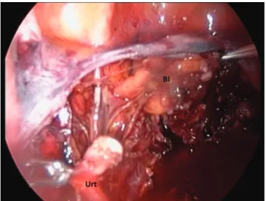
remained in the bladder and a drainage tube in the abdomen to prevent possible urinoma formation and to control bleeding. In the second case, a woman aged 59 years underwent TLH and BSO due to simple endometrial hyperplasia with atypia. TLH, BSO, and frozen pathology were planned with the suspicion of malignancy. Laparoscopy was performed using the same technique as in the first case. Frozen section was reported in favor of benign pathology. During the dissection of the left CL, the ureter was damaged approximately 10 cm distant from UVJ with the Halo™ bipolar cutting forceps (Gyrus ACMI,
Olympus) (Figure 2). The injured ureter was noticed during the dissection and repaired without converting to laparotomy or additional trocar insertion, similar to the first case. The duration of the ureteral repairs from the time of the injury to the end of the last anastomosis suture were 55 and 40 minutes for the first and second cases, respectively. Estimated blood loss during the ureteral repairs was negligible. The vesical catheters were withdrawn on 7 days day postoperatively and the ureteral stents were removed cystoscopically on the 21st
day. We did not use prophylactic antibiotics due to prolonged catheterization. Patients were closely monitored after discharge weekly for the first three weeks, then monthly up to 10-12 months using serial urinary system ultrasonography. Neither patient had any pelvicalyceal ectasia or visible ureters. The patients’ recoveries were uneventful with normal kidney and urinary function. Written informed consent was obtained by both patients.
Discussion
Ureter injuries can result from pelvic dissection or due to thermal injury by excessive use of energy adjacent to the ureter.In a retrospective review of 165 patients with iatrogenic ureteral injuries over a 20-year span, endourologic procedures
were responsible for most iatrogenic injuries with 42%, and gynecologic and general surgery procedures were 34% and 24%, respectively(3).Urinary tract injuries during LH are reported to
be more frequent than in abdominal or vaginal hysterectomies. The higher risk in LH compared with the abdominal approach is due to the use of electrocoagulation of uterine vessels during laparoscopic procedures(4). Extensive electrocoagulation of
uterine vessels and CLs near the ureter increases the risk of ureter injuries.
Intraoperative detection of ureter injuries has been reported in only 5-13% of cases(3,4). Among the acute injuries of the
urinary tract, ureter injuries are most difficult to recognize as there often may be few or with no symptoms(1). Intraoperative
iatrogenic ureteral injuries, if recognized during the procedure, should be repaired at that setting(5).In our cases, the transected
edges of the ureter were seen without any urine leakage during the dissection of the CLs.
Approximately 90% of the trauma to the ureter occurs in the lower portion, which extends from the inferior border of the sacroiliac joint to the UVJ(6).Transection injuries are repaired
depending on the severity of the injury and approximation to the UVJ. If the ureteral injury is more than 6 cm distant from the UVJ, a primary ureteral anastomosis is performed. Primary ureteroureterostomy is the optimal technique when the anastomosis can be performed without tension and if the initial approach was laparoscopic(7). Laparoscopic
ureteroureterostomy was preferred in our cases because the mid-ureter was transected approximately 8 cm and 10 cm distant from the UVJ, respectively.
Laparoscopic ureteral repair is technically feasible and an alternative to open repair for immediate, early, and delayed diagnosis of ureteral injury. In the literature, although long-term outcome of laparoscopic repair is limited to small series and case reports, the results are excellent if the diagnosis and repair is performed at the time of injury(8). A delay in diagnosis
worsens prognosis because of infection, hydronephrosis, abscess, and fistula formation. No postoperative complication occurred in our cases, the patients have continued their lives with normal kidney function and no urinary incontinence for 10 and 12 months follow-up, respectively.
Recommended techniques for reducing the risk of urinary tract injuries were agreed upon by experts in a Delphi consensus procedure: routine use of uterine manipulator, coagulation of uterine vessels close to the uterus with a perpendicular approach from the ipsilateral side, and visualization or dissection of the ureter in the case of distorted anatomy(9).Intraoperative routine
use of cystoscopy was proposed to assess the ureter flow as a part of all TLHs. Cystoscopy during TLH is well tolerated and can reassure surgeons of immediate urinary tract injuries; however, it is an additional time-consuming procedure that is not required in most patients. Cystoscopy during TLHs was postulated to be cost effective if the rate of ureter injury exceeded 2%(10).
Figure 2. The transected edges of the ureter at the level of left
cardinal ligament were seen
194
Turk J Obstet Gynecol 2017;14:191-4 Api et al. Laparoscopic repair of ureter injury
In the case of ureteral injury during laparoscopic gynecologic surgery, either a gynecologist or urologist who is experienced in suturing and knot-tying techniques can successfully perform a primary laparoscopic repair of the ureter. Visualization of the course of ureters at the beginning or end of the operation may prevent complications of the urinary tract and possible delayed diagnosis of ureteral injuries.
Ethics
Informed Consent: Written informed consents were obtained
from the patients for publication of these case reports and accompanying images.
Peer-review: External and internal peer-reviewed. Authorship Contributions
Surgical and Medical Practices: M.A., A.B., Concept: M.A., A.B., Design: M.A., A.B., Data Collection or Processing: B.B., Analysis or Interpretation: M.A., A.B., B.B., Literature Search: B.B., S.K., Writing: M.A., A.B., B.B., S.K.
Conflict of Interest: No conflict of interest was declared by
the authors.
Financial Disclosure: The authors declared that this study
received no financial support.
References
1. Teeluckdharry B, Gilmour D, Flowerdew G. Urinary Tract Injury at Benign Gynecologic Surgery and the Role of Cystoscopy: A Systematic Review and Meta-analysis. Obstet Gynecol 2015;126:1161-9.
2. Nieboer TE, Johnson N, Lethaby A, Tavender E, Curr E, Garry R, et al. Surgical approach to hysterectomy for benign gynaecological disease. Cochrane Database Syst Rev 2009;CD003677.
3. Delacroix SE Jr, Winters JC. Urinary tract injures: recognition and management. Clin Colon Rectal Surg 2010;23:104-12.
4. Donnez O, Jadoul P, Squifflet J, Donnez J. A series of 3190 laparoscopic hysterectomies for benign disease from 1990 to 2006: evaluation of complications compared with vaginal and abdominal procedures. BJOG 2009;116:492-500.
5. Redan JA, McCarus SD. Protect the ureters. JSLS 2009;13:139-41. 6. Brummer TH, Jalkanen J, Fraser J, Heikkinen AM, Kauko
M, Makinen J, et al. FINHYST, a prospective study of 5279 hysterectomies: complications and their risk factors. Hum Reprod 2011;26:1741-51.
7. McAninch Jack W. Injuries to the Genitourinary Tract, Injuries to the Ureter. In: Tanagho EA, McAninch JW, editors. Smith’s General Urology, 17th Ed, United States of America (USA): Mc Garw Hill Companies; 2011. p. 291-311.
8. Brandes S, Coburn M, Armenakas N, McAninch J. Diagnosis and management of ureteric injury: an evidence-based analysis. BJU Int 2004;94:277-89.
9. Janssen PF, Brolmann HAM, Huirne JAF. Recommendations to prevent urinary tract injuries during laparoscopic hysterectomy: a systematic Delphi procedure among experts. J Minim Invasive Gynecol 2011;18:314-32.
10. Visco AG, Taber KH, Weidner AC, Barber MD, Myers ER. Cost-effectiveness of universal cystoscopy to identify ureteral injury at hysterectomy. Obstet Gynecol 2001;685-92.