Case Reports Anatol J Cardiol 2018; 20: 130-3
132
An uncommon percutaneous treatment
of aortic pseudoaneurysm
Ahmet Arif Yalçın, Serkan Kahraman, Aydın Yıldırım*, Korhan Erkanlı**
Department of Cardiology, Mehmet Akif Ersoy Thoracic and Cardiovascular Surgery Training and Research Hospital; İstanbul-Turkey
*Departments of Cardiology, **Cardiovascular Surgery, İstanbul Medipol University Medipol Mega Hospital; İstanbul-Turkey
Introduction
Aortic pseudoaneurysm is a rare disease of the aorta and gen-erally occurs because of previous surgical procedure (1). It does not include all layers of the artery. Clinical presentation can vary from asymptomatic to catastrophic outcomes (2). Although surgi-cal management is the commonly accepted treatment, it can be re-lated with high morbidity and mortality (3); therefore, endovascular strategies, such as thrombin injection, coiling, endovascular stent-ing, and Amplatzer occluder devices, are emerging as alternative treatment modalities which have fewer complication rates (4). We herein present a case of aortic pseudoaneurysm, due to a previ-ous surgery, that was successfully treated using a transcatheter Amplatzer duct occluder device.
Case Report
A 62-year-old male patient was admitted to İstanbul University of Health Sciences Mehmet Akif Ersoy Thoracic and
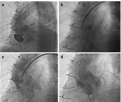
Cardiovas-cular Surgery Training and Research Hospital with mild dyspnea. He had undergone a coronary artery bypass surgery 10 years ago and aortic surgical supra coronary repair for acute type 1 aortic dissection 6 years ago. He had a mild diastolic murmur in the aortic area on physical examination. Echocardiographic imaging indicat-ed normal ejection fraction and mild aortic regurgitation. Contrast-enhanced computed tomography revealed 39×24-mm-size aortic pseudoaneurysm with 12-mm neck (Fig. 1a, 1b). Which was first identified 2 years ago. However; the patient had refused to undergo surgery when the pseudoaneurysm had been 36×20 mm in size. A 30-mm supracoronary aortic graft, settling above the pseudoaneu-rysm and ending before the arcus aorta was seen. Surgery was planned by our cardiology and cardiovascular surgery council. However, the risk of surgery was high because of two thoracoto-mies previously and also as the patient did not wish to undergo a third surgery. Accordingly, treatment using a transcatheter endo-vascular strategy was approved. Digital subtraction angiography was performed via the right common femoral artery using 7-French (F) femoral sheath. Images of the ascending aorta and pseudoan-eurysm were obtained (Fig. 2a). A 7-F destination sheath was posi-tioned into the pseudoaneurysm using a Supracore 300-cm guide wire. The Amplatzer duct occluder II with a 16-mm dimension was placed in the neck of the pseudoaneurysm via a destination sheath (Fig. 2b, 2c). The procedure was terminated with aortogra-phy, and the pseudoaneurysm was successfully treated with com-plete blood flow interruption (Fig. 2d). The patient was discharged from the hospital under 100 mg acetylsalicylic acid treatment and without any symptoms. One month and one year postoperatively, contrast-enhanced computed tomography revealed successful device closure of pseudoaneurysm (Fig. 1c, 1d), and the patient is still being followed up without any symptoms.
a b
c d
Figure 2. a: Digital subtraction angiographic imaging of the aortic pseu-doaneurysm. b, c: The Amplatzer duct occluder II with a 16-mm dimen-sion was placed to the neck of the pseudoaneurysm via a destination sheath. d: Terminating the procedure with an aortograph
a c
b d
Figure 1. a, b: Contrast-enhanced computed tomography of the aortic pseudoaneurysm (arrowhead). c, d: Contrast-enhanced computed to-mography of the aortic pseudoaneurysm treated with an Amplatzer duct occluder II (arrowhead)
Case Reports
Anatol J Cardiol 2018; 20: 130-3
133
Discussion
Aortic pseudoaneurysms are observed with a variety of aortic diseases with disruption of arterial wall layers and they are rare complications of post cardiothoracic surgeries because of weakness of the aortic wall, typically at the sites of graft anastomosis; needle/cannulation of the ascending aorta; or cross-clamping (1, 5). Clinical presentation can change from asymptomatic to catastrophic rupture (2). Even though conventional treatment of the pseudoaneurysm is surgical ligation or replacement with a graft, surgical procedures have higher morbidity and mortality rates (3). Transcatheter endovascular modalities, such as thrombin injection, coiling, endovascular stenting, and Amplatzer occluder devices, can be alternative treatments. However, they also have some limitations depending on the location of the pseudoaneurysm and the size of the neck of the pseudoaneurysm. Endovascular graft stenting is not suitable in the presence of inadequate landing zone (such as in our patient) and if the pseudoaneurysm is in the proximity of supra-aortic vessels. Thrombin injection and coil embolization may not be an option in cases of large-neck pseudoaneurysms and those in the proximity of the origin of great vessels (6). Amplatzer occluder devices seem to have broader use to treat these patients as they have different sizes and can be used in large-neck pseudoaneurysm. The first case of using an Amplatzer occluder device was described by Bashir et al. in 2005 (7). They were used in some cases with comorbidities and those that were unsuitable for surgery. The technique has a lower complication rate and shorter hospital stay. In light of foregoing data, it seems to be used more often as an alternative treatment option (4). Large-scale studies are needed for evaluating the efficiency and reliability of endovascular treatment of aortic pseudoaneurysms.
Conclusion
Aortic pseudoaneurysms are rare complications of post car-diothoracic surgery, and their surgical treatment has high
morbid-ity and mortalmorbid-ity rates. The Amplatzer occluder device is emerging as a better alternative endovascular transcatheter treatment of pseudoaneurysms with lower complication rates in suitable cases.
References
1. Sullivan KL, Steiner RM, Smullens SN, Griska L, Meister SG. Pseu-doaneurysm of the ascending aorta following cardiac surgery. Chest 1988; 93: 138-43. [CrossRef]
2. Agarwal M, Ray M, Pallavi M, Sen S, Ganguly D, Joshi P, et al. Device occlusion of pseudoaneurysm of ascending aorta. Ann Pediatr Car-diol 2011; 4: 195-9. [CrossRef]
3. Mulder EJ, van Bockel JH, Maas J, van den Akker PJ, Hermans J. Morbidity and mortality of reconstructive surgery of noninfected false aneurysms detected long after aortic prosthetic reconstruction. Arch Surg 1998; 133: 45-9. [CrossRef]
4. Kanani RS, Neilan TG, Palacios IF, Garasic JM. Novel use of the Am-platzer septal occlude device in the percutaneous closure of ascend-ing aortic pseudoaneurysms: A case series. Catheter Cardiovasc Interv 2007; 69: 146-53. [CrossRef]
5. Dhadwal AK, Abrol S, Zisbrod Z, Cunningham JN Jr. Pseudoaneu-rysms of the ascending aorta following coronary artery bypass sur-gery. J Card Surg 2006; 21: 221-4. [CrossRef]
6. Chapot R, Aymard A, Saint-Maurice JP, Bel A, Merland JJ, Houdart E. Coil embolization of an aortic arch false aneurysm. J Endovasc Ther 2002; 9: 922-5. [CrossRef]
7. Bashir F, Quaife R, Carroll JD. Percutaneous closure of ascending aortic pseudoaneurysm using Amplatzer septal occluder device: The first clinical case report and literature review. Catheter Cardiovasc Interv 2005; 65: 547-51. [CrossRef]
Address for Correspondence: Dr. Ahmet Arif Yalçın, Mehmet Akif Ersoy Kalp Hastanesi,
Kardiyoloji Bölümü, Turgut Özal Bulvarı No: 11 Halkalı, Küçükçekmece İstanbul-Türkiye
Phone: +90 505 489 47 75 E-mail: [email protected]
©Copyright 2018 by Turkish Society of Cardiology - Available online at www.anatoljcardiol.com