Pulmonary artery and right ventricle function in patients
with bicuspid aortic valve
Biküspit aort kapaklı hastalarda pulmoner arter ve sağ ventrikül fonksiyonları
1Department of Cardiology, Kartal Koşuyolu Yüksek İhtisas Training and Research Hospital, İstanbul, Turkey 2Department of Cardiology, İstanbul Medipol University Faculty of Medicine, İstanbul, Turkey
3Department of Cardiology, İstanbul Emsey Hospital, İstanbul, Turkey
Çetin Geçmen1, M.D., Gamze Babür Güler2, M.D.,Suzan Hatipoğlu3, M.D.,Muzaffer Kahyaoğlu1, M.D.,
Murat Çap1, M.D., Servet İzci1, M.D., Çağatay Önal1, M.D., Emrah Erdoğan1, M.D., Aykun Hakgör1, M.D.,
Özkan Candan1, M.D., Arzu Kalaycı1, M.D., Tuba Unkun1, M.D., İbrahim Akın İzgi1, M.D.
Objective: Bicuspid aortic valve (BAV) is a complex develop-mental anomaly caused by abnormal aortic leaflet formation during valvulogenesis. The present study is an assessment of the effects of BAV disease on the ascending aorta and pulmo-nary artery (PA), and an evaluation of the consequences for systolic and diastolic functioning of the left and right ventricles.
Methods: Total of 66 patients were eligible for inclusion. Pulmo-nary artery maximum diameter (PAD) was obtained 1 cm distal to the pulmonary annulus. Using pulsed-wave tissue Doppler imaging, left ventricular (LV) early diastolic velocity (E′) mea-surement was obtained at the annulus with placement of sam-ple volume. Right ventricle (RV) peak global strain rate during systole (RV-SRS), early diastole (RV-SRE), and late diastole (RV-SRA) were calculated.
Results: In this study, 40.9% (n=27) of patients were female and average age was 35±11years. RV-SRS values (β=-.781, t=-2.723; p=0.010) and log-LV tissue Doppler imaging E’ (β=-2.996, t=-5.405; p=<0.001) were negatively correlated, and log-PAD (β=4.861, t=3.052; p=0.005) was positively and inde-pendently correlated with ascending aortic diameter.
Conclusion: Ascending aorta diameter is positively correlat-ed with PA diameter in BAV patients, and RV strain rate and LV diastolic parameters are affected before development of the valve disease.
Amaç: Biküspit aort kapağı, valvulogenez sırasında anormal aorti kapakçık oluşumu sonucunda ortaya çıkan gelişimsel bir anomalidir. Bu çalışmada, biküspit aort kapak hastalığının pulmoner arter ve çıkan aortaya etkileri, bu durumun sağ ve sol ventrikül sistolik ve diyastolik fonksiyonları üzerine etkisi değerlendirildi.
Yöntemler: Çalışmaya 66 hasta dahil edildi. Pulmoner arter en yüksek çapı (PAD), pulmoner anulusun 1 cm distalinden ölçüldü. Sol ventrikül (LV) erken diyastolik hızı, örnek hacim anulusa yerleştirilerek elde edildi. Sol ventrikül (LV) erken diyastolik hız (E’), doku Doppler görüntüleme tekniği kullanı-larak ve örnek hacim anulusa yerleştirilerek elde edildi. Sağ ventrikül (RV) pik global strain hızı sistol (RV-SRS), erken di-yastol (RV-SRE) ve geç didi-yastolde (RV-SRA) ölçüldü.
Bulgular: Hastaların %40.9’u (n=27) kadın ve ortalama yaş-ları 35±11’di. Sağ ventrikül sistolik pik global strain hızı (β=-0.781, t=-2.723; p=0.010) ve log-LV E’ (β=-2.996, t=-5.405; p=<0.001) değerlerinin çıkan aorta çapı ile negatif, log-PAD (β=4.861, t=3.052; p=0.005) değerlerinin ise pozitif ve bağım-sız ilişkili olduğu saptandı.
Sonuç: Biküspit aort kapaklı hastalarda çıkan aort çapı ile pulmoner arter çapı arasında pozirtif bir korelason olduğunu ve RV strain hızı ve LV diyastolik parametrelerinin kapak has-talığı gelişmeden önce etkilendiğini saptadık.
Received:September 25, 2016 Accepted:January 26, 2017
Correspondence: Dr. Muzaffer Kahyaoğlu. Denizer Caddesi, Cevizli Kavşağı, No: 2, Cevizli, 34865 Kartal, İstanbul, Turkey.
Tel: +90 216 - 459 44 40 e-mail: mkahyaoglu09@hotmail.com
© 2017 Turkish Society of Cardiology
ABSTRACT ÖZET
B
icuspid aortic valve (BAV) is a complex devel-opmental anomaly caused by abnormal aortic leaflet formation during valvulogenesis.[1] BAV is themost commonly observed congenital heart
malforma-tion in adults, with prevalence of 1.3% in the general population.[2] Despite aortic stenosis and aortic
insuf-ficiency being the most widespread complications as-sociated with BAV, dilatation of the ascending aorta
from the root to the arch, known as bicuspid aortopa-thy, has been observed at rate of approximately 40% to 50%.[3,4] Presence of accelerated degeneration in
medial layer of aorta has been reported in patients with bicuspid aorta.[5] Some studies have shown that
elasticity of the ascending aorta is affected indepen-dently of the aorta’s dilatation and valve dysfunction.
[6] Having the same embryological origin, trunk of the
pulmonary artery (PA) has shown the same histologi-cal degeneration in BAV patients, especially after ap-plication of Ross procedure.[7] Some studies indicate
that BAV patients have drastic changes in both the ascending aorta and medial layer of the pulmonary trunk earlier than patients with tricuspid valve.[8]
Tak-ing this hypothesis as their startTak-ing point, Celik et al. demonstrated that elasticity of the pulmonary artery is affected in patients with BAV.[9]
The aim of the present study was to assess effects of BAV disease on the ascending aorta and PA, and to evaluate consequences of this condition for systolic and diastolic functioning of the left and right ventricles.
METHODS
This study was conducted between June and Decem-ber of 2015 with sequentially included patients older than 18 years of age and diagnosed with BAV. Total of 93 patients were assessed for inclusion in the study. Exclusion criteria included aortic stenosis of hemo-dynamic significance (peak velocity >3 m/seconds); aortic insufficiency (vena contracta >2 mm); exposure to angiotensin converting enzyme inhibitor, angioten-sin receptor blocker, or beta-blocker therapy; Marfan syndrome and similar or related congenital abnormal-ities related to connective tissue; moderate or severe valve pathology; history of aortic valve intervention; inappropriate echogenicity; diagnosis of coarctation of the aorta; diagnosis of coronary artery disease; and rhythm other than normal sinus. After initial evalua-tion, 66 of total 93 patients were eligible for inclusion.
The study was approved by the institutional ethics committee, oral and written informed consent was ob-tained from all study participants, and all procedures followed were in accordance with the Helsinki Decla-ration of 1975, as revised in 2008.
Clinical characteristics
Arterial hypertension was defined as present in pa-tients with blood pressure value >140/90 mmHg or
in patients receiving anti-hypertensive therapy. Dia-betes mellitus was defined as fasting blood glucose level >126 or >200 mg/dL 2 hours after oral glucose tolerance test, and as pres-ent in patipres-ents receiving permanent medical antidi-abetic therapy. Coronary artery disease was defined as history of myocardial infarction, coronary artery bypass grafting, percuta-neous coronary interven-tion, or an angiographic evidence of significant coronary artery stenosis (≥50%).
Conventional echocardiographic examination Echocardiographic examinations were performed offline by 2 experienced echocardiographers. Initial examination was performed by the first echocardiog-rapher, followed by blinded analysis completed of-fline by the second echocardiographer. Conventional 2-dimensional (2-D) echocardiographic examinations were performed utilizing the Vivid 7 (GE Healthcare, Inc., Chicago, IL, USA) echocardiography system in accordance with recommendations of the American Society of Echocardiography.[10] Left ventricular (LV)
diameter, interventricular septum (IVS), posterior wall (PW), and left atrial (LA) size and volume were measured. LV ejection fraction (LVEF) was calculat-ed using biplane Simpson’s method.[11]
i) Aortic measurement: Aortic diameter was
ob-tained in 2-D from the interior of 1 wall to the interior of the opposite wall at the level of the aortic valve, the widest point of the sinus of Valsalva, the sinotubular junction and the ascending aorta. Measurements were performed at end-systole. Aortic diameter measure-ment was indexed to body surface area. Aortic elas-ticity of the ascending aorta was measured echocar-diographically with M-mode at 3 cm above the aortic valve. Aortic systolic diameter was measured at the point of maximal movement of the anterior aortic wall toward the anterior. In order to define end-systole, measurement was performed at simultaneously re-corded peak point of R-wave on electrocardiography.
Abbreviations: A Late ventricular velocity A′ Late diastolic velocity BAV Bicuspid aortic valve E Early ventricular velocity E′ Early diastolic velocity GLS Global longitudinal strain IVS Interventricular septum LA Left atrial LV Left ventricle LVEF Left ventricular ejection fraction MMP Matrix metalloproteinase PA Pulmonary artery PAD Pulmonary artery diameter PW Posterior wall RV Right ventricle S′ Systolic velocity SRA Late diastolic strain rate SRE Early diastolic strain rate SRS Systolic strain rate TDI Tissue Doppler imaging
Arterial readings of systolic and diastolic blood pres-sure were meapres-sured brachially.
Aortic strain (%) observed while investigating aor-tic elasaor-ticity was calculated with the formula 100 x (aortic systolic diameter - aortic diastolic diameter) / aortic diastolic diameter.
Aortic stiffness index (β) was calculated using the formula (systolic blood pressure / diastolic blood pressure) / [(aortic systolic diameter - aortic diastolic diameter) / (aortic diastolic diameter.)]
Aortic distensibility index (cm-2 dyne-1 10-6) figure
was 2 x (aortic systolic diameter - aortic diastolic di-ameter) / [(aortic diastolic didi-ameter) x (systolic blood pressure - diastolic blood pressure)],[12] as described
by Nistri et al.
ii) Pulmonary artery stiffness was calculated with
recordings of pulmonary flow with pulse wave placed 1 cm distal to the pulmonary valve in parasternal short-axis view, and using formula of (kHz/sec) = maximal frequency shift of pulmonary flow / acceleration time.
[13] Pulmonary artery acceleration time and maximal
flow velocity values were measured with spectral Doppler. Maximum pulmonary artery diameter (PAD) was obtained 1 cm distal to the pulmonary annulus.
Fractional area change of the right ventricle (RV) was calculated through percentage area change of the RV at end-systole and end-diastole in apical 4-cham-ber view. Tricuspid annular plane systolic excursion was calculated by measuring distance of systolic movement of the RV annulus longitudinally with M-mode while passing the tricuspid annular plane and parallel to the lateral wall of the RV.
iii) RV and LV inflow measured with tissue
Dop-pler imaging: Early (E) and late (A) wave ventricular filling velocities, E/A ratio, and E deceleration time were measured from the mitral inflow profile. Isovol-umetric relaxation time was also measured with pulse wave Doppler using previously validated and recom-mended methods.[14] To acquire tissue Doppler
imag-ing (TDI) data, Nyquist limit was set at 15–20 cm/ second, and minimal optimal gain was used. Myocar-dial systolic (S′), early diastolic (E′), and late diastolic (A′) velocities were obtained at the septal and lateral mitral annulus with sample volume placement. E/E′ ratio was subsequently calculated for septal and lat-eral measurements and was also averaged. E, A, and DT measurements for tricuspid inflow were obtained
in similar fashion. TDI measurements of the S’, E’, and A’ velocities were performed by placing a sample volume on the free wall of the RV.
Speckle tracking echocardiography
Using dedicated software package (EchoPAC; GE Healthcare, Inc., Chicago, IL, USA) 2-D strain and strain rate were measured as previously described to obtain information on local myocardial function and velocity.[15]
i) LV: For speckle tracking analysis, 3 cycles were
recorded at frame rate ≥45 fps and were averaged for strain analysis. Aortic valve opening and closing times were measured from LV outflow Doppler profile and were incorporated into speckle tracking strain profile in order to exclude post-systolic components. From 3 manually selected landmark points (lateral and septal mitral annulus and LV apex) in apical views, LV en-docardial borders were automatically detected by the software. Subsequently, automatic tracking of myocar-dial speckles was performed throughout cardiac cycle. Manual corrections of border tracings were avoided as much as possible. Global longitudinal strain (GLS) and strain rate curves were obtained for apical 4-cham-ber, 3-cham4-cham-ber, and 2-chamber views, including all LV myocardial segments (6 segments per view). LV-GLS was calculated by averaging peak longitudinal strain of 16 segments. Similarly, peak global strain rate dur-ing systole (LV-SRS), early diastole (LV-SRE), and late diastole (LV-SRA) were calculated.
ii) RV: Using dedicated EchoPAC software, 2-D
strain and strain rate were measured as previously described, to obtain information on local myocar-dial function and velocity. After manual tracing of endocardial border of the RV (about 10 to 16 points) over 1 frame, endocardial borders were automatically tracked throughout cardiac cycles. Myocardial veloc-ity was derived as ratio between frame-to-frame dis-placement of speckles and time interval.[16]
After endocardial border of RV was manually traced, automatic tracing of entire endocardial bor-der was performed throughout cardiac cycle. GLS and time to peak strain were measured. RV-GLS was calculated by averaging the 6 segments of the lateral and interventricular septum. RV free wall strain was calculated by averaging the 3 segments of the lateral. Similarly, peak RV-SRS, RV-SRE, and RV-SRA were calculated.
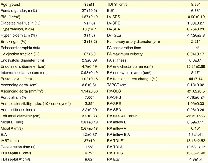
(n=13) of participants, and 7.6% (n=5) had diabetes mellitus. Mean LVEF was determined to be 67±5.9% and mean aortic diameter was 3.6±0.61.
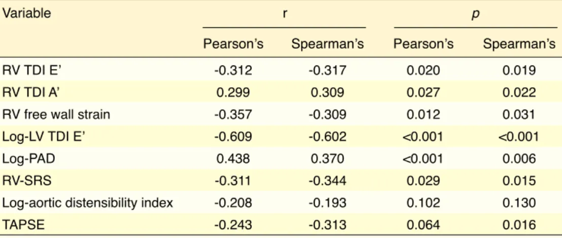
Correlation coefficients using Pearson’s and Spearman’s correlation analyses are presented in Table 2. Ascending aorta diameter was found by Pearson’s and Spearman’s, respectively, to be cor-related with RV-TDI E’ (r=-0.312, p=0.020; r=-317, p=0.019), RV-TDI A’ (r=0.299, p=0.027; r=0.309, p=0.022), RV-free wall strain (0.357, p=0.012; r=-0.309, p=0.031), log-LV TDI E’ (r=-0.609, p<0.001; r=-0.602, p<0.001), log-PAD (r=0.438, p=<0.001; r=0.370, p=0.006), RV-SRS (0.311, p=0.029; r=-0.344, p=0.015), log-aortic distensibility index (r=-0.208, p=0.102; r=-0.193, p=0.130), and tricuspid annular plane systolic excursion (r=-0.243, p=0.064; r=-0.313, p=0.016). Hypertension and age-adjusted multivariate linear regression analysis to determine relationship of ascending aorta diameter and echocar-diographic parameters is provided in Table 3. RV-SRS values (β=-0.781, t=-2.723; p=0.010) and log-LV TDI E’ (β=-2.996, t=-5.405; p:<0.001) were negatively correlated, and log-PAD (β=4.861, t=3.052; p=0.005) was positively and independently correlated with as-cending aortic diameter.
Reliability analysis
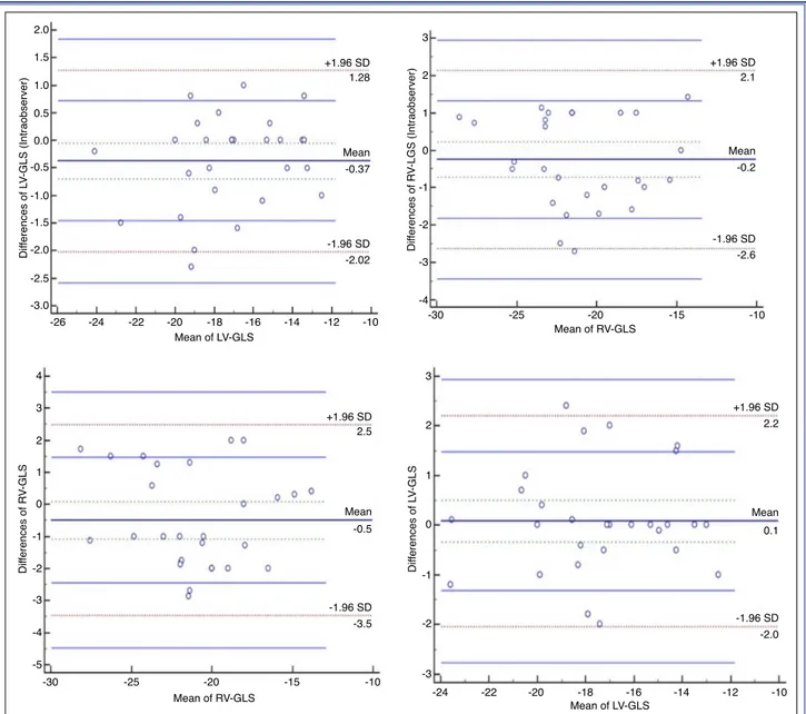
LV-GLS and RV-GLS were re-measured by first echocardiographer and second echocardiographer who was blinded to the first measurement in 28 ran-domly selected patients. Agreement analysis of inter- and intra-observer variability for LV-GLS measure-ments revealed high agreement; mean difference of 0.1 (95% limits of agreement [LoA], 2.2 to -2.0) and mean difference of -0.37 (95% LoA, 1.28 to -2.02), respectively. For intra-observer reliability, intraclass correlation coefficient was significant for LV-GLS at 0.95, (0.911–0.980). Agreement analyses of inter- and intra-observer reliability for RV-GLS revealed high agreement with mean difference of -0.5 (95% LoA, -3.5 to 2.5) and -0.2 (95% LoA, -2.6 to 2.1), respec-tively. Bland-Altman plotting of the 2 echocardiog-raphers’ results were within agreement limits of 1.96 (95% confidence interval) (Figure 1).
DISCUSSION
Our study led us to 3 essential conclusions. First, there was positive correlation between dilatation of the as-Statistical analysis
Data are presented as mean±SD and geometric mean for continuous variables, and as percentage (number of cases) for categorical variables. Normal distribu-tion of the data was tested with Kolmogorov-Smirnov test. Log-transformation was used for variables aortic strain, aortic distensibility index, deceleration time, LV-TDI E’, LV-TDI A’, LV-TDI S’, LV E/E’, PAD, PA acceleration time, RV end-systolic area and RV inflow A, which had skewed distribution. Pearson’s and Spearman’s correlation analyses were used to test possible associations between ascending aorta dimen-sion and study variables. Independent variables of as-cending aortic dilatation in bicuspid aorta disease was obtained with age and hypertension- adjusted mul-tiple linear regression analysis using stepwise model including significant variables at 25% level from uni-variate analysis as couni-variates. Multicollinearity was checked for variables used in regression analysis, since stiffness parameters were correlated with each other, which might cause bias in multivariate setting. Accordingly, variance inflation factor and tolerance values were used after regression analysis to check for multicollinearity. Tolerance value of 0.20 and above and/or variance inflation factor of 5 and less were ac-ceptable for our model to ignore multicollinearity. In-terobserver and intraobserver reliability were assessed with Bland-Altman plots. P value <0.05 was consid-ered statistically significant. Statistical analysis was performed using IBM SPPS Statistics 22 (IBM Corp., Armonk, NY, USA) and MedCalc 12.4.0 (MedCalc Software, Ostend, Belgium) software.
RESULTS
Total of 93 consecutive patients were evaluated. Eight patients with inappropriate echogenicity, 2 patients with diagnosis of coarctation of the aorta, 2 patients with coronary artery disease, 7 patients with con-comitant moderate to severe valvular disease, and 8 patients who were using beta-blocker or angiotensin converting enzyme inhibitor were excluded from the study. In all, 66 of total 93 patients were eligible for inclusion following initial evaluation.
Baseline demographic characteristics of the pa-tients are shown in Table 1. Of all papa-tients, 40.9% (n=27) were female and mean age of study group was 35±11 years. Hypertension was found in 19.7%
cending aorta and dilatation of the PA. The second and third are that RV-SRS and LV-TDI E’ negatively correlated with dilatation of the ascending aorta. No connection was established between dilatation of the aorta and parameters related to aortic and PA elastic-ity, or speckle tracking parameters of the LV.
BAV is a complex developmental anomaly caused by abnormal aortic leaflet formation at valvulogene-sis.[1] BAV is the most commonly observed congenital
heart malformation in adults, with prevalence of 1.3% in the general population.[2] Despite aortic stenosis
and aortic insufficiency being the most widespread complications associated with BAV, widening of the ascending aorta from the root to the arch, known as
bicuspid aortopathy, has been observed at rate of ap-proximately 40% to 50%.[3,4] Compared with those
with tricuspid aortic valve, BAV patients have higher rate of dilatation of the aorta and increased risk of aortic aneurysm, dissection, and rupture.[17,18]
Previ-ous studies of BAV patients have reported rates of dilatation of 8% to 59% at level of the aortic annulus, 16% to 78% at level of the sinus of Valsalva, 15% to 79% at level of the sinotubular junction, and 35% to 68% in the proximal ascending aorta.[5,19–21] BAV
can affect the PA and pulmonary valve alongside the aorta, ascending aorta, and aortic arch.[22–24] However
much the intrinsic pathologies of the aorta may be responsible for dilatation of the aorta, an increasing body of evidence shows that aortic flow anomalies
Table 1. Clinical and echocardiographic determinants of baseline
Age (years) 35±11 Female gender, n (%) 27 (40.9) BMI (kg/m2) 1.87±0.19 Diabetes mellitus, n (%) 5 (7.6) Hypertension, n (%) 13 (19.7) Hyperlipidemia, n (%) 3 (4.5) Smoking, n (%) 12 (18.2) Echocardiographic data LV ejection fraction (%) 67±5.9 Endsystolic diameter (cm) 2.9±0.39 Enddiastolic diameter (cm) 4.7±0.49 Interventricular septum (cm) 0.98±0.19 Posterior wall (cm) 1.02±0.18 Ascending aorta (cm) 3.6±0.61 Ascending aorta (mm/m2) 1.94±0.36 Aortic strain (%) 7.60*
Aortic distensibility index (10-6 cm-2 dyne-1) 3.35*
Aortic stiffness index 2.2±0.20 Left atrial diameter (cm) 3.2±0.33
Mitral E (m/s) 0.81±0.18
Mitral A (m/s) 0.67±0.18
E:A 1.2±0.37
IVRT (unit) 87±19
Deceleration time (s) 188*
TDI septal E’ cm/s 9.79*
TDI septal A’ cm/s 9.62*
TDI S’ cm/s 8.55* E:E’ 6.56* LV-SRS -0.90±0.19 LV-SRE 1.00±0.27 LV-SRA 0.76±0.23 LV -GLS -17.26±2.8
Pulmonary artery diameter (cm) 2.21*
PA acceleration time 114*
PA maximum velocity 0.94±0.17
PA stiffness 8.8±3.1
RV end-diastolic area (cm2) 15.81±2.88
RV end-systolic area (cm2) 8.47*
RV fractional area change (%) 44±7.14
TAPSE (cm) 2.13±0.32
RV-GLS -21.62±3.5
RV-SRS -1.18±0.24
RV-SRE 1.06±0.33
RV-SRA 0.96±0.26
RV free wall strain -26.32±5.97
RV inflow E 0.59±0.11 RV inflow A 0.40* RV inflow E:A 4.3±1.41 RV TDI E’ 13.16±2.52 RV TDI A’ 12.63±3.17 RV TDI S’ 13.85±1.98 RV E:E’ 4.3±1.4
*Geometric mean was given. A: Late ventricular velocity; A’: Late diastolic velocity; BMI: Body mass index; E: Early ventricular velocity; E’: Early diastolic velocity; GLS: Global longitudinal strain; IVRT: Isovolumetric relaxation time; LV: Left ventricle; PA: Pulmonary artery; RV: Right ventricle; SRA: Late diastolic strain rate; SRE: Early diastolic strain rate; SRS: Systolic strain rate; TAPSE: Tricuspid annular plane systolic excursion; TDI: Tissue Doppler imaging.
patients with or without dilated aortas.[29]
In our study, it was observed that aortic elasticity indexes were more affected than reported in the litera-ture. We attempted to explain cystic medial degenera-tion and intrinsic aortic pathologies in patients with isolated BAV. Similarly to results of Donato Aquaro et al., elasticity indexes were found to be abnormal independently of diameter of the ascending aorta. No difference was found between patients with dilated and undilated aortas in terms of elasticity indexes, suggesting that basic pathological agent in aortic dila-tation may be turbulent flow.
We have also demonstrated that LV-TDI E’ value decreased as diameter of the ascendant aorta increased. BAV group with normal valve functions showed dia-may also be responsible.[25,26] According to a study by
Bissell et al., the most important additional parameter in aortic dilatation is that of abnormal flow.[27]
Addi-tionally, they confirmed intrinsic pathologies of the medial layer, elastic tissue-collagen, and smooth mus-cle cell loss with accompanying accelerated degrada-tion and found increased wall stiffness and decreased aortic distensibility.[28] BAV patients were shown to
have abnormal aortic distensibility and stiffness.[17]
Parameters of aortic elasticity were reported to be in-dependent of aortic dilatation and valve pathology.[6]
Another study evaluated aortic maximum systolic dis-tension and maximum diastolic recoil with magnetic resonance imaging. Although they found that both were lower in BAV patients than in control group, they were unable to find any difference between BAV
Table 2. Correlation of ascending aorta diameter to echocardiographic measurements
Variable r p
Pearson’s Spearman’s Pearson’s Spearman’s
RV TDI E’ -0.312 -0.317 0.020 0.019
RV TDI A’ 0.299 0.309 0.027 0.022
RV free wall strain -0.357 -0.309 0.012 0.031
Log-LV TDI E’ -0.609 -0.602 <0.001 <0.001
Log-PAD 0.438 0.370 <0.001 0.006
RV-SRS -0.311 -0.344 0.029 0.015
Log-aortic distensibility index -0.208 -0.193 0.102 0.130
TAPSE -0.243 -0.313 0.064 0.016
A’: Late diastolic velocity; E’: Early diastolic velocity; LV: Left ventricle; PAD: Pulmonary artery diameter; RV: Right ventricle; SRS: Systolic strain rate; TAPSE: Tricuspid annular plane systolic excursion; TDI: Tissue Doppler imaging.
Table 3. Multivariate linear regression analysis to determine independent predictors of ascending aorta dilation in
bicuspid aortic disease
Unstandardized coefficients t p 95 % CI
First step
Log-LV TDI E’ -3.395 -5.167 <0.001 -4.730 – (-2.060)
Second Step
Log-LV TDI E’ -2.971 -4.904 <0.001 -4.203 – (-1.738)
Log-PAD 5.263 3.038 0.005 1.738 – 8.789
Third Step
RV-SRS -0.781 -2.723 0.010 -1.365 – (-.197)
Log-LV TDI E’ -2.996 -5.405 .<0.001 -4.126 – (-1.867)
Log-PAD 4.861 3.052 0.005 1.617 – 8.104
CI: Confidence interval; LV-TDI E’: Left ventricle tissue Doppler early diastolic velocity; PAD: Pulmonary artery diameter; RV-SRS: Right ventricle peak systolic strain rate.
may also be the cause of diastolic dysfunction. Pre-viously, study by Martos et al. reported that MMP-2 created myocardial fibrosis.[35] Yasmin et al. indicated
that MMP-2 was the cause of heart failure preserved ejection fraction.[36] As a result, elevated MMP-2
lev-els may cause diastolic dysfunction through increased fibrosis and stiffness.
Another finding was positive correlation between increased diameter of the ascending aorta and PA. Dilatation of the main PA has been demonstrated in BAV patients even in the absence of pulmonary valve disease.[23] Embryologically, the aorta and the PA
share their origin in the sole outflow tract, the cono-stolic dysfunction.[30] Kocabay et al. reported diastolic
dysfunction, abnormal left atrium strain parameters and volume index with increased LV E/E’ in isolated BAV patients.[31] Abnormal force parallel to the aortic
wall exerted by shear stress acts upon endothelial sur-face in terms of friction, which may activate cellular signaling cascades and result in overexpression of ex-tracellular matrix degenerating substances, including matrix metalloproteinases (MMPs) and their endog-enous tissue inhibitors.[32,33] Tzemos et al. have shown
that MMP2 levels are higher in BAV patients with dilated ascending aorta.[34] Increased MMP-2 levels
seen in patients with dilatation of the ascending aorta
Figure 1. Intraobserver and interobserver Bland-Altman plots of left ventricular global longitudinal strain (GLS) and right ven-tricular GLS, with representation of the limits of agreement (dotted line), -1.96s to +1.96s.
2.0 1.5 0.5 0.0 -0.5 -1.0 -2.0 -3.0 -26 Mean of LV-GLS Dif ferences of L V-GLS (Intraobserver) -24 -22 -20 -18 -16 -14 -2.02 -1.96 SD +1.96 SD -0.37 1.28 4 3 2 1 0 -1 -2 -3 -4 -5 -30 -25 -20 -15 -10 Mean of RV-GLS Dif ferences of R V-GLS +1.96 SD -1.96 SD Mean 2.5 -3.5 -0.5 Mean -12 -10 -2.5 -1.5 1.0 3 2 1 -1 -2 -3 -4 0 Mean of RV-GLS Dif ferences of R V-LGS (Intraobserver) -2.6 -1.96 SD +1.96 SD -0.2 2.1 Mean -30 -25 -20 -15 -10 -24 -22 -20 -18 -16 -14 -12 -10 Mean of LV-GLS Dif ferences of L V-GLS +1.96 SD -1.96 SD Mean 2.2 -2.0 0.1 3 2 1 0 -1 -2 -3
Limitations
The most important limitation of our study was rela-tively small number of patients. In addition, role of MMP-2, if any, was not investigated. Aortic elasticity was studied only with echocardiographic parameters. Another limitation was that, although small in num-ber, patients diagnosed with hypertension and diabe-tes mellitus were included in the study and an isolated group was not created by ruling out co-morbidities. As the other limiting parameters, left atrial volume and tricuspid regurgitant jet were not evaluated in definitions of diastolic dysfunction. Although numer-ous parameters were used for RV function, tricuspid regurgitant jet was not assessed and pulmonary arte-rial pressure was not specified in many patients due to missing data.
Conclusion
In this study, we concluded that ascending aorta diam-eter is positively correlated with PAD in BAV patients and that RV strain rate and LV diastolic parameters are affected before development of the valve disease. Financial support
None.
Conflict-of-interest issues regarding the authorship or article: None declared
REFERENCES
1. Evangelista A, Flachskampf FA, Erbel R, Antonini-Canterin F, Vlachopoulos C, Rocchi G, et al. Echocardiography in aor-tic diseases: EAE recommendations for clinical pracaor-tice. Eur J Echocardiogr 2010;11:645–58.
2. Verma S, Siu SC. Aortic dilatation in patients with bicuspid aortic valve. N Engl J Med. 2014;370:1920–9.
3. Verma S, Yanagawa B, Kalra S, Ruel M, Peterson MD, Ya-mashita MH, et al. Knowledge, attitudes, and practice pat-terns in surgical management of bicuspid aortopathy: a survey of 100 cardiac surgeons. J Thorac Cardiovasc Surg 2013;146:1033–40.
4. Girdauskas E, Rouman M, Borger MA, Kuntze T. Compar-ison of aortic media changes in patients with bicuspid aor-tic valve stenosis versus bicuspid valve insufficiency and proximal aortic aneurysm. Interact Cardiovasc Thorac Surg 2013;17:931–6.
5. Nistri S, Sorbo MD, Marin M, Palisi M, Scognamiglio R, Thiene G. Aortic root dilatation in young men with normally functioning bicuspid aortic valves. Heart 1999;82:19–22. 6. Oulego-Erroz I, Alonso-Quintela P, Mora-Matilla M, Gau-truncus.[37] Ross procedure and the complications that
may follow suggest that the aorta may be affected by the pulmonary bed beside it, especially in bicuspid valve patients. Ross procedure is a method in which the pulmonary valve is used as autograft replacement for the aortic valve. The most frequent problem after Ross procedure in BAV patients is late dilatation of the autograft. BAV patients are known to exhibit neo-aortic sinus dilatation and increase in diameter of the annulus after Ross procedure.[38] PA, which shares the
same embryological origin with the aorta, might have the potential to exhibit the same intrinsic pathologies in BAV patients. In a another study comparing iso-lated BAV patients with control group it was demon-strated that elasticity of the PA was as abnormal as that of the aorta.[9]
Using PA stiffness index as a new, non-invasive pa-rameter, it was determined that if distensibility of the PA is established as abnormal, ejection and accelera-tion times of the RV and PA would also be reduced.[39]
We did not find growth in diameter of the ascend-ing aorta and PA stiffness very meanascend-ingful. Despite increase in PAD, pulmonary stiffness didn’t change. This finding may have resulted from lack of standard cut-off value and not taking PAD into account when measuring stiffness. It is necessary to find a new, steady echocardiographic parameter for this area of focus. We considered that lack of change in PA stiff-ness values in our study might be connected to our quandary. Only new echocardiographic parameters that include PAD and longitudinal values would be able to shed light on this problem.
Another result of the present study was finding of decreased RV-SRS with increased dilatation of as-cending aorta. To the best of our knowledge, RV sys-tolic functions in the bicuspid aortic valve have not been evaluated so far. Santarpia et al. reported that compared to control group, BAV patients had lower LV longitudinal, circumferential, and radial strain.
[40] RV systolic dysfunction has been demonstrated
in patients with heart failure preserved ejection frac-tion.[41] It was suggested that increased LV pressure
results in elevated PA wedge pressure, which in turn causes pressure overload for RV.[42] Our findings
in-dicate that RV and PA parameters in BAV patients may be important in terms of clinical monitoring and that their prognostic value should also be inves-tigated.
73.
19. Hahn RT, Roman MJ, Mogtader AH, Devereux RB. Asso-ciation of aortic dilation with regurgitant, stenotic and func-tionally normal bicuspid aortic valves. J Am Coll Cardiol 1992;19:283–8.
20. Gurvitz M, Chang RK, Drant S, Allada V. Frequency of aortic root dilation in children with a bicuspid aortic valve. Am J Cardiol 2004;94:1337–40.
21. Ferencik M, Pape LA. Changes in size of ascending aorta and aortic valve function with time in patients with congenitally bicuspid aortic valves. Am J Cardiol 2003;92:43–6.
22. Warnes CA. The adult with congenital heart disease: born to be bad? J Am Coll Cardiol 2005;46:1–8.
23. Kutty S, Kaul S, Danford CJ, Danford DA. Main pulmonary artery dilation in association with congenital bicuspid aortic valve in the absence of pulmonary valve abnormality. Heart 2010;96:1756–61.
24. Tadros TM, Klein MD, Shapira OM. Ascending aortic dila-tation associated with bicuspid aortic valve: pathophysiol-ogy, molecular biolpathophysiol-ogy, and clinical implications. Circulation 2009;119:880–90.
25. Loscalzo ML, Goh DL, Loeys B, Kent KC, Spevak PJ, Dietz HC. Familial thoracic aortic dilation and bicommissural aor-tic valve: a prospective analysis of natural history and inheri-tance. Am J Med Genet A 2007;143:1960–7.
26. Girdauskas E, Borger MA, Secknus MA, Girdauskas G, Kun-tze T. Is aortopathy in bicuspid aortic valve disease a congen-ital defect or a result of abnormal hemodynamics? A critical reappraisal of a one-sided argument. Eur J Cardiothorac Surg 2011;39:809–14.
27. Bissell MM, Hess AT, Biasiolli L, Glaze SJ, Loudon M, Pitch-er A, et al. Aortic dilation in bicuspid aortic valve disease: flow pattern is a major contributor and differs with valve fu-sion type. Circ Cardiovasc Imaging 2013;6:499–507. 28. Grotenhuis HB, Ottenkamp J, Westenberg JJ, Bax JJ, Kroft
LJ, de Roos A. Reduced aortic elasticity and dilatation are as-sociated with aortic regurgitation and left ventricular hyper-trophy in nonstenotic bicuspid aortic valve patients. J Am Coll Cardiol 2007;49:1660–5.
29. Donato Aquaro G, Ait-Ali L, Basso ML, Lombardi M, Pin-gitore A, Festa P. Elastic properties of aortic wall in patients with bicuspid aortic valve by magnetic resonance imaging. Am J Cardiol 2011;108:81–7.
30. Kurt M, Tanboga IH, Bilen E, Isik T, Kaya A, Karakaş MF, et al. Abnormal left ventricular mechanics in isolated bicuspid aortic valve disease may be independent of aortic distensibility: 2D strain imaging study. J Heart Valve Dis 2012;21:608–14. 31. Kocabay G, Karabay CY, Kalkan S, Kalayci A, Efe SC,
Ak-gun T, et al. Relationship between left ventricular diastolic function and arterial stiffness in patients with bicuspid aortic valve. J Heart Valve Dis 2014;23:279–88.
32. Klein T, Bischoff R. Physiology and pathophysiology of ma-trix metalloproteases. Amino Acids 2011;41:271–90. treaux Minaya S, Lapeña-López de Armentia S. Ascending
aorta elasticity in children with isolated bicuspid aortic valve. Int J Cardiol 2013;168:1143–6.
7. Luciani GB, Barozzi L, Tomezzoli A, Casali G, Mazzucco A. Bicuspid aortic valve disease and pulmonary autograft root dilatation after the Ross procedure: a clinicopathologic study. J Thorac Cardiovasc Surg 2001;122:74–9.
8. De Sa M, Moshkovitz Y, Butany J, David TE. Histologic abnor-malities of the ascending aorta and pulmonary trunk in patients with bicuspid aortic valve disease: clinical relevance to the ross procedure. J Thorac Cardiovasc Surg 1999;118:588–94. 9. Celik M, Yuksel UC, Yalcinkaya E, Gokoglan Y, Yildirim
E, Bugan B, et al. Elasticity properties of pulmonary artery in patients with bicuspid aortic valve. Echocardiography 2014;31:759–64.
10. Lang RM, Bierig M, Devereux RB, Flachskampf FA, Foster E, Pellikka PA, et al. Recommendations for chamber quantifi-cation: a report from the American Society of Echocardiogra-phy’s Guidelines and Standards Committee and the Chamber Quantification Writing Group, developed in conjunction with the European Association of Echocardiography, a branch of the European Society of Cardiology. J Am Soc Echocardiogr 2005;18:1440–63.
11. Lang RM, Badano LP, Mor-Avi V, Afilalo J, Armstrong A, Er-nande L, et al. Recommendations for cardiac chamber quan-tification by echocardiography in adults: an update from the American Society of Echocardiography and the European As-sociation of Cardiovascular Imaging. J Am Soc Echocardiogr 2015;28:1–39.e14.
12. Nistri S, Grande-Allen J, Noale M, Basso C, Siviero P, Maggi S, et al. Aortic elasticity and size in bicuspid aortic valve syn-drome. Eur Heart J 2008;29:472–9.
13. Görgülü S, Eren M, Yildirim A, Ozer O, Uslu N, Celik S, et al. A new echocardiographic approach in assessing pulmonary vascular bed in patients with congenital heart disease: pulmo-nary artery stiffness. Anadolu Kardiyol Derg 2003;3:92–7. 14. Nagueh SF, Appleton CP, Gillebert TC, Marino PN, Oh JK,
Smiseth OA, et al. Recommendations for the evaluation of left ventricular diastolic function by echocardiography. Eur J Echocardiogr 2009;10:165–93.
15. Leitman M, Lysyansky P, Sidenko S, Shir V, Peleg E, Binen-baum M, et al. Two-dimensional strain-a novel software for real-time quantitative echocardiographic assessment of myo-cardial function. J Am Soc Echocardiogr 2004;17:1021–9. 16. Pirat B, McCulloch ML, Zoghbi WA. Evaluation of global
and regional right ventricular systolic function in patients with pulmonary hypertension using a novel speckle tracking method. Am J Cardiol 2006;98:699–704.
17. Nistri S, Sorbo MD, Basso C, Thiene G. Bicuspid aortic valve: abnormal aortic elastic properties. J Heart Valve Dis 2002;11:369–73.
18. Anagnostopoulos CE, Prabhakar MJ, Kittle CF. Aortic dissec-tions and dissecting aneurysms. Am J Cardiol 1972;30:263–
valve? Eur J Cardiothorac Surg 2010;38:333–9.
39. Mahfouz RA. Impact of pulmonary artery stiffness on right ventricular function and tricuspid regurgitation after success-ful percutaneous balloon mitral valvuloplasty: the importance of early intervention. Echocardiography 2012;29:1157–63. 40. Santarpia G, Scognamiglio G, Di Salvo G, D’Alto M,
Sa-rubbi B, Romeo E, et al. Aortic and left ventricular remod-eling in patients with bicuspid aortic valve without signifi-cant valvular dysfunction: a prospective study. Int J Cardiol. 2012;158:347–52.
41. Aschauer S, Kammerlander AA, Zotter-Tufaro C, Ristl R, Pfaffenberger S, Bachmann A, et al. The right heart in heart failure with preserved ejection fraction: insights from cardiac magnetic resonance imaging and invasive haemodynamics. Eur J Heart Fail 2016;18:71–80.
42. Tedford RJ, Hassoun PM, Mathai SC, Girgis RE, Russell SD, Thiemann DR, et al. Pulmonary capillary wedge pres-sure augments right ventricular pulsatile loading. Circulation 2012;125:289–97.
33. Kaunas R, Kang H, Bayless KJ. Synergistic Regulation of An-giogenic Sprouting by Biochemical Factors and Wall Shear Stress. Cell Mol Bioeng 2011;4:547–59.
34. Tzemos N, Lyseggen E, Silversides C, Jamorski M, Tong JH, Harvey P, et al. Endothelial function, carotid-femoral stiff-ness, and plasma matrix metalloproteinase-2 in men with bicuspid aortic valve and dilated aorta. J Am Coll Cardiol 2010;55:660–8.
35. Martos R, Baugh J, Ledwidge M, O’Loughlin C, Conlon C, Patle A, et al. Diastolic heart failure: evidence of increased myocardial collagen turnover linked to diastolic dysfunction. Circulation 2007;115:888–95.
36. Yasmin, McEniery CM, Wallace S, Dakham Z, Pulsalkar P, Maki-Petaja K, et al. Matrix metalloproteinase-9 (MMP-9), MMP-2, and serum elastase activity are associated with sys-tolic hypertension and arterial stiffness. Arterioscler Thromb Vasc Biol 2005;25:372.
37. Brown CB, Baldwin HS. Neural crest contribution to the car-diovascular system. Adv Exp Med Biol 2006;589:134–54. 38. Hanke T, Charitos EI, Stierle U, Robinson DR, Hemmer W,
Moritz A, et al. German Ross Registry. The Ross operation - a feasible and safe option in the setting of a bicuspid aortic
Keywords: Aortic valve; bicuspid; strain rate.


