Corresponding author: Mustafa GüVEN E-mail: [email protected]
Original Investigation
Mustafa guveN
1, davut ceYlaN
2, adem Bozkurt aras
1, ayla akBal
3, ferhat gOkMeN
3, Hatice resOrlu
3,
Bahadir alkaN
1, Mehmet TOkMak
4, Murat cOsar
11Canakkale Onsekiz Mart University, School of Medicine, Department of Neurosurgery, Canakkale, Turkey 2sakarya university, school of Medicine, Department of Neurosurgery, sakarya, Turkey
3Canakkale Onsekiz Mart University, School of Medicine, Department of Physical Therapy and Rehabilitation, Canakkale, Turkey 4İstanbul Medipol University, School of Medicine, Department of Neurosurgery, Istanbul, Turkey
Effect of Using High-Speed Drill in Anterior Cervical
Discectomy and Fusion
ABSTRACT
developed, particularly during the past two decades, and surgical success has increased. along with these techniques, allografts, hydroxyapatite grafts, ceramic grafts, titanium cages, carbon cages, polyetheretherketone cages, and plates were developed. These techniques were developed by focusing on the fusion and protection of cervical vertebra alignment (2, 3). although the outcomes are generally successful, rare major complications such as subsidence, pseudoarthrosis, and the replacement of fusion material have been reported (30). Nonunion and pseudoarthrosis rates have been reported to range between 0% and 20% in ACDF operations (9, 29, 35, 38). although some cases developing pseudoarthrosis
█
InTRODuCTIOn
Many surgical treatment approaches such as anterior cervical discectomy (ACD), anterior cervical discectomy and fusion (ACDF), anterior foraminotomy, keyhole foraminotomy, and arthroplasty have been applied for cervical disc hernia. Each of these treatment approaches has advantages and disadvantages (3, 24, 29-31, 33, 38).
The technique commonly used in surgical treatment of cervical disc hernia is anterior cervical discectomy and fusion (ACDF). Cervical lordosis and the height of the disc and foramen are protected by ACDF. Some surgical techniques have been
AIM: The objective of this study was to investigate the effect of using 2 different surgical techniques (curette or high-speed drill) in anterior cervical discectomy surgery on the healing of cases.
MATERIAl and METhODS: Fifty-four operated cervical disc hernia cases were retrospectively examined in 2 groups. Discectomy and osteophytectomy were carried out in Group A by using a high-speed drill, while a curette was used for group B. Preoperative and postoperative computerized tomography and direct radiography were performed. Cervical disc height, cervical and segmental lordotic angles were calculated. The visual analogue scale and Odom’s criteria were used in the assessment of pain and clinical healing. The fusion ratio of both groups was compared. The Mann-Whitney U test was used to compare data from the groups. RESulTS: Satisfactory results were obtained in the groups where high-speed drill and curette were used. Independently from the surgical technique, pain scores were significantly reduced in both groups after surgery. No radiologically significant differences were identified between the two groups within the postoperative period.
COnCluSIOn: Either high-speed drill or curette can be chosen for the osteophytectomy and discectomy stages of anterior cervical discectomy operations.
are asymptomatic, the absence of solid fusion causes poor functional outcomes and continuing pain (9, 29, 35, 38). The pseudoarthrosis ratio has been reported to be based on the number of levels at which the operation took place, the type of graft used, and the surgical technique (7, 29, 38).
The number of pseudoarthrosis cases has also increased with the rapidly increasing number of ACDF surgeries over the past twenty years. Therefore, it has become even more crucial to determine the causes of pseudoarthrosis. In the literature, the rates of pseudoarthrosis are reported to vary according to the type of graft used (7, 10, 11, 24, 25). Neural and osteoblastic injury based on the temperature increase was also reported in surgeries carried out using a high-speed drill (8, 14, 17, 21, 23, 37). However, no study was found that showed whether or not the use of a high-speed drill affects fusion when endplate and osteophyte surgical techniques are used.
In our retrospective study, we assessed and compared the changes observed after ACDF surgeries which were performed, either using a curette or a high-speed drill. The purpose of our study was to present and compare radiological and clinical findings of these two groups.
█
MATERIAl and METhODS
Thirty-one cases in the high-speed drill group (group a) and 23 cases in the curette group (group B) were retrospectively examined. The criteria for the patients’ inclusion in and selection for the study were as follows. Magnetic resonance images (MRI) and clinical findings were compatible, a single-level disc was affected, radiculopathy findings were present, surgery was performed for a cervical disc hernia for the first time, and either the surgical procedure was needed immediately or they did not respond to conservative treatment over a six-week period. The criteria for exclusion from the study included patients who had already undergone a percutaneous procedure due to cervical and/or radicular pain (nucleotomy, chemonucleolysis, epidural steroid injection, etc.) and who were experiencing multilevel disc herniation, significant degenerative spinal disease, fracture, infection, tumor, a spinal deformity, chronic systemic disease, or cervical kyphotic posture. No case was excluded from the study based on gender, age, or severe preoperative clinical findings. Preoperative plain and lateral cervical x-ray, computerized tomography (CT), and MRI were applied to all of the cases. group a consisted of 31 cases (13 males and 18 females, with a mean age of 45.4 years), where ACDF was carried out using a high-speed drill. group B consisted of 23 cases (9 males and 14 females, with a mean age of 51.5 years), where ACDF was carried out using a curette. In both groups, a PEEK (polyetheretherketone) cage (Titania, Izmir, Turkey or Medikon,
Ankara, Turkey) and bone graft into the cage (Osteotech, Eatontown, NJ-USA) were used for fusion.
Surgical Technique
Surgeries for both groups were performed under general anesthesia. In order to determine the operation level, C-arm fluoroscopy was used. After distraction of the space, ACD
and osteophytectomy were carried out through microsurgical technique. While endplate decortication and osteophytectomy in group A was performed using a 4 or 5 mm diamond ball cutter with the help of a high-speed drill (Anspach eMax2
plus, Synthes Inc., PA, USA), for group B, those procedures
were carried out by curette. Drilling was performed at 40,000 rpm with intervals lasting four to five seconds. Sufficient and constant cooling irrigation was done during drilling. The posterior longitudinal ligament for decompression and the spinal cord of the influenced nerve root was opened in both groups, and a part of it was resected. In all cases, the endplate around the cartilage was protected, while the endplates of the upper and lower spine were slightly decorticated. The disc fragment causing herniation was removed. Bone graft (Osteotech, Eatontown, NJ-USA) was injected into the pEEk cage, and a spacer was prepared. While distracting the spine, the spacer was placed. Following control of the graft position with C-arm fluoroscopy, the vertebra was redistracted. The wearing of a soft collar for three weeks following surgery was suggested for all cases in both groups.
Clinical Evaluation
Pain reflecting to the neck and arm of the cases was assessed with the Visual Analogue Scale (VAS) pain scoring the day before the operation and daily in the early postoperative period (32). Clinical evaluation also continued during the postoperative follow-up. Odom’s criteria were used in the last follow-up of clinical assessment, and patients were classified as excellent, good, fair, or poor (27).
Radiological Evaluation
For radiologic findings, the height of the level operated on, the cervical and segmental lordotic angles, and the fusion rates were determined for each case. For each case, an assessment was carried out five times, once preoperative, and postoperative at one day, one month, three months, and one year. The height of the disc was calculated using images of the mid-sagittal plane with the help of computerized tomography software (Toshiba, Prospeed Helical CT). The preoperative and postoperative segmental lordotic angle (SLA) and cervical lordotic angle (CLA) of both groups were measured by Katsuura’s method (10, 19).
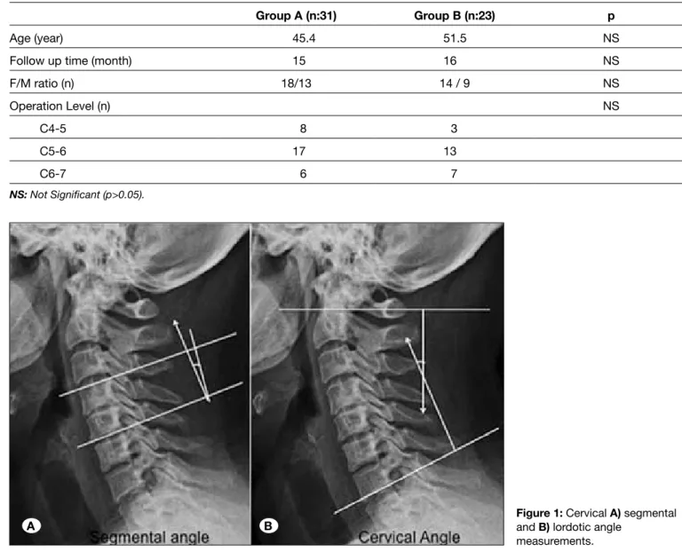
Lordosis of the cervical spine (CLA) was measured as the angle between the line parallel to the upper border of the C2 vertebral body and the line parallel to the lower border of the C7 vertebral body. In addition, segmental lordosis of the level operated on was measured as the angle between the line parallel to the upper border of the vertebral body proximal to the disc space involved and the line parallel to the lower border of the underlying vertebral body (Figure 1a,B) (10, 19). These values were noted as positive in lordosis, negative in kyphosis.
In both groups, fusion at the level operated on was assessed with x-ray and CT in the third and twelfth month follow-up. An assessment of excellent, good, average, or poor was given by a radiologist not involved in this study. Seeing trabecular bone bridges in at least one of the anterior and/or posterior regions of the PEEK cage was evaluated as fusion. The absence of
bridges or seeing a discontinuity in the anterior-posterior line in fusion was classified as non-fusion (12).
Statistical Analysis
The Mann-Whitney U test was performed using spss v.19 software (SPSS Inc., Chicago, IL, USA) to compare changes in the disc height, segmental and cervical lordosis angle, and fusion rates of both groups. Preoperative measurements and postoperative measurements at one day, one month, three months, and one year were assessed. The Mann-Whitney U
test was also used in comparisons of Odom’s criteria and VAS
scoring for each group. The values of p<0.05 were accepted as significant.
The study sample size power was found to be 86% (13). In our study, we complied with the Declaration of Helsinki, revised in 2008 and also accepted by the Çanakkale 18 Mart University Medical Ethical Committee. An informed consent form was received from all cases.
█
RESulTS
Clinical Results
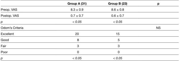
The demographical data of the cases are given in Table I, and the VAS scores of both groups are shown in Table II. While the mean VAS score of the high-speed drill group was 8.3 ± 0.9 preoperatively, it was detected to be 0.7 ± 0.7 in the twelfth month postoperative period. The reduction in VAS scores in both groups was considered significant (p<0.05). No significant difference was found in the preoperative and postoperative comparison of VAS scores between the groups (Table II).
Odom’s criteria were applied to clinically assess the improvement of symptoms at the twelfth month follow-up of both groups. Odom’s criteria were numbered as excellent: 4, good: 3, fair: 2, poor: 1. The “good” and “excellent” results, according to Odom’s criteria, were 93.3% and 89.9%, respectively, in group B. Satisfactory outcomes were observed
figure 1: Cervical A) segmental and B) lordotic angle
measurements. Table I: Demographic Distribution and Operation Levels of the Groups
Group A (n:31) Group B (n:23) p
age (year) 45.4 51.5 Ns
Follow up time (month) 15 16 Ns
F/M ratio (n) 18/13 14 / 9 Ns Operation level (n) Ns C4-5 8 3 C5-6 17 13 C6-7 6 7 NS: Not Significant (p>0.05). A B
follow-up is shown in Table IV. Change of the mean SLA measured in the preoperative and postoperative 1st day was found to be statistically significant (2.63 ± 4.41° vs. 5.14 ± 3.98°, respectively, p<0.05). Change of the mean SLA measured in the preoperative and postoperative 1st month and 3rd month was also found statistically significant (2.63 ± 4.41° vs. 4.64 ± 4.83° and 3.24 ± 5.20°, p<0.05 and p<0.05, respectively). Furthermore, a significant increase was identified between the preoperative and postoperative 12th month SLA (2.63 ± 4.41° vs. 3.48±5.39°, p<0.05 ) (Table IV).
Cervical Lordotic Angle (CLA) Group A (high-speed drill Group)
The mean group CLA measured at 5 different times during follow-up is shown in Table V. Change of the mean CLA measured in the preoperative and postoperative 1st day was found to be statistically significant (11.14 ± 5.92° vs. 10.42 ± 5.55°, respectively, p<0.05). Change of the mean CLA measured in the preoperative and postoperative 1st month and 3rd month was also found statistically significant (11.14 ± 5.92° vs. 14.68 ± 4.84° and 13.23 ± 10.11°, p<0.05 and p<0.05, respectively). Furthermore, significant increase was identified between the preoperative and postoperative 12th month CLA (11.14 ± 5.92° vs. 13.33±4.58°, p<0.05 ) (Table V).
Group B (Curette Group)
The mean group CLA measured at 5 different times during follow-up is shown in Table V. Change of the mean CLA measured in the preoperative and postoperative 1st day was found statistically significant (15.18 ± 11.56° vs. 7.69 ± 7.95°, respectively, p<0,05). Change of the mean CLA measured in the preoperative and postoperative 1st month and 3rd month was also found to be statistically significant (15.18 ± 11.56° vs. 11.82 ± 8.97° and 10.11 ± 9.74°, p<0.05 and p<0.05, respectively). Furthermore, a significant increase was identified between the preoperative and postoperative 12th month CLA (15.18 ± 11.56° vs. 9.06 ± 11.82°, p<0.05 ) (Table V).
Comparison of Fusion Rates
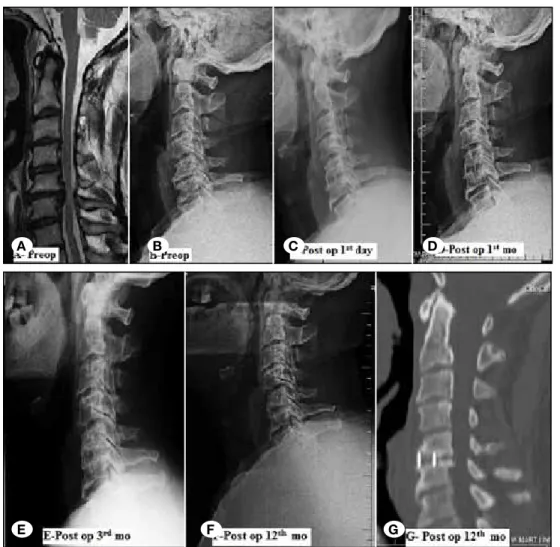
When the fusion rates at the level operated in the post-surgery 3rd and 12th months were compared, no significant difference was seen between the two groups. In the 3rd month follow-up of both grofollow-ups, a fusion rate of 41% was observed in high-speed drill group while 34% fusion was observed in the curette group. In the follow-up in the 12th months, a fusion rate of 100% was observed in all cases of both groups (Figure 2a-g).
Comparison of Data Between Groups
No significant difference was determined in terms of VAS scores, Odom’s criteria, disc heights, and preoperative mean segmental and cervical lordotic angles between the groups. Additionally, no significant differences were identified in terms of the postoperative 1st, 3rd, and 12th month VAS scores, Odom’s criteria, disc heights, and segmental and cervical angles between the groups.
all cases recovered uneventfully with good outcomes at their 1st year follow-up. No severe complications such as death, in both groups. No significant difference was identified in
intergroup comparison according to Odom’s criteria. However, an intragroup comparison of preoperative and postoperative values showed a significant difference (p<0.05) (Table II).
Radiological Results Disk Height
Group A (high-speed drill group)
The mean group disc height values measured at five different times during the follow-ups are shown in Table III.
Change of the mean disc height measured in the preoperative and postoperative 1st day was found to be statistically significant (3.91 ± 1.09 mm vs. 5.77 ± 0.93 mm, respectively, p<0.05). Change of the mean disc height measured in the preoperative and postoperative 1st month and 3rd month was also found to be statistically significant (3.91 ± 1.09 mm vs. 5.41 ± 0.94 mm and 5.15 ± 0.84 mm, p<0.05 and p<0.05, respectively). Furthermore, a significant increase was identified between the preoperative and postoperative 12th month disc heights (3.91 ± 1.09 mm vs. 6.30 ± 7.24 mm, p<0.05 ) (Table III).
Group B (Curette Group)
The mean group disc height values measured at 5 different times during follow-up are shown in Table III. Change of the mean disc height measured in the preoperative and postoperative 1st day was found statistically significant (4.1 ± 0.88 mm vs. 5.48 ± 1.20 mm, respectively, p<0.05). Changes of the mean disc height measured in the preoperative and postoperative 1st month and 3rd month were also found to be statistically significant (4.1 ± 0.88 mm vs. 5.31 ± 1.16 mm and 5.10 ± 1.07 mm, p<0.05 and p<0.05, respectively). Furthermore, a significant increase was identified between the preoperative and postoperative 12th month disc heights (4.1 ± 0.88 mm vs. 4.68 ± 1.14 mm, p<0.05 ) (Table III).
Subsidence was detected in 4 cases (12.9%) in the high-speed drill group and in 3 cases (13%) in the curette group at the end of the 1st year. No statistically significant difference was detected between the two groups (p>0.05).
Segmental Lordotic Angle (SLA) Group A (high-speed drill Group)
The mean group sla measured at 5 different times during follow-up is shown in Table IV. Change of the mean SLA measured in the preoperative and postoperative 1st day was found statistically significant (0.37 ± 3.87° vs. 4.89 ± 3.46°, respectively, p<0.05). Change of the mean SLA measured in the preoperative and postoperative 1st month and 3rd month was also found to be statistically significant (0.37 ± 3.87° vs. 2.47 ± 3.16° and 2.31 ± 2.53°, p<0.05 and p<0.05, respectively). Furthermore, significant increase was identified between the preoperative and postoperative 12th month sla (0.37 ± 3.87° vs. 1.98 ± 2.19°, p<0.05 ) (Table IV).
Group B (Curette Group)
No statistically significant difference was noted in the distribution of gender and age.
█
DISCuSSIOn
Today, the most common accepted gold standard technique in surgical treatment of cervical disc hernia is ACDF. ACDF can be made using bone graft (28), titanium (5), carbon fiber (6), and pEEk (4, 10, 28) cage. In an in vitro study, pEEk cages were reported not to have cytotoxicity and mutagenicity (26). reoperation, transient or persistent neurologic injury, Horner
syndrome, pseudoarthrosis, infection, or thromboembolic event occurred.
No misalignment was observed in the cages of cases in both groups. Moreover, no replacement or dislocation of cages was encountered up until the time of last follow-up.
all cases were allowed to return to mild activities within 4 weeks postoperatively, and to heavier work and to exercise within 3 months postoperatively.
Table II: The VAS Scores and Odom’s Criteria of the Groups
Group A (31) Group B (23) p Preop. VAS 8.3 ± 0.9 8.6 ± 0.8 Postop. VAS 0.7 ± 0.7 0.6 ± 0.7 p < 0.05 < 0.05 Odom’s Criteria Ns Excellent 20 15 good 8 5 Fair 3 3 poor 0 0 p < 0.05 < 0.05 NS: non-specific.
Table III: Disc Height Measurements of the groups
Preop. Postop. 1st day Early postop, (1st month) Early postop, (3rd month) late postop, (12th month) p
group a (mm) 3.91±1.09 5.77±0.93 5.41±0.94 5.15±0.84 6.30±7.24 <0.05 group B (mm) 4.1±0.88 5.48±1.20 5.31±1.16 5.10±1.07 4.68±1.14 <0.05 Table IV: segmental lordotic angle Measurements of the groups
Preop. Postop. 1st day Early postop. (1st month) Early postop. (3rd month) late postop. (12th month) p
Group A* 0.37±3.87 4.89±3.46 2.47±3.16 2.31±2.53 1.98±2.19 <0.05 Group B* 2.63±4.41 5.14±3.98 4.64±4.83 3.24±5.20 3.48±5.39 <0.05 * (plain [o] mean±SD).
Table V: Cervical Lordotic Angle Measurements of the Groups Preop. Postop. 1st day
Early postop. (1st month) Early postop. (3rd month) late postop. (12th month) p Group A* 11.14±5.92 10.42±5.55 14.68±4.84 13.23±10.11 13.33±4.58 <0.05 Group B* 15.18±11.56 7.69±7.95 11.82±8.97 10.11±9.74 9.06±11.82 <0.05 * (plain [o] mean±SD).
disc height measurements were compared in both groups, it was detected that significant increase was present in the postoperative 1st day and that minimal reduction was present in the postoperative 1st month, 3rd month, and at the end of the 1st year (p<0.05) (Table III). The rate of subsidence has been reported to be between 2 and 33% in the literature (1, 15). In addition, it was determined that the rate of subsidence is comparable with the literature and that no significant difference is present between the groups (7, 15, 18, 22). Adjacent segment disease is the main problem caused by fusion in the postoperative follow-up of cervical discectomy cases. In the meta-analysis study by Anderson et al., no significant difference was reported to be present among fusion options (pEEk cage, titanium cage, autograft, plate, and arthroplasty etc) (1). Therefore adjacent segment disease was not assessed in our study.
Study sample size power was found to be 86% (13). This information was added to the statistical analysis.
In our study, no statistically significant difference was identified in terms of disc height, segmental lordotic angle and cervical lordotic angle between groups (p<0.05) (Table IV, V). Preoperative and postoperative comparison of SLA and PEEK cages have been reported to be a reliable biomaterial in
spinal surgery due to their biocompatibility, non-absorbable and corrosion-resistant competence (20). pEEk elasticity has been reported to be similar to bone (34). In addition to these reasons, bone or demineralized bone matrix-filled PEEK cages are commonly used today in practice because they do not cause metallic artifacts or prevent postoperative imaging. Good and excellent outcomes according to Odom’s criteria were determined in group A at a rate of 93.3% while in group B the rate was 89.9%. Significant improvement in VAS scores was noted in both groups, and from this aspect, no difference was identified between groups. With these outcomes, no significant difference was detected in both techniques in terms of clinical recovery (p<0.05) (Table II). Similar clinical outcomes have been reported in many publications where ACDF was carried out using pEEk cage (4, 10, 28, 36).
Subsidence of the cage developing after surgery is a crucial problem. Subsidence occurs because of excessive curette of endplate, excessive distraction due to selection of a too-tall cage, non-appropriate cage geometry, and cages made of non-appropriate material. In consequence of subsidence, disc and foramen height decrease, cervical malalignment occurs. When the preoperative and postoperative 1st day
figure 2: A) preoperative MRI, B) preoperative, postoperative C) 1st day, D) 1st, E) 3rd and f) 12th months lateral plain radiograms and G) late postoperative sagittal CT scans of the patients (group A and B).
A B C D
7. Cosar M, Ozer AF, Iplikcioglu AC, Oktenoglu T, Kosdere S, Sasani M, Bozkus H, Khoo LT, Sarioglu AC: The results of beta-tricalcium phosphate coated hydroxyapatite (beta-TCP/HA) grafts for interbody fusion after anterior cervical discectomy. J Spinal Disord Tech 21: 436-441, 2008
8. Dolan EB, Haugh MG, Tallon D, Casey C, McNamara LM: Heat-shock-induced cellular responses to temperature elevations occurring during orthopaedic cutting. J R soc Interface 9: 3503-3513, 2012
9. Emery SE, Bolesta MJ, Banks MA, Jones PK: Robinson anterior cervical fusion comparison of the standard and modified techniques. spine (phila pa 1976) 19: 660-663, 1994
10. Faldini C, Chehrassan M, Miscione MT, Acri F, d’Amato M, Pungetti C, Luciani D, Giannini S: Single-level anterior cervical discectomy and interbody fusion using PEEK anatomical cervical cage and allograft bone. J Orthop Traumatol 12: 201-205, 2011 11. Faldini C, Miscione MT, Acri F, Leonetti D, Nanni M, Chehrassan
M, Giannini S: Single level cervical fusion by an anterior approach using autologous bone graft influences the adjacent levels degenerative changes: Clinical and radiographic results at 10-year minimum follow-up. Eur spine J 21 suppl 1: 90-93, 2012 12. Faldini C, Pagkrati S, Leonetti D, Miscione MT, Giannini S: Sagittal
segmental alignment as predictor of adjacent-level degeneration after a cloward procedure. Clin Orthop Relat Res 469: 674-681, 2011
13. Fleiss Jl, kingman a: statistical management of data in clinical research. Crit Rev Oral Biol Med 1: 55-66, 1990
14. Grunder U, Strub JR: Problems of temperature elevation during the treatment of bone with rotating instruments--a review of the literature. schweiz Monatsschr Zahnmed 96: 956-969, 1986 15. Ha sk, park JY, kim sH, lim DJ, kim sD, lee sk: Radiologic
assessment of subsidence in stand-alone cervical polyetheretherketone (pEEk) cage. J korean Neurosurg soc 44: 370-374, 2008
16. Harrison DE, Harrison DD, Cailliet R, Troyanovich SJ, Janik TJ, Holland B: Cobb method or Harrison posterior tangent method: Which to choose for lateral cervical radiographic analysis. spine (phila pa 1976) 25: 2072-2078, 2000
17. Hosono N, Miwa T, Mukai Y, Takenaka s, Makino T, Fuji T: potential risk of thermal damage to cervical nerve roots by a high-speed drill. J Bone Joint surg Br 91: 1541-1544, 2009
18. Kast E, Derakhshani S, Bothmann M, Oberle J: Subsidence after anterior cervical inter-body fusion. A randomized prospective clinical trial. Neurosurg Rev 32: 207-214; discussion 214, 2009 19. katsuura a, Hukuda s, saruhashi Y, Mori k: kyphotic malalignment
after anterior cervical fusion is one of the factors promoting the degenerative process in adjacent intervertebral levels. Eur Spine J 10: 320-324, 2001
20. Katzer A, Marquardt H, Westendorf J, Wening JV, von Foerster G: Polyetheretherketone--cytotoxicity and mutagenicity in vitro. Biomaterials 23: 1749-1759, 2002
21. Kondo S, Okada Y, Iseki H, Hori T, Takakura K, Kobayashi A, Nagata H: Thermological study of drilling bone tissue with a high-speed drill. Neurosurgery 46: 1162-1168, 2000
22. kulkarni ag, Hee HT, Wong Hk: solis cage (pEEk) for anterior cervical fusion: preliminary radiological results with emphasis on fusion and subsidence. Spine J 7: 205-209, 2007
23. Li S, Chien S, Branemark PI: Heat shock-induced necrosis and apoptosis in osteoblasts. J Orthop Res 17: 891-899, 1999 CLA measurements demonstrated that both techniques are
also useful in order to recover cervical sagittal alignment. In addition to high fusion rate, it was reported in the literature that successful treatment is based on disc height and protection of SLA and CLA (4, 16, 18, 22).
It has been reported that instead of fluted steel burr, diamond drill burr should be used due to its more sensitive drilling and less mechanical damage to bone tissue (21). During drilling with diamond drill burr, powdery fine bone dust has been reported to have hemostatic function (21). In our study, diamond drill burr was used to be compatible with this literature data. In the literature, it has been reported that the heat caused by using a high-speed drill causes necrosis in bone tissue and apoptosis in osteoblasts (8, 14, 21, 23). In consequence of osteoblast apoptosis and bone necrosis, no or less fusion is expected. In our study, the fusion of the level operated was evaluated in the post-surgery 3rd and 12th month. When the post-surgery 3rd and 12th month fusion rates were compared, no statistically significant difference was determined between the high-speed drill and curette group. In the 3rd month follow-up of both grofollow-ups, a fusion rate of 41% was observed in high-speed drill group and of 34% in the curette group. In the follow-up performed in the 12th month, a fusion rate of 100% was noted in cases from both groups. In the literature similar to our study, fusion rates after ACDF have been reported to be 95 to 100% (4, 7, 10, 12, 18, 22, 25, 28-31).
Both high-speed drill and curette group presented clinically and radiologically similar satisfactory outcomes at the end of a 1-year follow-up period. Both surgical techniques can be chosen according to surgeon’s choice and the technical infrastructure of hospital.
█
REfEREnCES
1. Anderson PA, Sasso RC, Hipp J, Norvell DC, Raich A, Hashimoto R: kinematics of the cervical adjacent segments after disc arthroplasty compared with anterior discectomy and fusion: a systematic review and meta-analysis. spine (phila pa 1976) 37: 85-95, 2012
2. Bertalanffy H, Eggert HR: Clinical long-term results of anterior discectomy without fusion for treatment of cervical radiculopathy and myelopathy. a follow-up of 164 cases. acta Neurochir (Wien) 90: 127-135, 1988
3. Brigham CD, Tsahakis PJ: Anterior cervical foraminotomy and fusion. surgical technique and results. spine (phila pa 1976) 20: 766-770, 1995
4. Brooke NS, Rorke AW, King AT, Gullan RW: Preliminary experience of carbon fibre cage prostheses for treatment of cervical spine disorders. Br J Neurosurg 11: 221-227, 1997
5. Chou YC, Chen DC, Hsieh WA, Chen WF, Yen PS, Harnod T, Chiou TL, Chang YL, Su CF, Lin SZ, Chen SY: Efficacy of anterior cervical fusion: Comparison of titanium cages, polyetheretherketone (PEEK) cages and autogenous bone grafts. J Clin Neurosci 15: 1240-1245, 2008
6. Colpan ME, Attar A, Sekerci Z, Tuna H, Egemen N: Cervical foraminal area and intervertebral height changes after titanium ring cage placement: Preliminary results. J Clin Neurosci 13: 228-232, 2006
31. savolainen s, Rinne J, Hernesniemi J: a prospective randomized study of anterior single-level cervical disc operations with long-term follow-up: surgical fusion is unnecessary. Neurosurgery 43: 51-55, 1998
32. Scott J, Huskisson EC: Graphic representation of pain. Pain 2: 175-184, 1976
33. Sonntag VK, Klara P: Controversy in spine care. Is fusion necessary after anterior cervical discectomy? spine (phila pa 1976) 21: 1111-1113, 1996
34. Toth JM, Wang M, Estes BT, scifert Jl, seim HB 3rd, Turner as: Polyetheretherketone as a biomaterial for spinal applications. Biomaterials 27: 324-334, 2006
35. Wang JC, McDonough PW, Endow K, Kanim LE, Delamarter RB: The effect of cervical plating on single-level anterior cervical discectomy and fusion. J spinal Disord 12: 467-471, 1999 36. Wenz lM, Merritt k, Brown sa, Moet a, steffee aD: In vitro
biocompatibility of polyetheretherketone and polysulfone composites. J Biomed Mater Res 24: 207-215, 1990
37. Yamamoto S, Kim P, Abe Y, Itoki K, Shingo T, Kurokawa R, Kawamoto T: Bone temperature elevation by drilling friction and neurological outcome in the cervical spino-laminoplasty. acta Neurochir (Wien) 155: 2321-2325, 2013
38. Zdeblick TA, Hughes SS, Riew KD, Bohlman HH: Failed anterior cervical discectomy and arthrodesis. analysis and treatment of thirty-five patients. J Bone Joint Surg Am 79: 523-532, 1997 24. Martins aN: anterior cervical discectomy with and without
interbody bone graft. J Neurosurg 44: 290-295, 1976
25. Mobbs RJ, Chau AM, Durmush D: Biphasic calcium phosphate contained within a polyetheretherketone cage with and without plating for anterior cervical discectomy and fusion. Orthop surg 4: 156-165, 2012
26. Niu CC, Liao JC, Chen WJ, Chen LH: Outcomes of interbody fusion cages used in 1 and 2-levels anterior cervical discectomy and fusion: Titanium cages versus polyetheretherketone (pEEk) cages. J spinal Disord Tech 23: 310-316, 2010
27. Odom GL, Finney W, Woodhall B: Cervical disk lesions. J Am Med assoc 166: 23-28, 1958
28. Pechlivanis I, Thuring T, Brenke C, Seiz M, Thome C, Barth M, Harders a, schmieder k: Non-fusion rates in anterior cervical discectomy and implantation of empty polyetheretherketone cages. spine (phila pa 1976) 36: 15-20, 2011
29. Phillips FM, Carlson G, Emery SE, Bohlman HH: Anterior cervical pseudarthrosis. Natural history and treatment. spine (phila pa 1976) 22:1585-1589, 1997
30. Riley LH Jr, Robinson RA, Johnson KA, Walker AE: The results of anterior interbody fusion of the cervical spine. Review of ninety-three consecutive cases. J Neurosurg 30:s127-133, 1969