Full Terms & Conditions of access and use can be found at
http://www.tandfonline.com/action/journalInformation?journalCode=ines20
ISSN: 0020-7454 (Print) 1543-5245 (Online) Journal homepage: http://www.tandfonline.com/loi/ines20
PROJECTED COLOR SLIDES AS A METHOD FOR
MASS SCREENING TEST FOR COLOR VISION
DEFICIENCY (A PRELIMINARY STUDY)
NİMET ÜNAY GÜNDOGAN, NEZİH DURMAZLAR, KORAY GÜMÜŞ, PINAR GEYİK
ÖZDEMİR, AYŞE GÜL ALTINTAŞ, IRMAK DURUR & GÖLGE ACAROGLU
To cite this article: NİMET ÜNAY GÜNDOGAN, NEZİH DURMAZLAR, KORAY GÜMÜŞ, PINAR GEYİK ÖZDEMİR, AYŞE GÜL ALTINTAŞ, IRMAK DURUR & GÖLGE ACAROGLU (2005) PROJECTED COLOR SLIDES AS A METHOD FOR MASS SCREENING TEST FOR COLOR VISION DEFICIENCY (A PRELIMINARY STUDY), International Journal of Neuroscience, 115:8, 1105-1117, DOI: 10.1080/00207450590914365
To link to this article: https://doi.org/10.1080/00207450590914365
Published online: 07 Jul 2009.
Submit your article to this journal
Article views: 83
1105
Copyright 2005 Taylor & Francis Inc. ISSN: 0020-7454 / 1543-5245 online DOI: 10.1080/00207450590914365
PROJECTED COLOR SLIDES AS A METHOD FOR MASS SCREENING TEST FOR COLOR VISION DEFICIENCY (A PRELIMINARY STUDY)
NI.MET ÜNAY GÜNDOG∨∨∨∨∨ AN NEZI.H DURMAZLAR
Department of Physiology
Hacettepe University, Faculty of Medicine Ankara, Turkey
KORAY GÜMÜS¸
Department of Ophthalmology
Hacettepe University, Faculty of Medicine Ankara, Turkey
PINAR GEYI.K ÖZDEMI.R
Department of Biostatistics
Hacettepe University, Faculty of Medicine Ankara, Turkey AYS¸E GÜL ALTINTAS¸ Department of Ophthalmology Traffic Hospital Ankara, Turkey Received 18 October 2004.
The authors thank Dr. James P. Ganley, MD, for his kind communication and express gratitude to the Ethical Committee of Hacettepe University and to the students for their help with this project. This study was supported by Hacettepe University Research Institute grant 01 01 101 014.
Address correspondence to Nimet Ünay Gündog∨an, MD, Professor and Chairman of Physiology and Pharmacology Departments, Bas¸kent University, Faculty of Medicine, Departments of Physiol-ogy and PharmacolPhysiol-ogy, Eskis¸ehir Yolu 20.km Bag∨hca kampusü 06815, Etimesgut, Ankara, Turkey. E-mail: [email protected]
IRMAK DURUR
Department of Radiology
Atatürk University, Faculty of Medicine Erzurum, Turkey
GÖLGE ACAROG∨∨∨∨∨ LU, MD
SSK Eye Hospital
Department of Ophthalmology Ankara, Turkey
This article compared the efficiency of the mass screening test with projected color slides in detecting color-blindness with the authentic classic method of Ishihara. The study was conducted in a randomly selected lecture room with 104 students aged between 19–25 years (median 21). Using Ishihara projected slides, performed mass screening test. Re-testing was done individually with printed Ishihara plates. Six male and one female with color-blindness were detected. The frequency of color-blindness was 13.6% among males, with a total incidence of 6.7%. The results of two testing methods were compared statistically. Sensitivities and specificities of both tests were 100%. Using pro-jected slides of Ishihara plates instead of the authentic method is an effective and timesaving method for detecting color-blindness. This method can be sug-gested as a mass-screening test and might be beneficial in detecting color-blindness in large populations such as students, soldiers, and so on.
Keywords a new method for Ishihara color vision test, mass screening test for color vision deficiencies, projected Ishihara color slides, projected slides, timesaving test for detecting color-blindness
INTRODUCTION
The Ishihara plates have been widely used as a test for color vision originally designed for the purpose of detecting red/green (r/g), congenital color vision deficiency (CCVD) and it is the most effective test for detecting CCVD (Birch, 1997; Sloan & Habel, 1956; Belcher et al., 1958). It has been used confidently for a long time and it has become a standard for testing color vision deficiency. Nevertheless, this individual testing consumes a considerably long time in screening larger groups. In this regard, the Ishihara test should be improved
and standardized. For this purpose, the authors modified the Ishihara test as a mass screening test and produced its color slide projections.
The goal of this preliminary study was to find out the sensitivity and the specificity of projected slides of Ishihara plates, as a mass screening method by comparing the results with that of the individual authentic classic method of Ishihara. The aim was to show the potential benefits of projected slides as a mass screening method for detecting CCVD individuals in short time.
MATERIALS AND METHODS
This study was carried out at the Physiology Department of Hacettepe Uni-versity with permission from Hacettepe UniUni-versity Ethical Committee and with the written consent of the subjects. The study was conducted in a ran-domly selected lecture room with 104 students aged between 19–25 years (median 21). None of the students had a known history of ocular pathology, ocular operations, and occlusion or penalization therapy. Except five stu-dents, none had refractive disorders and a usage history of corrective color-less glasses. The visual acuity of all students was 20/20 in both eyes. Ishihara’s pseudo-isochromatic test plates (1990, 38-plate edition), numbered 1 to 25, were used (Ishihara, 1990). Thirty-five mm-projected slides were prepared with Kodak Ektachrome Professional PC 100 film from the Ishihara test plates. All slides were exposed by compact automatic 35 mm slide projector (Leica P 150, Typ 627 lamp 24 V/150W), which had an automatic focusing function. It was kept perpendicular to the screen. The test color slides were projected on a clean good-quality screen.
The printed forms of the same Ishihara test plates (numbered 1 to 25) were used for re-testing the subjects with the classical individual method. Because the frequency of blue-yellow color vision deficiency in the general population is very low (0.0002%), it is found impractical to test this abnor-mality in the present study.
The inability to recognize at least 9 plates out of 24 projected color plate slides or color printed plates was accepted as CCVD.
Application of the Mass Screening Test
First, the students were seated in the lecture room, and then the rational for the test and the importance of color vision were clearly explained to all subjects. It was explained that this test was just like a mirror from which they would learn their real sense of color vision. These explanations aimed to
build an interest in their minds for color vision, and also to ensure a curiosity for their own ability accompanied by arousing questions in their minds such as: “How do I see the colors?,” ”Could I see colors like the others?,” and “Is my color vision normal?” They were directed to write whatever they saw on the projected slide. To increase the chance of getting reliable results for the diagnosis of color vision ability, the subjects were told not to look at others’ answers. Following all these preparations, the color vision test examination form was given (Table 1). Before starting the test, they filled out some de-mographic characteristics such as name, date of birth, sex, and whether they were known to be color-blind or not. It was explained that when the slide was flashed on the screen they might have seen Arabic numbers. In a case of not seeing any number on the screen, they were directed to put down an “X” sign for that color plate.
All the lights of the lecture room were dimmed in order to provide enough overhead lighting to allow the students to record their written re-sponses. After the introductory plate seen by all subjects was projected, it was checked whether they could see or not for establishing equal vision quality in the classroom. Twenty-four Ishihara slides were projected in se-quence. The subjects were told to close their eyes after each slide and to open them again for the consequent protection. A directive was given such as “open your eyes,” which meant look at the plate and write, and then “close your eyes” in order to sweep the effect of retinal image and cortical stimula-tion for avoiding the contrasts in the visual image.
The students were allowed 4 s for each plate and elapsed total time was 25 × 4 = 100 s. Total time to complete the test, including the explanatory remarks, was about 15–20 min.
Application of the Classical Individual Method
A few weeks after mass screening, all the same subjects were re-tested with Ishihara test plates in classic individual method in a well-illuminated room with sunlight between 13.00–15.00 PM. Ishihara test plates, holding them perpendicular to their eyes, were shown to the subjects from a distance of 1 meter. They were asked to record the numbers on the answer form whatever they saw. During the test, it was impossible to record the test time for each test plate and also for the total test time of all the test plates. Nevertheless, it could not be possible to establish test time equality among the students dur-ing individual tests with Ishihara printed color plates.
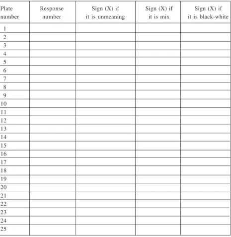
Table 1. Examination form for detecting color vision deficiency
HACETTEPE UNIVERSITY FACULTY OF MEDICINE DEPARTMENT OF PHYSIOLOGY COLOR VISION TEST FORM
Name: ……… Date of Birth: …. / …. / 19… Gender: Male ( ) / Female ( ) City: ……… Faculty: ………
Do you have color vision problem? No ( ) / Yes ( )
This is a color vision test answer form. You should write whatever you see at the color plates in the corresponding box. Your answer time for each of color plate is only four seconds. If you see numbers on the color plates please write this number on the number response colon; if the color plates seems unmeaning to you the mix or black-white please put the symbol (X) to the colon seen below. Please follow carefully the plate numbers during the whole period of test.
Plate Response Sign (X) if Sign (X) if Sign (X) if number number it is unmeaning it is mix it is black-white
1 2 3 4 5 6 7 8 9 10 11 12 13 14 15 16 17 18 19 20 21 22 23 24 25
Statistical Analysis
Chi-square test (Fisher’s Exact Test) was used to determine the relationship between sex and the color vision status of the students, and Kappa statistics were used for the concordance between Mass Screening test and classic indi-vidual Ishihara Test. The classical indiindi-vidual method was accepted as the gold standard in identifying the red/green color-blind subjects.
RESULTS
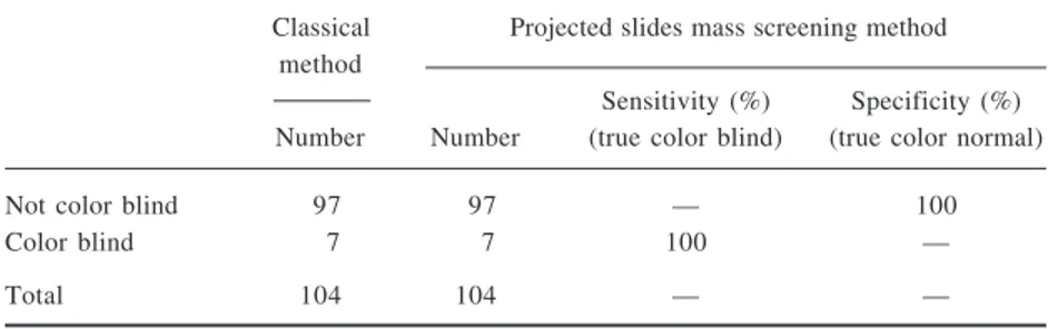
There were seven (6M, 1F) blind students. The incidence of color-blindness was 13.6% (6/44) among male population and 6.7% (7/104) for the whole population (Table 2). There was complete concordance between the mass screening test and the classical method (κ = 1.00, p = .000). The sensi-tivity and the specificity of the mass-screening test were both found to be 100% (Table 3).
Table 2. The response of classical individual method of Ishihara Detection of color sense ability Not color-blind individuals Color-blind individuals
Number % Number % Total
Male 38 86.4 6 13.6 44
Female 59 98.3 1 1.7 60
Total 97 93.3 7 6.7 104
Table 3. The specificity and sensitivity of projected mass screening test method (according to the individual classic method of Ishihara, as a gold standard).
Classical Projected slides mass screening method method
Sensitivity (%) Specificity (%) Number Number (true color blind) (true color normal)
Not color blind 97 97 — 100
Color blind 7 7 100 —
All subjects were able to see the introductory plate in two methods as was expected. Four normal subjects made errors during the mass-screening test. One of them made three errors, one student made two errors, and the remaining two students made one error in different plates. In the classical authentic individual method, only one normal subject made three errors. The errors made by these subjects were similar to the errors of color-blind sub-jects’ errors listed in Table 4.
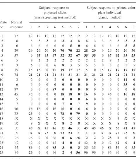
The results of color-blind students obtained from two different methods were compared in Table 5 and the numbers of wrong answers of two tests are shown in Figure 1.
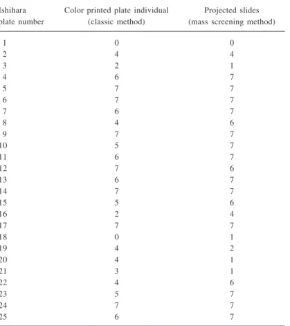
Table 4. The number of wrong answers of color-blind students for each of Ishihara test plates according to numeral range of Ishihara plates in both methods
Ishihara Color printed plate individual Projected slides plate number (classic method) (mass screening method)
1 0 0 2 4 4 3 2 1 4 6 7 5 7 7 6 7 7 7 6 7 8 4 6 9 7 7 10 5 7 11 6 7 12 7 6 13 6 7 14 7 7 15 5 6 16 2 4 17 7 7 18 0 1 19 4 2 20 4 1 21 3 1 22 4 6 23 5 7 24 7 7 25 6 7
DISCUSSION
Color blindness (color vision deficiency) is a condition in which certain col-ors cannot be distinguished, and is most commonly due to an inherited con-dition (Swanson & Cohen, 2003; Tagarelli et al., 1999).Red/Green (r/g) color blindness is by far the most common form, and causes problems in distinguish-ing reds and greens (Tagarelli et al., 1999).There is no known treatment for
Table 5. Comparing the results of color-blind students in two different methods Ishihara’s pseudoisochromatic color plate tests
Subjects response to Subject response to printed color projected slides plate individual (mass screening test method) (classic method) Plate Normal no. response 1 2 3 4 5 6 7 1 2 3 4 5 6 7 1 12 12 12 12 12 12 12 12 12 12 12 12 12 12 12 2 8 8 3 3 8 3 3 8 8 3 3 8 3 3 3 3 6 6 6 6 6 6 5 0 6 6 6 6 6 5 5 4 29 29 20 70 20 70 70 22 28 20 0 29 70 20 70 5 57 0 65 35 67 32 32 67 35 35 0 67 32 35 25 6 5 8 2 2 2 2 2 2 2 2 2 8 2 2 2 7 3 6 5 0 6 8 3 5 5 5 0 0 6 5 5 8 15 15 15 0 15 19 17 17 15 0 15 17 17 17 17 9 74 21 21 21 21 21 21 21 21 21 21 21 21 21 21 10 2 2 0 0 2 0 0 0 0 0 0 0 0 14 0 11 6 6 0 0 0 0 0 0 0 0 0 0 0 35 0 12 97 0 0 0 87 0 0 0 0 0 0 0 0 0 0 13 45 45 0 0 0 18 18 0 16 0 0 46 0 16 18 14 5 0 0 0 0 0 0 0 6 0 0 0 0 0 0 15 7 0 0 0 0 7 0 7 9 0 0 0 0 0 0 16 16 16 16 0 16 16 0 16 16 0 0 0 0 0 0 17 73 23 0 0 0 78 0 79 0 0 0 0 0 0 0 18 X X X X X X X X X X X X 9 X X 19 X 8 X X X 8 2 8 X X X X 8 X 8 20 X 45 X 45 46 X 46 X 45 45 46 X 66 41 45 21 X X X 73 X 73 23 X X X X X 72 23 X 22 26 26 0 0 26 0 6 26 2 2 0 28 2 0 2 23 42 42 0 0 42 4 0 4 42 0 0 42 62 0 4 24 35 86 0 0 85 3 0 3 35 35 0 86 36 0 35 25 96 26 0 0 96 2 4 56 96 96 0 96 96 0 9
color blindness, nor is it usually the cause of any significant disability. Being colorblind keeps one from performing certain jobs and makes others difficult (Poole et al., 1997; Spalding, 1993). Consequently, it is crucial to detect color-blind individuals earlier, especially before they choose their profes-sions, enabling the prevention of serious problems, possibly to be faced throughout their professional life. In order to do this, the best way is to organize screen-ing programs, especially among the students. However, it entails such an effective, timesaving method, which has high specificity and sensitivity for detecting color-blind individuals.
In the large-size classrooms with more than one hundred students, indi-vidual testing with color plates consumes considerable lecture time. It is essential to establish a color vision testing method that could screen a large population at one session. It should be timesaving, sensitive, and easy to perform. The classical individual application of Ishihara method has some handicaps: At first, it is not suitable for mass screening because it is a time-consuming individual method. Second, as test plates are kept together in the form of a book and the examiner who allows time for the subject to identify the figures turn the pages, the recognition time for each plate varies individu-ally, depending on the color perception ability of every subject. The recogni-tion time of a subject with normal color vision is shorter than the one with
Figure 1. The number of wrong answers detected in two methods.
Projected slides Printed color plates
N u mb e r o f wr ong a n s w e rs Number of plates
CCVD. Therefore, the test time shows considerable individual variation and leads to a time-dependant heterogeneity in the evaluation of the subjects. On the other hand, a projected slide method solved this time-dependant hetero-geneity problem. Reply time was kept constant for each color plate and for also total test time. Third, in this method, the practitioner would prevent the confounding variables arising from the place, lighting, and testing-time dif-ferences in large groups. Moreover, color hue variances in the printed test book editions also affect the number of misreadings. The printed color plates have a tendency to fade in time for testing large groups. But with projected slides, a mass-screening test can be administered to a large group at once as presenting study; the CCVD examination test took only 15–20 min for 104 students.
Another issue is the correction of eye refraction by wearing colored lenses. A pair of greenish sunglasses or contact lenses lets the green but not the red light pass through. Thus, a red digit on green background will appear dark on a bright background. It will become easily readable despite a color vision defect. A green digit on a red background will be just as easily read. It will appear bright on a darker background (Linksz, 1971). So in the present study, students were not allowed to wear colored sunglasses and colored contact lenses.
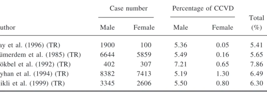
It is so important to eliminate the confounding effects of aging factors on the color vision deficiencies (Lakowski, 1962). In the current study, the subjects consisted of young individuals (median age 21). All of the students were Caucasians. The prevalence of red-green CCVD in Caucasian males changes from 8% to 10% (Neitz et al., 2001; Pokorny et al., 1979; Pokorny & Smith, 1987).In the present study it was found that the prevalence of R/G CCVD among male students was 13.6% in both of the methods. It may be attributed to the widely seen consanguinity relation in the Turkish population (Tunçbilek, 2001). This may be a factor for such a high prevalence of CCVD in the present study group. However, the R/G CCVD prevalence was re-ported to be as high as this study by other authors (Barna et al., 1981; Tocantins & Jones, 1993; Poole et al., 1997). On the other hand, the incidence of CCVD for whole population in the present study was found as 6.7%. This finding is in line with other studies (Ganley & Lian, 1997; Is¸ikli et al., 1999). The reports on the prevalence of R/G CCVD in Turkey show that Ishihara color plates is the only test used by the researchers to determine color vision defects for mass screening, although their results are controversial. Gökbel et al. (1992) reported findings approximately close to the present ones. On the other hand, as can be seen in Table 6, there are several contradictory reports
in Turkey (Is¸ikli et al., 1999; Say et al., 1996; Tümerdem et al., 1985; Ayhan et al., 1994). However, the error scores in these studies are not clear. A consensus for the interpretation of the test results should be established among investigators. Although the performance of the subject in reading the Ishihara plates is evaluated to determine color-blindness, there is not a standard crite-rion for this. Some investigators diagnosed color deficiency by using trans-formation plates (plates 2–9) (Is¸ikli et al., 1999), whereas others used hidden digit plates (plates 18–21) to determine color deficiency (Birch, 1997). Obvi-ously, consensus among the investigators is very important, in order to avoid controversial results. For example, Birch defined a color deficient subject as the person making 8 or more mistakes in reading plates(Birch, 1997), al-though another researcher used a different definition such as the subject making 5 or more mistakes in reading 14 plates is color deficient (Ganley & Lian, 1997). Determination of error score plays a crucial role in comparing the findings and determining the efficiency of the test. If misreading some plates causes uncertainty, further color vision tests are recommended. Standardiza-tion of the error scores for the diagnosis of CCVD is very important. Conse-quently, the current authors agree with the view of Alwis, who suggested rearrangement of Ishihara plates combined with a new recording chat and scoring strategy enhances the usefulness of the test (Alwis & Kon, 1992).
The efficiency of the screening test can be assessed in terms of sensitiv-ity and specificsensitiv-ity. Sensitivsensitiv-ity is the percentage of abnormal subjects is cor-rectly identified as abnormal. Specificity in the percentage of the normal subjects is correctly identified as normal. According to our results, the sensi-tivity and specificity of the projected slide mass-screening test and classical Ishihara test methods were both found to be 100% (Table 3). The present results were similar to the results of Ganley and Lian (1997), who reported
Table 6. The studies about the prevalence of R/G CCVD in Turkish population Case number Percentage of CCVD
Total
Author Male Female Male Female (%)
Say et al. (1996) (TR) 1900 100 5.36 0.05 5.41
Tümerdem et al. (1985) (TR) 6644 5859 5.49 0.16 5.65
Gökbel et al. (1992) (TR) 402 307 7.21 0.65 7.86
Ayhan et al. (1994) (TR) 8382 7413 5.19 1.30 6.49 Is¸ikli et al. (1999) (TR) 3345 2606 5.50 0.80 6.30
that Ishihara projected slide mass screening test sensitivity was 100% and the specificity was 98.1%. The present study used classical individual Ishihara test with printed color plates as the reference or gold standard test for com-paring the results of projected slides. There was complete accordance with the authentic Ishihara classic method. The Ishihara projected slides test can be replaced with the classical Ishihara test method. Based on these results, projected slide could be used for detecting CCVD in large groups, under well-designed stipulations as proposed by Ganley and Lian (1997).
CONCLUSION
The present study detected a relatively high prevalence of color-blindness among a randomly selected group of students. Proper guidance and counsel-ing of the students require early detection of color vision defect. This new screening projected slide method is quite promising to become a widely used color vision test in practice because it screens large groups in a short time and it is in complete accordance with the application of classical Ishihara test method. The authors developed and applied this mass-screening test for the first time in Turkey.
REFERENCES
Alwis, D. V., & Kon, C. H. (1992). A new way to use the Ishihara Test. Journal of Neurology, 239, 451–454.
Ayhan, B., Tümerdem, Y., & Özsüt, H. (1994). Dyschromatopsia ve genetik yapi (epidemiyolojik aras¸tirma). Istanbul Tip Fakültesi Mecmuasi, 57, 13–15. Barna, G. J., Taylor, J. W., King, G., & Pelleu, G. B. (1981). The influence of
se-lected light intensities on color perception within a color range of natural teeth. Journal of Prosthetic Dentistry, 46, 450–453.
Belcher, S. J., Greenshields, K. W., &Wright, W. D. (1958). A color vision survey. British Journal of Ophthalmology, 42, 355 –359.
Birch, J. (1997). Efficiency of the Ishihara test for identifying red-green color defi-ciency. Ophthalmic and Physiological Optics, 17, 403–408.
Ganley, J. P., & Lian, M. C. (1997). Projected color slides as a method for mass screening of red-green color deficient individuals. Ophthalmic Epidemiology, 4, 213–221.
Gökbel, H., S¸emin, I., Güvel, H., Yalaz, G., Özgönül, H., & Özen, B. (1992). 4–6 yas¸ grubu çocuklarda renk körlüü sikligi. Dokuz Eylül Üniversitesi Tip Fakültesi Dergisi, 6, 55–57.
Ishihara, S. (1990). Ishihara’s tests for color-blindness, 38 plate ed. Tokyo/Kyoto: Kanehara, Shuppan Co. Ltd.
Is¸ikli, B., Kalyoncu, C., Metintas¸, S., Aslantas¸, D., & Ünsal, A. (1999). 6–17 yas¸ okul çocuklarinda kirma kusuru ve renk körlügü. Türkiye Tip Dergisi, 6, 29–32. Lakowski, R. (1962). Is the deterioration of color discrimination with age due to lens
or retina changes? Farbe, 11, 69–86.
Linksz, A. (1971). Color vision test in clinical practice. Transactions-American Academy of Ophthalmology and Otolaryngology, 75, 1078–1090.
Neitz, J., Carrol, J., & Neitz, M. (2001). Color vision almost reason enough for hav-ing eyes. Optic & Photonic News, 12, 29–33.
Pokorny, J., Smith, V. C., Verriest, G., & Pinckers, A. J. L. G. (1979). Congenital and acquired color defects. New York: Grune & Stratton.
Pokorny, J., & Smith, V. C. (1987). New observations concerning red-green color defects. Color Research and Application, 7, 159–164.
Poole, C. J. M., Hill, D. J., Christie, J. L., & Birch, J. (1997). Deficient color vision and interpretation of histopathology slides: Cross sectional study. British Medi-cal Journal, 315, 1279–1281.
Say, B., Güngör, E., & Altay, Ç. (1996). Türk halkinda renk körlügü sikligi ve bu herediter defektlerle G6PD eksikligi arasinda münasebet. Hacettepe Çocuk Sagligi ve Hastaliklari Dergisi, 9, 96–101.
Sloan, L. L., & Habel, A. B. (1956). Test for color deficiency based on the pseudo-isochromatic principle. Archives of Ophthalmology, 55, 229–239.
Spalding, J. A. B. (1993). The doctor with an inherited defect of color vision: Effect on clinical skills. British Journal of General Practise, 43, 32–33.
Swanson, W. H., & Cohen, J. M. (2003). Color vision. Ophthalmology Clinics of North America, 16, 179–203.
Tagarelli, A., Piro, A., & Tagarelli, G. (1999). Genetic, epidemiologic and social features of color blindness. Community Genetics, 2, 30–35.
Tocantins, L. M., & Jones, J. W. (1993). Defective color vision and handicaps in medicine. American Journal of Science, 293, 243–249.
Tunçbilek, E. (2001). Clinical outcomes of consanguineous marriages in Turkey. The Turkish Journal of Paediatrics, 43, 277–279.
Tümerdem, Y., Alpay, T., & Ayhan, B. (1985). Türk çocuklarinda dogumsal renk körlükleri (Epidemiyolojik bir aras¸irma II). Istanbul Tip Fakültesi Mecmuasi, 48, 611–617.