EP & Arrythmia
Heart Rate Recovery as a Novel Test for
Predicting Cardiac Involvement in
Beta-Thalassemia Major
Selcuk Kucukseymen,1Isa Oner Yuksel,1Goksel Cagirci,1Erkan Koklu,1Volkan Karakus,2Serkan Cay,3 Gorkem Kus,1Erdal Kurtoglu4and Sakir Arslan1
Background: Abnormal heart rate recovery (HRR) is predictive of cardiac mortality. Autonomic abnormalities in beta-thalassemia major (TM) patients have been reported in previous studies. However, the importance of low HRR in exercise stress test in TM patients has not yet been ascertained. Therefore, this study will be the first of its kind in the literature.
Methods: Exercise stress test was performed on 56 TM patients who were being treated at the Thalassemia Center of our hospital, along with 46 non-TM iron deficiency anemia (IDA) patients as a control group. Values for HHR were recorded at 1, 2, 3, 4 and 5 min, and HRR was calculated by the difference of heart rate at peak exercise and at a specific time interval following the onset of recovery.
Results: All HRR values were found to be lower in TM patients compared to those in the IDA group. Exercise capacity [metabolic equivalents (METs)] was also found to be low in these patients (p < 0.001) as well. Total exercise time was significantly lower in the TM group compared to the IDA group (8.40± 1.7 min vs. 11.17 ± 1.51 min, p < 0.001). Exercise capacity (METs) was also lower in the TM group compared to the IDA group. Mean T2* value was 28.3± 13.7 ms in TM patients on magnetic resonance imaging (MRI). In addition, there are 18 TM patients with T2* value was < 20 ms. Conclusions: This study found that TM was independently associated with low HRR. Such a condition is an indicator of autonomic dysfunction in TM patients, since abnormal HRR is related to impaired autonomic response. In addition, impaired HRR may be a marker of early cardiac involvement in patients, whose T2* value is high on MRI. Modifying HRR with a cardiac rehabilitation program in TM patients with impaired HRR is a field open for further investigation.
Key Words: Beta thalassemia major· Exercise stress test · Heart rate recovery · Magnetic resonance imaging
INTRODUCTION
Iron cardiomyopathy is a leading cause of death in
transfusion-dependent thalassemia major (TM) patients, and magnetic resonance imaging T2* (MRI T2*) can rec-ognize preclinical cardiac iron overload. However, the technology is unavailable to many medical centers. In thalassemic patients, cardiac T2* has been recently used to evaluate myocardial iron content and has been correlated to left ventricular ejection fraction reduction. Cardiac involvement in thalassemia affects mainly the myocardium: iron overload of the myocytes reduces left ventricular distensibility.1,2Also, heart failure is the most frequent manifestation of cardiac involvement. How-ever, early diagnosis and iron depletion improve survival by reducing organ iron overload, especially in the
myo-Original Article doi: 10.6515/ACS20161104A
Received: May 1, 2016 Accepted: November 4, 2016
1
Department of Cardiology, Antalya Education and Research Hospital, Antalya;2Department of Hematology, Mula Sòtkò Koçman University, School of Medicine, Mula; 3Department of Cardiology, Türkiye Yüksek¤htisas Training and Research Hospital, Ankara;4Department of Hematology, Antalya Education and Research Hospital, Antalya, Turkey.
Corresponding author: Dr. Selcuk Kucukseymen, Department of Cardiology, Antalya Education and Research Hospital, Muratpasa, 07100 Antalya, Turkey. Tel: +90 507 431 5691; Fax: +90 242 244 4900; E-mail: [email protected]
cardium. Elevated iron could affect autonomic nervous system activity, possibly by impairing neuronal function or interfering with other elements of the baroreceptor reflex pathways external to the heart. Alternatively, in-trinsic electrical activity of the specialized pacemaker myocytes located in the sinoatrial node could also be disrupted by iron as a result of modulation of a number of membrane currents, Ca2+ handling or intracellular signalling.3
Heart rate recovery (HRR) is an expression of de-creased heart rate in a specified time, in the recovery period at the end of an exercise test. In healthy individu-als, the heart rate rapidly decreases through increased parasympathetic tone and decreased catecholamine levels at the sino-atrial node at the end of exercise. Ab-normal HRR is defined as ablunted heart rate decrease after exercise, which is independently associated with increased mortality and morbidity.4-8HRR is a strong in-dicator of fitness and partial mediator of the autonomic nervous system.4,5,8,9In addition, HRR has been shown to be similar to traditional risk factors and related to clinical outcomes, even in individuals with no evident cardiovascular disease.6,10HRR is related to increased mortality, independent from the systolic function, the angiographic severity of disease or exercise capacity. Therefore, assessment of HRR is recommended as a non-invasive method for assessment of cardiovascular risk in children and adults.11-13However, the mechanism and clinical correlation of abnormal HRR has not yet been fully understood.
Presence of autonomic dysfunction in TM patients has been demonstrated in an earlier study.14The influ-ence of autonomic dysfunction on HRR and clinical events was not known for these patients. In the present study, we aimed to investigate the exercise properties of TM patients, and the relationship between HRR and car-diac involvement by comparing T2 scores.
METHODS
Patients population
Our study enrolled 56 TM patients who were being treated at the Thalassemia Center of our hospital be-tween January and December 2014, along with 46 iron deficiency anemia (IDA) patients as a control group
without TM. A two-unit blood transfusion was given as necessary to TM patients in terms of hemodynamics (physical activity, occupation) every three to four weeks. The average of the last three measurements was used to determine Hb values. On the other hand, there were no differences in hemoglobin values among the study groups.
The following were exclusion criteria for patients in our study: left ventricular ejection fraction (LVEF) of less than 50% in echocardiography, any active inflammatory process, below 18 years of age, diabetes mellitus, hyper-tension, and previous ischemic heart disease. Informed consent was obtained from all the patients, and our study was approved by the Ethics Committee of the Antalya Education and Research Hospital.
Echocardiographic measurements and data
Echocardiographic imaging of patients and IDA group were obtained at the Antalya Education and Re-search Hospital Cardiology Clinic using a PhilipsEPIQ 7 echocardiography device (Philips Healthcare, 3000 Minuteman Road Andover, MA, USA), and a 2.5 MHz probe. All images were recorded to include 3 consecu-tive heart beats for later evaluation. Echocardiographic evaluation was done with patients in the left lateral decubitus position accompanied by ECG monitoring consistent with recommendations of the American Ec-hocardiography Society.15 LVEF was calculated using modified Simpson’s method, with end-diastole and sys-tole volumes measured with echocardiography.
Exercise stress test
Patients waited for 10 minutes in a supine position to stabilize the heart rate before starting the exercise test. They were asked not to use substances that affect-ing heart rate like cigarettes or caffeine in the 48 hours prior to testing. The symptom-limited treadmill exercise test was performed on patients to evaluate exercise time, exercise capacity [metabolic equivalents (Mets)], and heart rare recovery using the standard multistage Bruce protocol.16The exercise testing began with sub-jects walking slowly for 3 min at 1.7 m/h at a 10% grade; thereafter, speed and grade increased every 3 min until patient exhaustion. Patient electrocardiogram, heart rate and blood pressure were recorded during the last minute of each stage of exercise. This exercise data in-cluded duration on treadmill, age-predicted heart rate
achieved and capability measurements in terms of METs. Starting heart rate, peak heart rate, heart rate at 1-5 min in recovery, and blood pressure measurements were taken during established protocol exercise stage intervals to calculate pressure rate product (PRP). Exer-cise was terminated when the participants reached their target HR (85% of their age- and sex-predicted maximal HR), and the participants immediately got off the tread-mill and rested in a supine position. Exercise testing was terminated prematurely for the following reasons: limit-ing chest discomfort, dyspnea, fatigue, or leg discom-fort. All exercise testing was performed according to American College of Cardiology/American Heart Associ-ation (ACC/AHA) practice guidelines1,7and hospital eth-ics committee approval was obtained for this prospec-tive study.
Heart rate recovery
Heart rate data during the exercise were obtained from the continuously recording electrocardiographic monitor. Recovery data at the end of the exercise were obtained when the patient was resting in a supine posi-tion for 10 min. HR data were recorded at the end of every min during recovery period, and defined as HRR 1, HRR 2, HRR 3, HRR 4 and HRR 5. HRR was calculated by subtracting the heart rate value at that moment from the peak heart rate during exercise (e.g., HRR 1 = peak HR during exercise – HR at recovery min 1). Chrono-tropic response was evaluated in both groups and ob-tained using the following formula employing HR re-serve:
Chronotropic index = (HR peak – HR baseline) / (220-age-HR baseline)´ 10018,19
Chronotropic incompetence was defined as < 0.8 of chronotropic index.19,20
MRI T2*
MRI was performed by Magneto Symphony Gra-niand 32, 1.5 Tesla (Siemens, Germany, 2009) in Antalya Education and Research Hospital (Antalya, Turkey). Each scan lasted about 20 minutes and included measure-ment of cardiac T2*. Cut-off points using this MRI instru-ment were as follows: cardiac: normal > 20 ms, mild: 14-20 ms, moderate: 10-14 ms, severe < 10 ms.21
Statistical analysis
In this study, data were expressed as mean± stan-dard deviation for continuous variables, with counts and percentages for categorical variables. Data were tested for normal distribution using the Kolmogorov-Smirnov test, and frequencies, means and standard deviations were calculated by descriptive statistics. Independent Samplest Test (parametric) and Mann Whitney U-test (non-parametric) were used to assess the differences between the variables in patients with beta-thalassemia and IDA individuals. Correlations of variables were eva-luated using the Pearson or Spearman’s correlation an-alysis; a correlatiaon coefficient between 0-0.3 indicated a weak correlation, and 0.7-1.0 indicated significant cor-relation. A p value < 0.05 was considered statistically significant. Statistical analyses were conducted with a commercially available software package (SPSS version 16.0, SPSS, Chicago, IL, USA).
RESULTS
The symptom-limited exercise test was done on a total of 102 patients (56 TM patients, 46 IDA patients). There was no significant difference between the two groups regarding physiological variables such as body mass index (BMI), hemoglobin level, basal heart rate, age, and gender. However, there was a significant differ-ence in serum iron (SI), ferritin and transferrin satura-tion (TS) (p < 0.001). Baseline clinical properties and ex-ercise characteristics of the TM and the IDA patients are shown in Table 1 and 2. The mean patient age was 24.5 ± 6 years in TM group, and 24.1 ± 6 years in the IDA group (p = 0.70).
Due to exercise exhaustion, 13 patients with TM (23.2%) and 5 IDA patients (10.8%) left the exercise test before reaching the target heart rate. Overall, total exer-cise time was significantly lower in the TM group com-pared to the IDA group (8.40± 1.7 min vs. 11.17 ± 1.51 min, p < 0.001). Exercise capacity (METs) was also lower in the TM group compared to the IDA group (10.13± 1.81 METs vs. 13.10± 1.88 METs, p < 0.001). The mean peak HR was 170.43± 13.49 bpm during exercise in the TM group and 181.65± 15.85 bpm in the IDA group, and the difference was statistically significant (p < 0.001). HR reserve (220-age-baseline HR) was 107 ± 11 in the
thalassemia group and 111± 17 in the IDA group, and the difference was not statistically significant (p = 0.30). The mean chronotropic index was 0.76± 0.11 in the TM group, and 0.84± 0.11 in the IDA group, with a statisti-cally significant difference (p = 0.001).
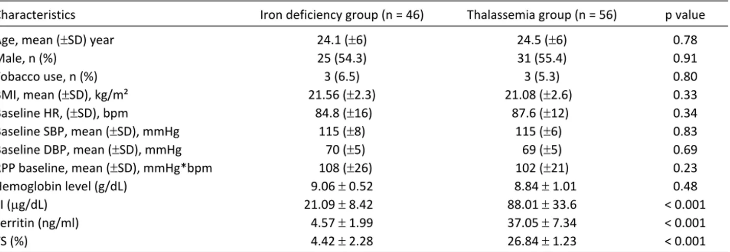
HRR 1, HRR 2, HRR 3, HRR 4, and HRR 5 values were
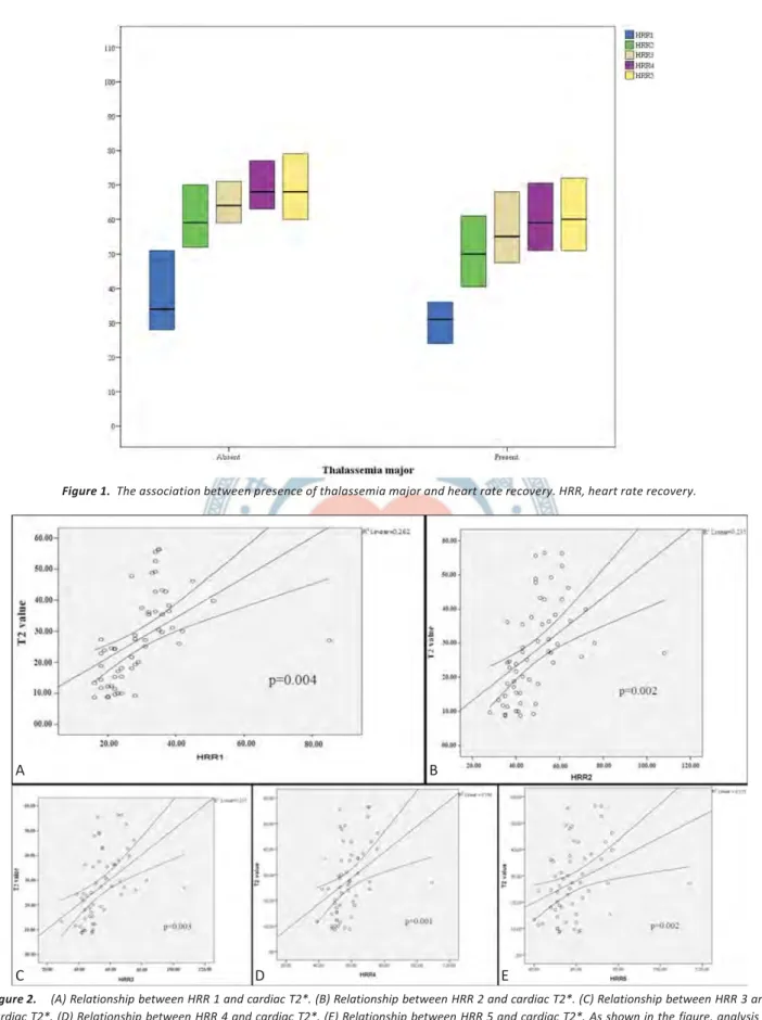
statistically significantly lower in the TM group com-pared to the IDA group, independent from exercise time (p = 0.004, p = 0.002, p = 0.003, p = 0.001, p = 0.002, re-spectively) (Figure 1, Table 2). There was a consistent and significant decline in HRR and T2* values. Figure 2A, Figure 2B, Figure 2C, Figure 2D and Figure 2E showed a Table 1. Baseline clinical and exercise characteristics according to thalassemia major and iron deficiency group
Characteristics Iron deficiency group (n = 46) Thalassemia group (n = 56) p value
Age, mean (±SD) year 24.1 (±6) 24.5 (±6) 0.78
Male, n (%) 25 (54.3) 31 (55.4) 0.91 Tobacco use, n (%) 3 (6.5) 3 (5.3) 0.80 BMI, mean (±SD), kg/m² 21.56 (±2.3) 21.08 (±2.6)0 0.33 Baseline HR, (±SD), bpm .84.8 (±16) 87.6 (±12). 0.34 Baseline SBP, mean (±SD), mmHg 115 (±8) 115 (±6)0 0.83 Baseline DBP, mean (±SD), mmHg 070 (±5) 69 (±5) 0.69 RPP baseline, mean (±SD), mmHg*bpm 0108 (±26) 102 (±21) 0.23 Hemoglobin level (g/dL) 9.06± 0.52 08.84± 1.01 0.48 SI (mg/dL) 21.09± 8.420 88.01± 33.6 < 0.001 Ferritin (ng/ml) 4.57± 1.99 37.05± 7.34 < 0.001 TS (%) 4.42± 2.28 26.84± 1.23 < 0.001
Values are expressed as mean and standard deviation. p < 0.05 value indicates significance. In all cases, differences were not statistically significant. BMI, body mass index; HR, heart rate; RPP, rate pressure product; SBP, systolic blood pressure; SI, serum iron; TS, transferrin saturation. (RPP = HR * SBP). One way analysis of variance, Independent samples t-test, Mann-Whitney U-test.
Table 2. Exercise characteristics after exercise according to thalassemia major and iron deficiency anemia group
Characteristics
(mean± SD or median 25th-75th%) Iron deficiency group (n = 46) Thalassemia group (n = 56) p
HR Stage 1, bpm 107 (±11) 121 (±14) < 0.001 HR Stage 2, bpm 121 (±14) 138 (±16) < 0.001 HR Stage 3, bpm 142 (±16) 159 (±16) < 0.001 HR Stage 4, bpm 173 (±16) 171 (±10) 0.48 RPP Stage 1 146 (±31) 156 (±31) 0.10 RPP Stage 2 178 (±40) 187 (±39) 0.25 RPP Stage 3 204 (±28) 217 (±46) 0.17 RPP Recovery 154 (±32) 141 (±25) 0.03 Peak HR, bpm 181 (±15) 170 (±13) < 0.001 HR reserve 111 (±17) 107 (±11) 0.29 Chronotropic index 84 (±11) 076 (±11) 0.001
Exercise capacity (METs) 13.1 (±1.8) 10.1 (±1.8) < 0.001
Exercise time, minute 11.1 (±1.5) 08.4 (±1.7) < 0.001
1-min HRR 34 (28-51) 31 (24-36)0. 0.004
2-min HRR 59 (52-70) 50 (40.3-61) 0.002
3-min HRR 64 (59-71) 55 (47.3-68) 0.003
4-min HRR 68 (63-77) 0.59 (50.5-70.8) 0.001
5-min HRR 68 (60-79) 60 (51-72)0. 0.002
Values are expressed as mean± standard deviation or median 25th-75th%. p < 0.05 value is significant. HR, heart rate; METs, metabolic equivalents; RPP, rate pressure product; SD, standard deviation. (Rate Pressure Product = Heart Rate * Systolic Blood Pressure), One way analysis of variance, Independent samples t-test, Mann-Whitney U-test.
Figure 1. The association between presence of thalassemia major and heart rate recovery. HRR, heart rate recovery.
Figure 2. (A) Relationship between HRR 1 and cardiac T2*. (B) Relationship between HRR 2 and cardiac T2*. (C) Relationship between HRR 3 and cardiac T2*. (D) Relationship between HRR 4 and cardiac T2*. (E) Relationship between HRR 5 and cardiac T2*. As shown in the figure, analysis is statically significant according to regression and average lines.
A B C D E
I
I
Tlmln•seml . "mnjor p==0.004 00.00 60.00 80,00relationship between HRR and cardiac T2*.
Correlation analysis between HRR and exercise time was performed on the whole group, with no correlation found between HRR 1 min and exercise time (Spear-man’s correlation coefficient was 0.162 and p = 0.10); however, there was a weak positive correlation between HRR 2, HRR 3, HRR 4, HRR 5 and exercise time. Spear-man’s correlation coefficient was 0.222 (p = 0.03) for HRR 2, and exercise time 0.216 (p = 0.03) for HRR 3, 0.250 (p = 0.01) for HRR 4, 0.249 (p = 0.01) for HRR 5.
When the groups were analyzed separately, in the TM group, there was a positive correlation between HRR 2 min and exercise time (r = 0.282, p = 0.04). A cor-relation was not detected between other HRR values and exercise time in TM patients. However, in the IDA group, there was no correlation found between HRR values (include all HRR values) and exercise time.
Correlation analysis between exercise capacity (METs) and HRR values was done in the whole group. A correla-tion was not detected between HRR 1 value and exer-cise capacity (Spearman’s correlation coefficient 0.149 and p = 0.14); however, a weak positive correlation was detected between HRR 2, HRR 3, HRR 4, HRR 5 values and exercise capacity (p = 0.02, 0.03, 0.01 and 0.01, re-spectively; r = 0.229 and 0.217, 0.244, 0.253, respec-tively). In addition, when the groups were analyzed sep-arately, in the TM group, there was a positive correla-tion between HRR 2 min and exercise capacity (r = 0.274, p = 0.04). However, in the IDA group, there was no correlation between HRR values (include all HRR val-ues) and exercise capacity.
Mean T2* value was 28.3± 13.7 ms in the TM pa-tients; T2* value was < 20 ms in 18 patients (33%). In TM patients with T2* values < 20 ms and > 20 ms, HRR values were compared and the results are illustrated in Table 3.
Correlation analysis between HRR and T2 scores was done in TM patients, with no correlation detected be-tween HRR values and T2 scores. Moreover, a correla-tion was not detected between exercise time, exercise capacity and T2* value. Pearson’s correlation coefficient for HRR 1 and T2 score was -0.024 (p = 0.86), for HRR 2 and T2 score was 0.053 (p = 0.70), for HRR 3 and T2 score was 0.079 (p = 0.57), for HRR 4 and T2 score was 0.096 (p = 0.49), and for HRR 5 and T2 the score was 0.099 (p = 0.48).
There were no statistical significance in the rate pressure product (RPP) stages and recovery, RPP is a measure of the stress put on the cardiac muscle based on the number of times it needs to beat per minute and the arterial blood pressure it is pumping against (SBP). It will be a direct indication of the energy demand of the heart and thus a good measure of the energy consump-tion of the heart. Rate pressure product allows you to calculate the internal workload or hemodynamic re-sponse.22
No correlation was detected between the T2* value and exercise time (p = 0.65), and similarly no correlation was found between T2* value and exercise capacity (p = 0.62).
DISCUSSION
HR response after exercise test has been shown to be predictive and prognostic in many studies.11,23,24HRR is a strong indicator of fitness and partial mediator of autonomic nervous system. Autonomic dysfunction at a certain level has been shown in TM patients;14however, the influence of this on HRR values is not yet known.
Iron is an essential element which forms a necessar-ily component of biological systems. However, when this element is present in extremes, it can produce tissue damage due to oxidative stress.25Excess body iron can be collected in the heart, liver, spleen, bone marrow, pi-tuitary, pancreas, and the central nervous system, caus-ing damage to these organs. Iron overload cardiopathy results from the accumulation of iron in the myo-cardium, and it is the leading cause of morbidity and Table 3. HRR in patients with T2* < 20 ms vs. those with > 20 ms
Characteristics T2* < 20 ms (n = 18) T2* > 20 ms (n = 38) p HRR 1 30.5± 6.00 31.5± 12.4 0.70 HRR 2 49.6± 12.6 52.7± 14.9 0.42 HRR 3 54.7± 12.1 58.5± 14.6 0.80 HRR 4 57.6± 11.7 61.0± 13.2 0.33 HRR 5 57.8± 12.0 62.2± 14.3 0.23
Values are expressed as mean± standard deviation. p < 0.05 value is significant. In all cases, differences were not statistically significant. HRR, heart rate recovery. One way analysis of variance, Mann-Whitney U-test, Independent samples t-test.
mortality in patients receiving recurrent blood transfu-sion therapy.26The incidence of cardiomyopathy due to iron overload is increasing and usually managed by cardiologists.
In this study, we calculated HRR values at the first, second, third, fourth, and fifth minutes during the re-covery period after a submaximal exercise test in pa-tients with TM and healthy individuals. We found that all HRR values were impaired in the patients with TM, compared to the IDA group that was matched for age, smoking, BMI, and sex.
Autonomic function is impaired in patients with heart failure, and abnormal HRR is related to decreased exercise capacity.27,28Cardiac failure remains as the ma-jor cause of death in beta-thalassemia mama-jor. In our study, decreased HRR response was observed, and de-creased exercise capacity was detected in TM patients compared to the IDA group. These findings may be an indicator of autonomic dysfunction. We also found in our investigation that the peak heart rate during exer-cise test was lower in TM patients compared to the IDA group. This observation might be associated with de-creased sympathetic stimulus and autonomic dysfunc-tion in TM patients.
There are a small number of studies investigating the association between HRR value, aerobic fitness, and mortality. Kokkinos et al.29 evaluated HRR (recovery at min 1 and 2), aerobic fitness and all causes of mortality on the basis of a follow-up of 6.2 years, and found that low aerobic fitness and low HRR increased mortality risk by 7-fold compared to those who had a good aerobic fit-ness and high HRR. In our study, exercise capacity, exer-cise time and HRR values were found lower in TM pa-tients compared to the IDA group. Further studies are required to investigate the influence of these findings on mortality in TM patients.
In our study, all HRR values (HRR 1, HRR 2, HRR 3, HRR 4, HRR 5) were found to be lower in TM patients compared to the IDA group. Low HRR was detected to be associated with poor aerobic fitness in an earlier study.30Besides, baseline abnormal HRR is associated with increased mortality and can predict mortality.31 Jolly et al. achieved 41% recovery and improved survival in patients with abnormal baseline HRR through cardiac rehabilitation program.31 However, sufficient data are not available regarding the influence of cardiac
rehabili-tation program on exercise parameters in TM patients and its clinical significance. Some other studies have shown that exercise training could modify HRR; how-ever, the clinical benefit of this modification on HRR and whether it reflects to endpoints is not still known.32,33 Studies are required investigating the influence of car-diac exercise rehabilitation program on HRR in TM pa-tients with abnormal HRR.
In this study, we also detected that exercise capacity (METs) and exercise time was lower in patients with TM than in the IDA group. Hao et al.34provided a significant improvement in METs values through cardiac rehabilita-tion program. However, the influence of this exercise training on exercise capacity and its association with clinical outcomes is not yet known.
In a study of Hai et al.,35 the some patients under-went and other patients bypassed a cardiac rehabilita-tion program after acute myocardial infarcrehabilita-tion, which were later compared. While both METs and HRR values were observed to improve in patients who underwent a cardiac rehabilitation program, only METs values im-proved but HRR did not change in the IDA group, which did not undergo a cardiac rehabilitation program. Given these studies, it seem that improved HRR is related with only exercise training.
Abnormal HRR reflects abnormal vagal tone8,9and prognostic value of abnormal HRR has been shown in many patient populations in many studies.10,11,36 Exer-cise training was also shown to improve autonomic tone,37,38endothelial dysfunction39and positively af-fect all cause mortality in patients with coronary heart disease.40Useful autonomic effects of exercise train-ing are realized through increased vagal reactivation, and the influence on HRR is explained with this mech-anism. However, the influence of exercise training on HRR and whether it is useful on clinical endpoints is not known.
There are many methods that define iron load in TM patients. Cardiac MRI is the most important among them and is only presently available noninvasively with the potential to assess quantitatively myocardial iron load.12,41MRI constructs images from transmitted micro-wave signals induced by exciting protons in the body in a high magnetic field. In noniron-overloaded hearts, these signals are homogenous, and relaxation time (time to fade excited signals) lasts for a longer duration
(brighter over time). In iron-overloaded hearts, how-ever, the iron paramagnetic effect produces changes in MR signal intensity and susceptibility, and shortens the relaxation time and darkens the image more quickly.42 MRI scanning can refocus the signals returning from the tissues, using a special radiofrequency pulse [spin echo (SE)] or by using special small magnetic fields called gra-dients [gradient echo (GE)] at specific time intervals [echo time (TE)].43 MRI measures a parameter called “T2*”, which is defined as the rate of loss of signal in tis-sues that are iron-loaded.44 Increased myocardial iron accumulation on MRI leads to decreased T2*. While T2* value < 20 ms shows iron load in cardiac tissues and an absence of cardiac dysfunction, T2* < 6 ms indicates se-vere cardiac siderosis and predicts development of 50% cardiac dysfunction within one year.45,46In addition, ac-cording to November 2009 American Heart Association report, cardiac T2* < 10 ms predicts cardiac event devel-opment within one year with 98% sensitivity and 86% specificity.12,45 In our study, a correlation was not de-tected between T2* values and HRR values, which could be an outcome of small number of subjects. The associ-ation between HRR and T2* can be revealed more ex-plicitly in a larger cohort. In addition, considering that mean T2* value of our patients is 28.3 ms and only 18 patients had a T2* value of < 20 ms, impaired HRR may be an early marker of cardiac dysfunction in TM patients. In TM patients, there was no correlation between HRR and T2*, here may also be influenced by factors that small number of patients, also HRR could be affected in the early stages before showing involvement with MRI T2*. Because in our study, HRR values were found signifi-cantly lower in TM patients than IDA group. These early findings may reflect autonomic dysfunction in TM pa-tients. This case is an open spectrum to be searched, with further studies to be performed in the future.
Impaired HRR may be an early marker of cardiac dysfunction. Impaired HRR is an indicator of autonomic dysfunction. We consider that impaired HRR in patients with TM shows autonomic dysfunction, on this basis au-tonomic dysfunction may be an early sign of cardiac involvement.
The value of HRR as a marker to show early cardiac involvement may be increased through more compre-hensive studies, showing that T2* values decrease dur-ing follow up of TM patients with impaired HRR.
Limitations
This study had several limitations. First, the study was a single center study. It was not large enough to de-tect HRR in patients whom T2 score was high and low, or in other words, the number of patients was limited. Other parameters such as heart rate variability (HRV) and gas analysis were not evaluated during exercise test. With all these limitations, our country is not enough wealthy to perform MRI for T2 score in patients with iron deficiency anemia. Thus, we were unable to pro-vide the data about the T2 in both groups. In our study, exercise capacity was measured as METs and exercise time as min, while symptom-limited exercise time was taken rather than maximum exercise time.
CONCLUSIONS
Low HRR in an exercise test reflects cardiovascular fitness and/or abnormal autonomic status in TM pa-tients. Abnormal HRR is an indicator of autonomic dys-function and may be a predictor of early cardiac involve-ment in this group of patients. Future studies may inves-tigate the other parameters affecting HRR and the influ-ence of impaired HRR on clinical endpoints in TM pa-tients.
ACKNOWLEDGEMENT
This study has been accepted as abstract number as 90302 in ESC Congress 2016 in Rome, Italy.
DECLARATION OF CONFLICT OF INTEREST
All the authors declare no conflict of interest.REFERENCES
1. Detterich J, Noetzli L, Dorey F, et al. Electrocardiographic conse-quences of cardiac iron overload in thalassemia major. Am J
Hematol 2012;87:139-44.
2. Anderson LJ, Holden S, Davis B, et al. Cardiovascular T2-star (T2*) magnetic resonance for the early diagnosis of myocardial iron overload. Eur Heart J 2001:22:2171-9.
3. Rose RA, Sellan M, Simpson JA, et al. Iron overload decreases CaV1.3-dependent L-type Ca2+ currents leading to bradycardia, altered electrical conduction, and atrial fibrillation. Circ
Ar-rhythm Electrophysiol 2011;4:733-42.
4. Kligfield P, Lauer MS. Exercise electrocardiogram testing: beyond the ST segment. Circulation 2006;114:2070-82.
5. Fletcher GF, Balady GJ, Amsterdam EA, et al. Exercise standards for testing and training: a statement for healthcare professionals from the American Heart Association. Circulation 2001;104: 1694-740.
6. Cole CR, Foody JM, Blackstone EH, Lauer MS. Heart rate recovery after submaximal exercise testing as a predictor of mortality in a cardiovascularly healthy cohort. Ann Intern Med 2000;132: 552-5.
7. Vivekananthan DP, Blackstone EH, Pothier CE, Lauer MS. Heart rate recovery after exercise is a predictor of mortality, independ-ent of the angiographic severity of coronary disease. J Am Coll
Cardiol 2003;42:831-8.
8. Imai K, Sato H, Hori M, et al. Vagally mediated heart rate recovery after exercise is accelerated in athletes but blunted in patients with chronic heart failure. J Am Coll Cardiol 1994;24:1529-35. 9. Buyukterzi Z, Ozeke O, Ozlu MF, et al. Heart rate acceleration and
recovery indices are not related to the development of ventricu-lar premature beats during exercise test. Acta Cardiol Sin 2014; 30:259-65.
10. Shetler K, Marcus R, Froelicher VF, et al. Heart rate recovery: vali-dation and methodologic issues. J Am Coll Cardiol 2001;38: 1980-7.
11. Cole CR, Blackstone EH, Pashkow FJ, et al. Heart-rate recovery im-mediately after exercise as a predictor of mortality. New Eng J
Med 1999;341:1351-7.
12. Yuksel IO, Koklu E, Kurtoglu E, et al. The association between se-rum ferritin level, tissue Doppler echocardiography, cardiac T2* MRI, and heart rate recovery in patients with beta thalassemia major. Acta Cardiol Sin 2016;32:231-8.
13. Singh TP, Rhodes J, Gauvreau K. Determinants of heart rate re-covery following exercise in children. Med Sci Sports Exerc 2008; 40:601-5.
14. Stamboulis E, Vlachou N, Voumvourakis K, et al. Subclinical auto-nomic dysfunction in patients withb-thalassemia. Clin Auton Res 2012;22:147-50.
15. Douglas PS, Garcia MJ, Haines DE, et al. ACCF/ASE/AHA/ASNC/ HFSA/HRS/SCAI/SCCM/SCCT/SCMR 2011 appropriate use crite-ria for echocardiography. J Am Soc Echocardiogr 2011;24:229-67. 16. Bruce RA, Kusumi F, Hosmer D. Maximal oxygen intake and nomographic assessment of functional aerobic impairment in cardiovascular disease. Am Heart J 1973;85:546-62.
17. Gibbons RJ. ACC/AHA guidelines for exercise testing. A report of the American College of Cardiology/American Heart Association Task Force on practice guidelines (Committee on Exercise Test-ing). J Am Coll Cardiol 1997;30:260-315.
18. Wilkoff BL, Miller RE. Exercise testing for chronotropic assess-ment. Cardiol Clin 1992;10:705-17.
19. Lauer MS, Francis GS, Okin PM, et al. Impaired chronotropic re-sponse to exercise stress testing as a predictor of mortality. JAMA 1999;281:524-9.
20. Lauer MS, Mehta R, Pashkow FJ, et al. Association of chrono-tropic incompetence with echocardiographic ischemia and prog-nosis. J Am Coll Cardiol 1998;32:1280-6.
21. Eghbali A, Taherahmadi H, Shahbazi M, et al. Association be-tween serum ferritin level, cardiac and hepatic T2-star MRI in pa-tients with majorb-thalassemia. Iran J Ped Hematol Oncol 2014; 4:17-21.
22. Clausen JP, Trap-Jensen J. Heart rate and arterial blood pressure during exercise in patients with angina pectoris. Effects of train-ing and of nitroglycerin. Circulation 1976;53:436-42.
23. Yanagisawa S, Miki K, Yasuda N, et al. The prognostic value of treadmill exercise testing in very elderly patients: heart rate re-covery as a predictor of mortality in octogenarians. Europace 2011;13:114-20.
24. Leino J, Minkkinen M, Nieminen T, et al. Combined assessment of heart rate recovery and T-wave alternans during routine exercise testing improves prediction of total and cardiovascular mortal-ity: the Finnish Cardiovascular Study. Heart Rhythm 2009;6: 1765-71.
25. Liu P, Olivieri N. Iron overload cardiomyopathies: new insights into an old disease. Cardiovasc Drugs Ther 1994;8:101-10. 26. Olivieri NF, Nathan DG, MacMillan JH. Survival in medically
treated patients with homozygous beta-thalassemia. N Engl J
Med 1994;331:574-8.
27. Borlaug BA, Melenovsky V, Russell SD, et al. Impaired chrono-tropic and vasodilator reserves limit exercise capacity in patients with heart failure and a preserved ejection fraction. Circulation 2006;114:2138-47.
28. Phan TT, Shivu GN, Abozguia K, et al. Impaired heart rate re-covery and chronotropic incompetence in patients with heart failure with preserved ejection fraction. Circ Heart Fail 2010; 3:29-34.
29. Kokkinos P, Myers J, Doumas M, et al. Heart rate recovery, exer-cise capacity, and mortality risk in male veterans. Eur J Prev
Cardiolog 2012;19:177-84.
30. Trevizani GA, Benchimol-Barbosa PR, Nadal J. Effects of age and aerobic fitness on heart rate recovery in adult men. Arq Bras
Cardiol 2012;99:802-10.
31. Jolly MA, Brennan DM, Cho Leslie. Impact of exercise on heart rate recovery. Circulation 2011;124:1520-6.
32. Tiukinhoy S, Beohar N, Hsie M. Improvement in heart rate recovery after cardiac rehabilitation. J Cardiopulm Rehabil 2003; 23:84-7.
33. Adams BJ, Carr JG, Ozonoff A, et al. Effect of exercise training in supervised cardiac rehabilitation programs on prognostic vari-ables from the exercise tolerance test. Am J Cardiol 2008;101: 1403-7.
34. Hao SC, Chai A, Kligfield P. Heart rate recovery response to symp-tom limited treadmill exercise after cardiac rehabilitation in pa-tients with coronary artery disease with and without recent
events. Am J Cardiol 2002;90:763-5.
35. Hai JJ, Siu CW, Ho HH, et al. Relationship between changes in heart rate recovery after cardiac rehabilitation on cardiovascular mortality in patients with myocardial infarction. Heart Rhythm 2010;7:929-36.
36. Nishime EO, Cole CR, Blackstone EH, et al. Heart rate recovery and treadmill exercise score as predictors of mortality in patients referred for exercise ECG. JAMA 2000;284:1392-8.
37. Goldsmith RL, Bloomfield DM, Rosenwinkel ET. Exercise and au-tonomic function. Coron Artery Dis 2000;11:129-35.
38. Liu JL, Irvine S, Reid IA, et al. Chronic exercise reduces sympa-thetic nerve activity in rabbits with pacing-induced heart failure: a role for angiotensin II. Circulation 2000;102:1854-62. 39. DeSouza CA, Shapiro LF, Clevenger CM, et al. Regular aerobic
ex-ercise prevents and restores age-related declines in endothe-lium-dependent vasodilation in healthy men. Circulation 2000; 102:1351-7.
40. Oldridge NB, Guyatt GH, Fischer ME, Rimm AA. Cardiac rehabili-tation after myocardial infarction: combined experience of
ran-domized clinical trials. JAMA 1988;260:945-50.
41. Modell B, Khan M, Darlison M, et al. Improved survival of thalas-saemia major in the UK and relation to T2* cardiovascular mag-netic resonance. J Cardiovasc Magn Reson 2008;10:42. 42. Wood JC. Magnetic resonance imaging measurement of iron
overload. Curr Opin Hematol 2007;14 :183-90.
43. Kaltwasser JP, Gottschalk R, Schalk KP, et al. Non-invasive quan-titation of liver iron-overload by magnetic resonance imaging. Br
J Haematol 1990;74:360-3.
44. Fragasso A, Ciancio A, Mannarella C, et al. Myocardial iron over-load assessed by magnetic resonance imaging (MRI) T2* in multi-transfused patients with thalassemia and acquired ane-mias. Eur J Intern Med 2011;22:62-5.
45. Wood JC. History and current impact of cardiac magnetic reso-nance imaging on the management of iron overload. Circulation 2009;120:1937-9.
46. Wood JC. Impact of iron assessment by MRI. Hematology Am Soc