doi:10.30569.adiyamansaglik.717688
Bu eser, Creative Commons Atıf-GayriTicari 4.0 Uluslararası Lisansı ile lisanslanmıştır. Telif Hakkı © 2020 Adıyaman Üniversitesi Rektörlüğü
Research Article/Özgün Araştırma
Effect of tumor location and lymph node involvement on prognosis and survival
in gastric cancer patients
Mide kanseri hastalarında tümör lokalizasyonu ve lenf nodu tutulumunun
prognoz ve sağkalıma etkisi
Cihan GÖKLER1 , Oktay İRKÖRÜCÜ2 , Enver REYHAN2 , Hilmi BOZKURT3 ,
Mustafa GÖRÜR2
1Adıyaman Training and Research Hospital, 02040, Adıyaman-Turkey 2Adana City Education and Research Hospital, 01330, Adana-Turkey 3İstanbul Haseki Training and Research Hospital, 34130, İstanbul-Turkey
Atıf gösterme/Cite this article as: Gökler C, İrkörücü O, Reyhan E, Bozkurt H, Görür M. Effect of tumor location and
lymph node involvement on prognosis and survival in gastric cancer patients. ADYÜ Sağlık Bilimleri Derg. 2020;6(2):248-257. doi:10.30569.adiyamansaglik.717688
Abstract
Aim: The present study evaluates the effect of tumor
localization and lymph node involvement on prognosis and survival in patients undergoing surgery for gastric cancer.
Materials and Methods: The clinical and histopathological characteristics of patients who underwent surgery in our clinic were evaluated to determine the prognostic factors.
Results: No difference was observed in the survival
rates of the groups in terms of tumor locations and metastatic lymph nodes (Log Rank p=0.255 and 0.188). A significant difference was found in the survival rates of the groups based on stage and age over 60 years (p=0.001, p=0.003). The number of metastatic lymph nodes dissected was high in gastric cancers located in the upper-third of the stomach (p=0.026, 0.036).
Conclusion: No effect of tumor localization or lymph
node involvement was determined on survival in patients with gastric cancer; however, age over 60 years and stage III were found to be poor prognostic factors.
Keywords: Gastric Cancer; Gastrectomy; Metastatic Lymph Nodes; Advanced Age, Prognosis.
Öz
Amaç: Mide kanseri nedeniyle ameliyat edilen
hastalarda tümör yerleşimi ve lenf nodu tutulumunun prognoz ve sağkalım üzerine etkisini değerlendirmeyi amaçladık.
Gereç ve Yöntem: Prognostik faktörleri belirlemek
için kliniğimizde ameliyat edilen hastaların klinik ve histopatolojik özelliklerini araştırdık.
Bulgular: Tümör lokalizasyon grupları arasında ve
metastatik lenf noduna göre sağkalım farkı gözlenmedi (Log Rank p=0,255 ve 0,188). Evreye ve 60 yaş üstü olma durumuna göre anlamlı sürvi farkı vardı (p=0,001, p=0,003). Üst 1/3 yerleşimli gastrik kanserde diseke edilen metastatik lenf nodu sayısı fazlaydı (p=0,026, 0,036)
Sonuç: Mide kanseri hastalarında tümör lokalizasyonu
ve lenf nodu tutulumunun sürviye etkisi saptanmamışken, 60 yaş üzeri olma ve Evre-III kötü prognostik faktörler olarak saptandı.
Anahtar Kelimeler: Mide Kanseri; Gastrektomi
Metastatik Lenf Nodu; İleri yaş; Prognoz.
Yazışma Adresi/Address for Correspondence: Cihan GÖKLER, Adıyaman Training and Research Hospital, 02040, Adıyaman-Turkey, E-mail: [email protected]
Geliş Tarihi/Received:10.04.2020 Kabul Tarihi/Accepted:17.06.2020 Yayım Tarihi/Published online:30.08.2020 https://dergipark.org.tr/tr/pub/adiyamansaglik
Bu makale araştırma ve yayın etiğine uygun hazırlanmıştır. intihal incelemesinden geçirilmiştir.
249 Introduction
Gastric cancer is currently the fifth most common cancer worldwide and third among cancer-related deaths1. Gastric cancer is the
fifth common cancer type in males and sixth in females, and third among cancer related deaths in our country. Unfortunately, 46% of gastric cancer patients are metastatic at the time of diagnosis2. Five-year survival in
gastric cancer is approximately 27% (range; 9-94%) and higher survival rates are only seen in early-diagnosed patients3-7. Survival
rate varies between countries and several prognostic factors are considered responsible for this variation. Consequently, different management approaches that may affect prognosis are brought forward. Among them, extended lymph node (LN) dissection or spleen-pancreas preserving D2 LN dissections which are adopted as a surgical approach that may have an impact on prognosis are performed as standart therapy8-13. In this
study, we aimed to evaluate prognostic factors and impact of tumor localization and LN involvement on prognosis and survival rates in gastric cancer patients.
Materials and Methods
The prospectively recorded clinical data, pathology reports and operation notes related to 95 patients who underwent gastrectomy for gastric adenocarcinoma between January 2011 and July 2014 in the General Surgery Clinic of the Adana Numune Training and Research Hospital were evaluated retrospectively. All procedures performed in studies involving human participants were in accordance with the ethical standards of the institutional and/or national research committee and with the 1964 Helsinki declaration and its later amendments or comparable ethical standards. The Institutional Review Board of the Adana City Hospital approved the present study (2014/61). In order to evaluate the prognostic factors affecting survival in these surgical patients, gender, age, duration of follow-up, patient status (survivor or exitus), location and dimensions of the tumor, operation type, T, N, M stages according to the IUCC 2010 TNM Classification, stage, number of dissected and metastatic lymph nodes,
positivity of the surgical margin, hemoglobin and albumin levels, platelet and leucocyte counts and status of adjuvant treatment were defined as prognostic parameters, and an analysis was made accordingly. The tumor locations were divided into four groups as upper-third gastric cancer (UTGC), middle-third GC (MTGC), lower-middle-third GC (LTGC) and diffuse (≥two-thirds) GC (DGC), and their effect on early survival and other prognostic parameters was evaluated. Furthermore, the patients were also divided into four groups in accordance with their baseline N status, as N0, N1, N2 and N3, and prognostic factors and survival were evaluated.
A total gastrectomy was applied in tumors with proximal or diffuse locations, and a subtotal gastrectomy was applied in distally located tumors. The surgeries were evaluated from the operation reports written by the surgeon who carried out the operation.
After scrutinizing the operation notes, patients with a pathological diagnosis of adenocarcinoma with D2 dissection and who underwent a curative resection were included in the study. Patients with metastatic stage 4 cancer, gastric malignancies other than adenocarcinoma, and patients with a synchronous malignancy in addition to gastric cancer were excluded from the study. Furthermore, patients who receive neoadjuvant treatment, who had undergone a previous abdominal operation, who had a D1 dissection or other surgical procedure, and those with a fatal outcome in the postoperative 30 days were excluded from the study.
Statistical Analysis
The SPSS 15.0 for Windows software package was used for the statistical analysis. Descriptive statistics were presented as numbers and percentages for categorical variables, while quantitative variables were presented as mean, standard deviation, minimum, maximum and median values.
The independent numerical values in more than two groups was analyzed with a One Way ANOVA test, and a Kruskal Wallis Test in groups with normal distribution and
non-250
normal distribution, respectively. Subgroup analyses were made with a Mann-Whitney U test, and the results were interpreted using a Bonferroni correction.
Survival was analyzed with a Kaplan Meier Analysis. Risk factors were evaluated with a Cox Regression Analysis. The alpha level of statistical significance was accepted as p<0.05.
Results
A total of 95 patients had undergone a gastrectomy for gastric cancer, of which 17 who had undergone palliative operations due to metastasis or who had a fatal outcome in the first 30 days following the operation were excluded from the study. Consequently, 78 patients with a D2 dissection were included in the study. Among the patients, 54 (69.2%) were male and 24 (30.8%) were female. The mean age of the patients was 63.4±12.8 years. The number of patients who were found to have LTGC, MTGC, UTGC and DGC was 39 (50%), 17 (21.8%), 19 (24.4%) and three (3.8%), respectively. The N status of the patients was N0, N1, N2 and N3 in 26, 16, 18 and 18, respectively according to the number of metastatic LNs. The majority of patients were stage III. The patients were evaluated in four groups, depending on their tumor localization (Table 1).
Statistically significant differences were noted in the gender ratio depending on the tumor localization (p=0.019). The ratio of females was lower in the LTGC and DGC localizations, and the ratio of males was lower in the MTGC and UTGC localizations. A statistically significant difference was found in the mean number of LNs dissected according to tumor localization (p=0.026). The number of LNs dissected was statistically significantly higher in the UTGC group than in the DGC group. Furthermore, the ratio of “two or more metastatic LNs” was significantly different between different localizations (p=0.036). The rate of two or more metastatic LNs was higher in the UTGC localization when compared to other localizations. That said, no statistically significant difference was noted in the overall survival rates of patients with LN
involvement of “<2” and “2 or more” (Log Rank p=0.331).
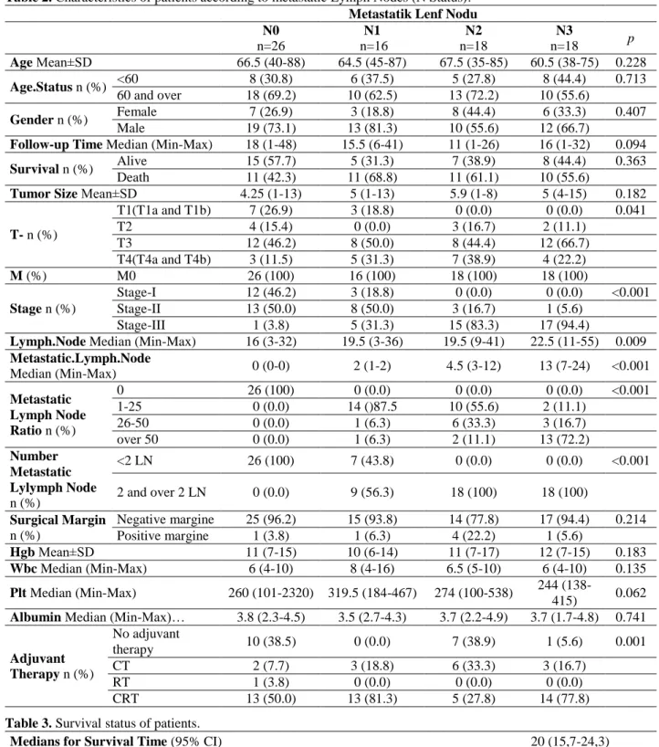
The evaluation of metastatic LN was carried out in accordance with the N (metastatic LN count) in the TNM staging system. T stages were statistically significantly different between the N stages (p=0.041). The rate of T1 tumors was high among tumors with N0 and N1, and T3 tumors were high among those with tumors with N3 involvement. The stages were also statistically significantly different depending on the different N status (p<0.001). Stage I-II rates were high among tumors with N0 and N1, and stage III was high among tumors with N2-N3. Furthermore, the total number of lymph nodes dissected was high among patients with N-positive tumors (p=0.009). The mean number of lymph nodes dissected was statistically significantly lower in N0 tumors when compared to N3 tumors (p=0.001). The rates of adjuvant treatment were statistically significantly different, since the N stage was important in adjuvant treatment decisions (p=0.001). The administration of no treatment was high in tumors with N0 status, while CT and CRT rates were higher in the N2, and N1 and N3 tumors, respectively (Table 2).
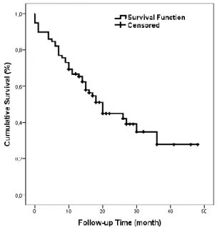
The median survival of the patients was 20 months (95% CI 15.7–24.3). The 1-year, 2-year and 3-2-year survival rates of the patients was 66.6%, 44.9% and 27.8%, respectively (Table 3). Overall survival data is given in Figure 1.
No statistically significant difference was found in survival rates according to tumor localization among the patients (Log Rank
p=0.255) (Table 4) (Figure 2-a). No
statistically significant difference was found in the survival rates according to the metastatic LN groups (p=0.188). (Figure 2-b)
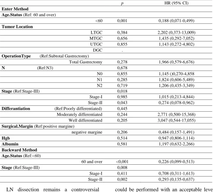
“Age <60 years” compared to “age 60 years and above”, and “stage II” compared to “stage III” were identified as the most significant prognostic factors affecting mortality with the enter and backward method in a multivariate Cox Regression Analysis Model that was formed through the addition of tumor localizations to the model (Model;
251
Age.Status, N, Stage, Differantiation, Surgical.Margin, Hgb, Albumin) composed of variables with p<0.250 among univariate
analysis (p<0.001 p=0002) (Table 5-7) (Figure 3).
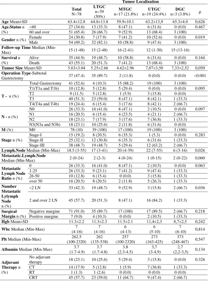
Table 1. Patient characteristics according to Tumor Localization
Tumor Localization Total N=78 LTGC n=39 (50%) MTGC n=17 (21.8%) UTGC n=19 (24.4%) DGC n=3 (3.8%) p Age Mean±SD 63.4±12.8 64.8±13.8 59.9±10.1 63.2±13,9 65.3±4.0 0,626 Age.Status n (%) <60 27 (34.6) 13 (33.3) 8 (47.1) 6 (31.6) 0 (0.0) 0.467 60 and over 51 (65.4) 26 (66.7) 9 (52.9) 13 (68.4) 3 (100) Gender n (%) Female 24 (30.8) 7 (17.9) 7 (41.2) 10 (52.6) 0 (0.0) 0.019 Male 54 (69.2) 32 (82.1) 10 (58.8) 9 (47.4) 3 (100)
Follow-up Time Median
(Min-Max) 15 (1-48) 15 (2-48) 16 (2-41) 12 (1-30) 15 (13-16)
Survival n
(%)
Alive 35 (44.9) 19 (48.7) 10 (58.8) 6 (31.6) 0 (0.0) 0.164
Death 43 (55.1) 20 (51.3) 7 (41.2) 13 (68.4) 3 (100)
Tumor Size Mean±SD 5.63±3.04 5.25±2.66 4.62±2.96 7,07±3,50 7,33±2,52 0,050
Operation Type-Subtotal Gastrectomy 37 (47.4) 35 (89.7) 2 (11.8) 0 (0.0) 0 (0.0) <0.001 Total Gastrectomy 41 (52.6) 4 (10.3) 15 (88.2) 19 (100) 3 (100) T - n (%) T1(T1a and T1b) 10 (12.8) 5 (12.8) 5 (29.4) 0 (0.0) 0 (0.0) 0.095 T2 9 (11.5) 5 (12.8) 1 (5.9) 3 (15.8) 0 (0.0) T3 40 (51.3) 23 (59.0) 8 (47.1) 8 (42.1) 1 (33.3) T4(T4a and T4b) 19 (24.4) 6 (15.4) 3 (17.6) 8 (42.1) 2 (66.7) N - n (%) N0 26 (33.3) 16 (41.0) 8 (47.1) 2 (10.5) 0 (0.0) 0.097 N1 16 (20.5) 6 (15.4) 4 (23.5) 4 (21.1) 2 (66.7) N2 18 (23.1) 7 (17.9) 3 (17.6) 7 (36.8) 1 (33.3) N3(N3a and N3b) 18 (23.1) 10 (25.6) 2 (11.8) 6 (31.6) 0 (0.0) M (%) M0 78 (10) 39 (100) 17 (100) 19 (100) 3 (100) Stage n (%) Stage-I 15 (19.2) 8 (20.5) 6 (35.3) 1 (5.3) 0 (0.0) 0.283 Stage-II 25 (32.1) 12 (30.8) 6 (35.3) 6 (31.6) 1 (33.3) Stage-III 38 (48.7) 19 (48.7) 5 (29.4) 12 (63.2) 2 (66.7)
Lymph.Node Median (Min-Max) 18.5 (3-55) 17 (3-41) 20 (4-39) 22 (7-55) 6 (3-16) 0,026
Metastatic.Lymph.Node Median (Min-Max) 2 (0-24) 2 (2-3) 4 (0-24) 1 (0-15) 2 (0-22) 0,060 Metastatic Lymph Node Ratio n (%) 0 26 (33.3) 16 (41.0) 8 (47.1) 2 (10.5) 0 (0.0) 0.063 1-25 26 (33.3) 9 (23.1) 7 (41.2) 9 (47.4) 1 (33.3) 26-50 10 (12.8) 6 (15.4) 0 (0.0) 3 (15.8) 1 (33.3) over 50 16 (20.5) 8 (20.5) 2 (11.8) 5 (26.3) 1 (33.3) Number Metastatic Lylymph Node n (%) <2 LN 33 (42.3) 19 (48.7) 9 (52.9) 3 (15.8) 2 (66.7) 0.036 2 and over 2 LN 45 (57.7) 20 (51.3) 8 (47.1) 16 (84.2) 1 (33.3) Surgical Margin n (%) Negative margine 71 (91.0) 35 (89.7) 17 (100) 17 (89.5) 2 (66.7) 0.218 Positive margine 7 (9.0) 4 (10.3) 0 (0.0) 2 (10.5) 1 (33.3) Hgb Mean±SD 11.3±2.2 11.3±2.3 11.5±2.1 11,4±2,3 8,7±2,5 0,242 Wbc Median (Min-Max) 7 (4-16) 7 (4-16) 6 (4-13) 6 (5-10) 7 (6-10) 0,814 Plt Median (Min-Max) 262.5 (100-2320) 262 (135-538) 257 (100-2320) 273 (163-425) 373 (248-467) 0,547
Albumin Median (Min-Max) 3.7
(1.7-4.9) 3.7 (1.7-4.8) 3.8 (2.3-4.5) 3,7 (3-4,9) 2,7 (2,2-3,5) 0,134 Adjuvant Therapy n (%) No adjuvant therapy 18 (23.1) 10 (25.6) 5 (29.4) 3 (15.8) 0 (0.0) 0.326 CT 14 (17.9) 5 (12.8) 1 (5.9) 7 (36.8) 1 (33.3) RT 1 (1.3) 1 (2.6) 0 (0.0) 0 (0.0) 0 (0.0) CRT 45 (57.7) 23 (59.0) 11 (64.7) 9 (47.4) 2 (66.7)
252 Table 2. Characteristics of patients according to metastatic Lymph Nodes (N Status).
Metastatik Lenf Nodu
N0 n=26 N1 n=16 N2 n=18 N3 n=18 p Age Mean±SD 66.5 (40-88) 64.5 (45-87) 67.5 (35-85) 60.5 (38-75) 0.228 Age.Status n (%) <60 8 (30.8) 6 (37.5) 5 (27.8) 8 (44.4) 0.713 60 and over 18 (69.2) 10 (62.5) 13 (72.2) 10 (55.6) Gender n (%) Female 7 (26.9) 3 (18.8) 8 (44.4) 6 (33.3) 0.407 Male 19 (73.1) 13 (81.3) 10 (55.6) 12 (66.7)
Follow-up Time Median (Min-Max) 18 (1-48) 15.5 (6-41) 11 (1-26) 16 (1-32) 0.094
Survival n (%) Alive 15 (57.7) 5 (31.3) 7 (38.9) 8 (44.4) 0.363
Death 11 (42.3) 11 (68.8) 11 (61.1) 10 (55.6)
Tumor Size Mean±SD 4.25 (1-13) 5 (1-13) 5.9 (1-8) 5 (4-15) 0.182
T- n (%) T1(T1a and T1b) 7 (26.9) 3 (18.8) 0 (0.0) 0 (0.0) 0.041 T2 4 (15.4) 0 (0.0) 3 (16.7) 2 (11.1) T3 12 (46.2) 8 (50.0) 8 (44.4) 12 (66.7) T4(T4a and T4b) 3 (11.5) 5 (31.3) 7 (38.9) 4 (22.2) M (%) M0 26 (100) 16 (100) 18 (100) 18 (100) Stage n (%) Stage-I 12 (46.2) 3 (18.8) 0 (0.0) 0 (0.0) <0.001 Stage-II 13 (50.0) 8 (50.0) 3 (16.7) 1 (5.6) Stage-III 1 (3.8) 5 (31.3) 15 (83.3) 17 (94.4)
Lymph.Node Median (Min-Max) 16 (3-32) 19.5 (3-36) 19.5 (9-41) 22.5 (11-55) 0.009
Metastatic.Lymph.Node Median (Min-Max) 0 (0-0) 2 (1-2) 4.5 (3-12) 13 (7-24) <0.001 Metastatic Lymph Node Ratio n (%) 0 26 (100) 0 (0.0) 0 (0.0) 0 (0.0) <0.001 1-25 0 (0.0) 14 ()87.5 10 (55.6) 2 (11.1) 26-50 0 (0.0) 1 (6.3) 6 (33.3) 3 (16.7) over 50 0 (0.0) 1 (6.3) 2 (11.1) 13 (72.2) Number Metastatic Lylymph Node n (%) <2 LN 26 (100) 7 (43.8) 0 (0.0) 0 (0.0) <0.001 2 and over 2 LN 0 (0.0) 9 (56.3) 18 (100) 18 (100) Surgical Margin n (%) Negative margine 25 (96.2) 15 (93.8) 14 (77.8) 17 (94.4) 0.214 Positive margine 1 (3.8) 1 (6.3) 4 (22.2) 1 (5.6) Hgb Mean±SD 11 (7-15) 10 (6-14) 11 (7-17) 12 (7-15) 0.183 Wbc Median (Min-Max) 6 (4-10) 8 (4-16) 6.5 (5-10) 6 (4-10) 0.135 Plt Median (Min-Max) 260 (101-2320) 319.5 (184-467) 274 (100-538) 244 (138-415) 0.062
Albumin Median (Min-Max)… 3.8 (2.3-4.5) 3.5 (2.7-4.3) 3.7 (2.2-4.9) 3.7 (1.7-4.8) 0.741
Adjuvant Therapy n (%) No adjuvant therapy 10 (38.5) 0 (0.0) 7 (38.9) 1 (5.6) 0.001 CT 2 (7.7) 3 (18.8) 6 (33.3) 3 (16.7) RT 1 (3.8) 0 (0.0) 0 (0.0) 0 (0.0) CRT 13 (50.0) 13 (81.3) 5 (27.8) 14 (77.8)
Table 3. Survival status of patients.
Medians for Survival Time (95% CI) 20 (15,7-24,3)
Cumulative Proportion Surviving at the Time n (%) 6 months 82,1%
1 year 66,6%
2 years 44,9%
3 years 27,8%
Discussion
Prognostic factors, survival and the treatment strategy associated with gastric adenocarcinoma vary between Western and Eastern countries in the world14,15; and so studies in this region are also important. The
median survival and overall rate of survival of the patients in this study was 20 months and 27.8%, respectively, while tumor stage and age above 60 years were found to be prognostic factors affecting survival. Although no significant differences were identified in the survival rates of the groups in
253
terms of tumor localizations and metastatic LNs the number of LNs dissected was lower in the DGC group than in the UTGC group, and the rate of two or more metastatic LNs in the UTGC localization was higher when compared to other localizations.
One limitation of this study is the low number of patients and short median duration of follow-up; although its findings can still be considered important, since we reached some conclusions.
Survival has been found to be better in LTGC in some studies in literature5,14, while
others3,15,16 that are compatible with this present study report that the site of involvement alone has no significant effect on survival. In addition, as expected, the number of LNs dissected and the number of metastatic LNs was high in patients with UTGC in the present study. This might be attributed to the
fact that we dissected more LN stations and made larger dissections in patients with UTGC and who needed a total gastrectomy while performing a D2 dissection.
Figure 1. General Survival Table 4. Survival of patients according to Tumor Location
LTGC MTGC UTGC DGC
Medians for Survival Time (95% CI) 27 (11,3-42,7) - 20 (9,1-30,9) 15 (11,8-18,2)
Cumulative Proportion Surviving at the Time
(%)
6 ay 87,2% 76,5% 84,2% 100%
1 yıllık 64,1% 76,5% 57,4% 100%
2 yıllık 50,4% 53,5% 38,3% 33,3%
3 yıllık 43,2% 53,5% - -
Figure 2. a) Survival according to tumor localization. b) Survival according to metastatic Lymph Nodes.
Where do we stand according to the Western and Eastern countries in the world in terms of prognostic and certain histopathological properties in gastric cancer? Jung Ho Shim et al15. investigated the effects of tumor localization on prognosis in patients
with gastric cancer in two different centers in Korea and the United States, and found that the rate of patients with UTGC was 8.8%, with mostly undifferentiated, diffuse type and advanced stage cancers when compared to the Korean patients with LTGC, MTGC and
254
UTGC. The rate of UTGC and LTGC was 25.7% and 40.9%, respectively, in the United States, and T stage was more significantly distributed according to tumor localization. Furthermore, the independent predictors affecting survival were found to be T stage, tumor size, retrieved and positive lymph node counts, and age in the Korean center, and only T stage and a positive lymph node count in the US center. In short, significant differences were noted between the tumor characteristics of tumors in different localizations between these two countries. When the patients in the region covered by the present study were evaluated in terms of tumor localization and
characteristics, half had LTGC and 24.4% had UTGC, and the distribution of localization was similar to those reported for US patients. The ratio of T3-T4 tumors was 75.7%, and 48.7% were stage-III and had poorer histological findings in a comparison in both two groups of patients. Furthermore, the number of Stage-IV patients who underwent palliative operations due to metastasis and were excluded from the study was not low. An additional finding in the present study when compared to the above-mentioned study was that age over 60 years was a poor prognostic factor.
Table 5. Survival effect according to multivariate Cox Regression Analysis.
p HR (95% CI)
Enter Method
Age.Status (Ref: 60 and over)
<60 0,001 0,188 (0,071-0,499) Tumor Location LTGC 0,384 2,202 (0,373-13,009) MTGC 0,656 1,435 (0,292-7,052) UTGC 0,855 1,143 (0,272-4,802) DGC .
OperationType (Ref:Subtotal Gastrectomy)
Total Gastrectomy 0,278 1,966 (0,579-6,676) N (Ref:N3) 0,678 N0 0,855 1,145 ()0,270-4,858 N1 0,285 1,824 (0,606-5,489) N2 0,719 1,206 (0,435-3,349) Stage (Ref:Stage-III) 0,018 Stage-I 0,985 1,015 (0,213-4,844) Stage-II 0,043 0,274 (0,078-0,962)
Differantiation (Ref:Poorly differentiated) 0,445
Moderately differentiated 0,244 2,771 (0,500-15,368)
Well differentiated 0,205 3,047 (0,544-17,055)
Surgical.Margin (Ref:positive margine)
negative margine 0,206 0,484 (0,157-1,491) Hgb 0,514 0,947 (0,806-1,114) Albumin 0,581 1,197 (0,632-2,266) Backward Method Age.Status (Ref:<60) 60 and over <0,001 0,226 (0,099-0,513) Stage (Ref:Stage-III) 0,008 Stage-I 0,411 0,708 (0,311-1,613) Stage-II 0,002 0,293 (0,135-0,637)
LN dissection remains a controversial issue. While the discussions of this subject are continuing worldwide, some studies in Western countries have reported that D2 dissection without a pancreato-splenectomy
could be performed with an acceptable level of mortality and morbidity, although the number of dissected lymph nodes is considered more important17,18. In parallel to this, the addition of paraaortic LN dissection
255
to D2 was found to have no effect on survival in a prospective randomized Japanese study19. Finally, the development of a universal TNM
system and the dissection of at least 15 LNs is a widely accepted approach.
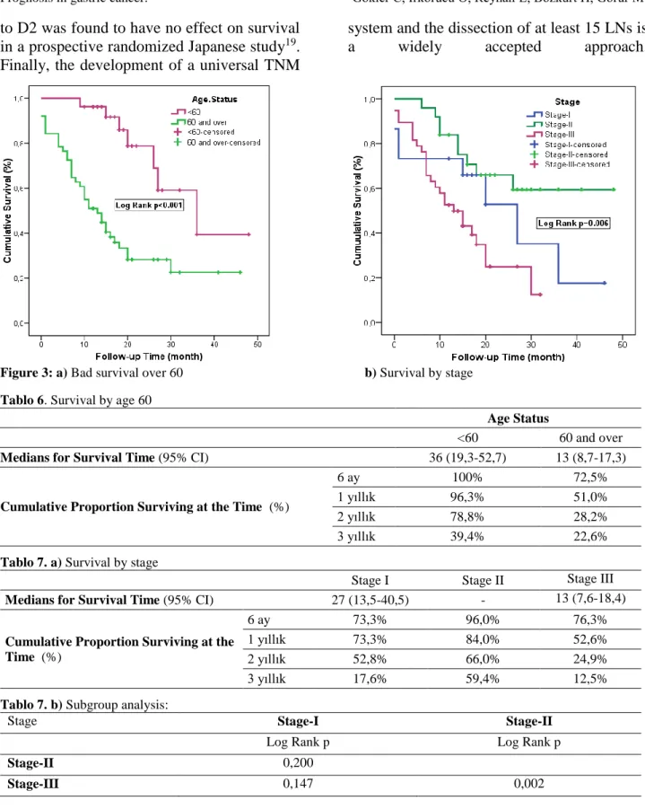
Figure 3: a) Bad survival over 60 b) Survival by stage Tablo 6. Survival by age 60
Age Status
<60 60 and over
Medians for Survival Time (95% CI) 36 (19,3-52,7) 13 (8,7-17,3)
Cumulative Proportion Surviving at the Time (%)
6 ay 100% 72,5%
1 yıllık 96,3% 51,0%
2 yıllık 78,8% 28,2%
3 yıllık 39,4% 22,6%
Tablo 7. a) Survival by stage
Stage I Stage II Stage III
Medians for Survival Time (95% CI) 27 (13,5-40,5) - 13 (7,6-18,4)
Cumulative Proportion Surviving at the Time (%)
6 ay 73,3% 96,0% 76,3%
1 yıllık 73,3% 84,0% 52,6%
2 yıllık 52,8% 66,0% 24,9%
3 yıllık 17,6% 59,4% 12,5%
Tablo 7. b) Subgroup analysis:
Stage Stage-I Stage-II
Log Rank p Log Rank p
Stage-II 0,200
Stage-III 0,147 0,002
Japanese guidelines also recommends performing N stage according to the number of LNs, concurring with the International Union Against Cancer (UICC)/TNM staging system20. The Japanese D1-D2 LN dissection has also changed, with, for example, LN station 7 being included in the extent of D1 dissections21. In conclusion, D2 LN dissection
is preferred in our clinic for patients who
undergo curative resections, although a mean 19 LNs were dissected in those patients. Adjuvant treatment administered at our clinic is based on the International Union Against Cancer (UICC)/TNM staging system. However, when the stages of the patients in this present series is considered, it is apparent that neoadjuvant therapy is not yet standardized in our clinic. Accordingly, the
256
number of patients who received neoadjuvant therapy and were thus excluded from the study was low.
Should D2 LN dissection be performed in all patients? Can prognostic factors offer predictions in this subject? What important achievement can be provided by knowing the prognostic factors and their effect on survival? Ozer I et al.22, in a study at a
high-volume hospital specializing in gastric cancer surgery , evaluated the causes of postoperative early phase mortality in patients over 70 years with gastric cancer, and identified age, albumin levels lower than 3 mg/dl, higher American Society of Anesthesiologists Advanced scores, palliative resections and resections of two or more additional organs as independent risk factors for mortality. They concluded that a more limited surgery can be performed considering not only advanced age, but all risk factors. In addition, Zhou C-J et al.23, in their manuscript evaluating the applicability of radical gastrectomy in elderly patients, identified high comorbidity and TNM stage III as strong predictive factors. The authors emphasized the importance of making surgical decisions taking into account the postoperative complications and low survival associated with this group of patients. Age over 60 years and stage III tumors were found to be poor prognostic factors in the present study. That said, larger and more extensive, randomized and controlled studies are required to clarify the effect of these findings on patient management.
Preoperative serum albumin level is a significant prognostic factor in gastric cancers, especially in intensive care patients, and also in APACHE scoring and in the determination of treatment. In their study involving patients from Mexico, which emphasized such findings, Onate-Ocana, LF et al.24 found the prognostic value of serum albumin. Albumin is a parameter that is used to determine the nutritional condition of the patient, with low albumin levels being associated with severe nutritional risk. The provision of nutritional support prior to major surgery in patients at significant perioperative nutritional risk, and even delaying surgery,
has been reported to be indicated25-27. Nutritional risk is especially high in esophagus, stomach and pancreas malignancies, and nutrition, and even immunonutrition, is recommended especially in such cases28. The mean albumin level was
found to be 2.7 in DGC and 3.7 in the remaining three groups in the present study of patients who were mostly at an advanced stage, although the difference was found not to be statistically significant. That said, statistics indicate that this may be due to the low number of patients with DGC. Related to this issue, preoperative and postoperative nutritional support is generally provided to patients with gastric cancer in our hospital.
The median survival time and the three-year overall survival in patients with gastric cancer was found to be 20 months and 27.8%, respectively in this center, which is a reference center for most of the southern cities in the country. This result is underwhelming. Nevertheless, variable survival rates have been reported related to gastric cancer worldwide. Median survival was reported to be 32.8 months and 18.5 months in stage IIIB and stage IIIC, respectively in a review of 45,411 patients treated at 59 centers in 15 countries, including Japan, Korea and some eastern and western countries, within the International Gastric Cancer Association Staging Project. Survival was found to be 64.4%, 48.2% and 27.7% in patients with stage IIIA, stage B and stage C tumors, respectively29.
In conclusion, stage III tumor and age over 60 years were found to be poor prognostic factors affecting survival. Although no significant difference was found in the survival of those with different tumor localizations and metastatic LNs, the number of dissected LNs was found to be lower in the DGC group than in the UTGC group, and the rate of two or more metastatic LNs was found to be higher in the UTGC localization when compared to other localizations. Median survival and overall survival were found to be 20 months and 27.8%, respectively, in this center for patients with gastric cancer.
257
Ethics committee approval was received for this study from the hospital (2014/61).
Informed Consent
Requirement for informed consent was waived by the hospital ethics committee.
Author Contributions
Conception–C.G., O.I.; Design–C.G., E.R.; Supervision–O.I:, H.B.; Materials– C.G., O.I., E.R.; Data Collection and/or Processing–C.G., M:G.; Analysis and/or Interpretation–C.G., E.R.; Literature review– C.G., H.B., M.G.; Writer–C.G., E.R.; Critical Review–C.G., O.I., M.G.
Conflict of Interest
No conflict of interest was declared by the authors.
Financial Disclosure
The authors declared that this study has received no financial support.
References
1. Torre LA, Bray F, Siegel RL, Ferlay J, Lortet‐Tieulent J, Jemal A. Global cancer statistics, 2012. CA Cancer journal for clinicians 2015; 65(2): 87-108.
2. Sencan I, Ince GN ed. Turkish Ministry of Health Public Health Agency Cancer Statistics. 2016; 1-60.
3. Siewert JR, Böttcher K, Stein HJ, Roder JD. Relevant prognostic factors in gastric cancer: ten-year results of the German Gastric Cancer Study. Ann Surg. 1998;228(4):449-61. 4. Sert OZ, Bozkurt H, Bulut IC, et al.C-Reactive Protein to
Albumin Ratio:A Reliable Marker in Gastric Surgery. Indian J Surg. (2020) https://doi.org/10.1007/s12262-020-02310-y. 5. Park JC, Lee YC, Kim JH, et al. Clinicopathological aspects
and prognostic value with respect to age: an analysis of 3,362 consecutive gastric cancer patients. J Surg Oncol.
2009;99(7):395‐401.
6. Kim JP, Lee JH, Kim SJ, Yu HJ, Yang HK. Clinicopathologic characteristics and prognostic factors in 10 783 patients with gastric cancer. Gastric Cancer. 1998 ;1(2):125-133.
7. Jemal A, Siegel R, Xu J, Ward E. Cancer statistics. 2010. CA Cancer J Clin. 2010;60:277–300.
8. Schwarz RE, Smith DD. Clinical impact of lymphadenectomy extent in resectable gastric cancer of advanced stage. Ann Surg Oncol. 2007;14(2):317-28.
9. Sert OZ, Bozkurt H, Ozlem T, et al. Clinical research Clinicopathologic and immunohistochemical features of gastrointestinal stromal tumors: a single-center experience. Arch Med Sci Civil Dis. 2020; 5: 8–13.
10. Seevaratnam R, Bocicariu A, Cardoso R, et al. How many lymph nodes should be assessed in patients with gastric cancer? A systematic review. Gastric Cancer. 2012 ; 15(1): 70-88. 11. Songun I, Putter H, Kranenbarg EM, Sasako M, van de Velde
CJ. Surgical treatment of gastric cancer: 15-year follow-up results of the randomised nationwide Dutch D1D2 trial. Lancet Oncol. 2010 May;11(5):439-49.
12. Sasako M, Sano T, Yamamoto S, et al. D2 lymphadenectomy alone or with para-aortic nodal dissection for gastric cancer. N
Engl J Med. 2008;359(5):453‐462.
13. Seevaratnam R, Bocicariu A, Cardoso R, et al.. A meta-analysis of D1 versus D2 lymph node dissection. Gastric Cancer. 2012 ;15(1):60-9.
14. Liu X, Cai H, Wang Y. Prognostic significance of tumor markers in T4a gastric cancer. World J Surg Oncol. 2012;10:68.
15. Shim JH, Song KY, Jeon HM, et al. Is gastric cancer different in Korea and the United States? Impact of tumor location on prognosis. Ann Surg Oncol. 2014;21(7):2332‐2339.
16. Qiu MZ, Wang ZQ, Zhang DS, et al. Clinicopathological characteristics and prognostic analysis of gastric cancer in the young adult in China. Tumour Biol. 2011;32(3):509‐514.
17. Songun I, Putter H, Kranenbarg EM, Sasako M, van de Velde CJ. Surgical treatment of gastric cancer: 15-year follow-up results of the randomised nationwide Dutch D1D2 trial. Lancet
Oncol. 2010;11(5):439‐449.
18. Cuschieri A, Weeden S, Fielding J, et al. Patient survival after D1 and D2 resections for gastric cancer: long-term results of the MRC randomized surgical trial. Surgical Co-operative Group. Br J Cancer. 1999;79(9-10):1522‐1530.
19. Sano T, Sasako M, Yamamoto S, et al. Gastric cancer surgery: morbidity and mortality results from a prospective randomized controlled trial comparing D2 and extended para-aortic lymphadenectomy--Japan Clinical Oncology Group study 9501. J Clin Oncol. 2004;22(14):2767‐2773.
20. Sano T, Aiko T. New Japanese classifications and treatment guidelines for gastric cancer: revision concepts and major revised points. Gastric Cancer. 2011;14(2):97‐100.
21. Japanese Gastric Cancer Association. Japanese gastric cancer treatment guidelines 2010 (ver. 3). Gastric Cancer.
2011;14(2):113‐123.
22. Ozer I, Bostanci EB, Koc U, et al. Surgical treatment for gastric cancer in Turkish patients over age 70: early postoperative results and risk factors for mortality. Langenbecks Arch Surg. 2010;395(8):1101‐1106.
23. Zhou CJ, Chen FF, Zhuang CL, et al. Feasibility of radical gastrectomy for elderly patients with gastric cancer. Eur J Surg
Oncol. 2016;42(2):303‐311.
24. Oñate-Ocaña LF, Aiello-Crocifoglio V, Gallardo-Rincón D, et al. Serum albumin as a significant prognostic factor for patients with gastric carcinoma. Ann Surg Oncol. 2007;14(2):381‐389.
25. Tegels JJ, De Maat MF, Hulsewé KW, Hoofwijk AG, Stoot JH.
Improving the outcomes in gastric cancer surgery. World J
Gastroenterol. 2014;20(38):13692‐13704.
26. Shim H, Cheong JH, Lee KY, Lee H, Lee JG, Noh SH. Perioperative nutritional status changes in gastrointestinal cancer patients. Yonsei Med J. 2013;54(6):1370‐1376.
27. Rey-Ferro M, Castaño R, Orozco O, Serna A, Moreno A. Nutritional and immunologic evaluation of patients with gastric cancer before and after surgery. Nutrition. 1997;13(10):878‐ 881.
28. Weimann A, Braga M, Harsanyi L, et al. ESPEN Guidelines on Enteral Nutrition: Surgery including organ transplantation. Clin
Nutr. 2006;25(2):224‐244.
29. Sano T, Coit DG, Kim HH, et al. Proposal of a new stage grouping of gastric cancer for TNM classification: International Gastric Cancer Association staging project. Gastric Cancer. 2017;20(2):217‐225.