192
ÖZ
GİRİŞ ve AMAÇ: İntraoperatif nöromonitörizasyon, hem primer hem de sekonder gergin omurilik operasyonlarında, operasyonun daha güvenli gerçekleşmesi ve kompleks operasyonlarda cerraha yol göstermesi adına bir gereklilik olarak kabul edilir. Bu makalede, çocuklarda iki farklı operasyonun nöromonitörizasyon sonuçları ve postoperatif klinik değişimleri yer almaktadır: okült spinal disrafizm ve geçmiş myelomeningosel onarımına sekonder gergin omurilik.
YÖNTEM ve GEREÇLER: Yirmi dört operasyon üç modalite ile monitörize edildi: transkraniyal motor uyarılmış potansiyeller (TcMEP), sürekli elektromiyografi (EMG) ve direkt sinir
stimülasyonu (DNS). Grup 1’de okült spinal disrafizme bağlı gergin omurilik operasyonu geçirmiş 14 hasta, grup 2’de geçmiş myelomeningosel onarımına sekonder gergin omurilik operasyonu geçirmiş 10 hasta bulunuyordu.
BULGULAR: Alt ekstremitelerden TcMEP yanıt elde etme oranı grup 1’de %92, grup 2’de %80 olarak bulundu. Anal sfinkterden TcMEP yanıt elde etme oranı grup 1’de %83, grup 2’de %60 olarak bulundu. Her iki grupta da operasyonlar esnasında TcMEP değişikliği görülmedi. Her iki grubun postoperatif ürodinami sonuçları 1 yıl periyodu içerisinde iyileşti (grup 1’de %78, grup 2’de %43). Hipoaktif mesaneli hastalarda TcMEP’te anal sfinkter yanıtları alınamadı. Yeni ya da kötüleşmiş postoperatif nörolojik defisit görülmedi.
TARTIŞMA ve SONUÇ: Hem asemptomatik okült spinal disrafizm hem de MMC’ye sekonder TCS’de spinal kordun serbestleştirilmesi bozulmuş ürodinami sonuçlarını iyileştirebilir. İntraoperatif nöromonitörizasyon ve direkt stimülasyon daha güvenli bir ameliyat için bilgi sağlayabilir ve özellikle sekonder serbestleştirmede cerrahi manevraları yönlendirebilir. İntraoperatif nörofizyolojik monitörizasyon, kalan motor fonksiyonları korumak adına nörolojik defisitli MMC hastalarının operasyonlarında yararlıdır. Anal sfinkter fonksiyonları ile mesane fonksiyonları arasında korelasyon olduğundan hasta hipoaktif mesaneye sahipse anal sfinkterden TcMEP yanıtı alınamayabilir.
Anahtar Kelimeler: İntraoperatif nöromonitörizasyon, gergin omurilik sendromu, myelomeningosel, motor uyarılmış potansiyeller
ABSTRACT
INTRODUCTION: Using intraoperative neuromonitoring in both primary and secondary tethered cord operations is accepted as a necessity for a safer operation and guiding surgeon in complex surgeries.
METHODS: Twenty four operations which were monitored with three modalities; transcranial motor evoked potentials (TcMEP), free-run electromyography and direct nerve stimulations. In group 1, there were 14 patients underwent tethered cord operations due to occult spinal dysraphism, in group 2 there were 10 patients underwent tethered cord operations secondary to previous myelomeningocele repair. RESULTS: TcMEP responses of lower extremity were elicited in 92 % in group 1, 80 % in group 2. TcMEP responses of anal sphincter were elicited in 83 % in group 1, 60 % in group 2. No TcMEP change was observed during the surgeries in both group. Postoperative urodynamic results of both group were improved in 1 year period (78 % in group 1, 43 % in group 2). In patients with hypoactive bladder, we could not take anal sphincter responses in TcMEP.
DISCUSSION and CONCLUSION: Untethering of spinal cord both in asymptomatic occult spinal dysraphism and TCS secondary to MMC, can improve impaired urodynamic results. Intraoperative neuromonitoring and direct stimulation provides information for a safer surgery and guide surgical maneuvers especially in secondary untethering. Intraoperative
neurophysiological monitoring is beneficial for operations of MMC patients with neurological deficits, to preserve their residual motor functions. Since anal sphincter functions are correlated with bladder functions, it is possible to get no anal sphincter TcMEP response if patient has hypoactive bladder.
Keywords: Intraoperative neuromonitoring, tethered spinal cord, myelomeningocele, motor evoked potentials
İletişim / Correspondence: Dr. Hüseyin Canaz
İstanbul Bilim Üniversitesi, Nöroşirurji Anabilim Dalı, İstanbul, Türkiye E-mail: [email protected]
Başvuru Tarihi: 03.05.2018, Kabul Tarihi: 03.06.2018
İ
İn
nt
tr
ra
a
op
o
pe
e
ra
r
at
t
if
i
f
N
Nö
ör
ro
om
mo
on
ni
it
t
ör
ö
ri
iz
z
a
a
s
s
yo
y
on
n
i
il
le
e
B
B
ir
i
rl
li
ik
k
te
t
e
S
Se
er
rb
be
e
st
s
t
le
l
eş
ş
ti
t
ir
rm
me
e
İ
İş
ş
le
l
em
ml
le
e
ri
r
in
n
in
i
n
B
B
ul
u
lg
gu
ul
la
ar
rı
ı:
:
O
Ok
k
ül
ü
lt
t
S
Sp
p
in
i
na
al
l
D
Di
is
s
ra
r
af
fi
iz
zm
m
v
v
e
e
M
M
ye
y
e
lo
l
om
me
en
ni
in
ng
g
os
o
s
e
e
le
l
e
S
Se
e
ko
k
on
n
de
d
er
r
G
Ge
e
rg
r
gi
in
n
O
Om
mu
ur
ri
il
li
ik
k
S
Se
e
nd
n
dr
ro
om
mu
u
T
Th
h
e
e
R
Re
es
su
u
lt
l
ts
s
o
o
f
f
U
Un
nt
te
e
th
t
h
er
e
ri
in
ng
g
P
P
ro
r
oc
c
e
e
du
d
u
re
r
es
s
w
wi
it
th
h
I
In
n
tr
t
ra
a
op
o
p
er
e
ra
at
ti
iv
ve
e
N
Ne
eu
u
ro
r
om
mo
o
ni
n
it
to
or
ri
in
n
g
g
:
:
O
Oc
cc
c
ul
u
lt
t
S
S
pi
p
in
na
a
l
l
D
Dy
ys
sr
r
a
a
ph
p
h
is
i
s
m
m
a
a
nd
n
d
T
T
et
e
th
he
e
re
r
ed
d
S
S
pi
p
in
na
a
l
l
C
Co
or
rd
d
S
Se
e
co
c
on
n
da
d
a
ry
r
y
t
to
o
M
M
y
y
e
e
l
l
o
o
m
m
e
e
n
n
i
i
n
n
g
g
o
o
c
c
e
e
l
l
e
e
Hüseyin Canaz1, Ezgi Tuna Erdoğan2, İbrahim Alataş1 1 İstanbul Bilim Üniversitesi, Nöroşirurji Anabilim Dalı, İstanbul, Türkiye 2 İstinye Üniversitesi Tıp Fakültesi, Fizyoloji Anabilim Dalı, İstanbul, Türkiye
INTRODUCTION
Intraoperative neurophysiological monitoring (IONM) is a continuously progressing area for today’s neurosurgery. It is not proven that intraoperative neuromonitoring decreases postoperative neurological deficits in tethered cord surgery because it is difficult to conduct a prospective study to compare with a similar control group. On the other hand, the benefit of neuromonitoring on surgical strategy and low postoperative neurological complications in tethered cord operations was shown in many studies (1-6). Therefore, using intraoperative neuromonitoring is accepted as a necessity for a safer surgery during tethered cord operations today. Myelomeningocele (MMC) is the most common (1per /1000 births) form of neural tube defects which requires a closure operation immediately after birth for preventing infection and trauma to open spinal cord (7). In postoperative years, many patients develop tethering of spinal cord due to previous MMC operation which is known as secondary tethered spinal cord (8). Untethering operations of secondary tethered spinal cords due to MMC are more challenging because of the scar tissue, attachments and abnormal neuroanatomy. Both in primary and secondary tethered cord operations, surgeons need to differentiate functional neural tissue from non-functional structures. Intraoperative transcranial motor evoked potentials (TcMEP) provide a continuous information about spinal cord integrity and direct nerve stimulation guides the manipulations of surgeon to find and protect functional nerves in surgical area. In our institution, we are performing intraoperative neuromonitoring together with mapping techniques in all type of tethered cord operations.
In this paper, we presented the results of IONM in two different type of operations in children: TCS due to occult spinal dysraphism and TCS secondary to previous myelomeningocele repair. Monitoring results and postoperative clinical changes compared between two groups of patients.
METHODS
Twenty four operations which were performed by same neurosurgeon, were monitored with three modalities; TcMEP, free-run EMG and direct nerve
stimulations. We divided surgical procedures into 2 subgroup, group 1: tethered cord operations of occult spinal dysraphism, group 2: tethered cord operations of secondary tethered cord due to previous myelomeningocele repair. Group 1 includes 14 patients 9 female, 5 male with occult spinal dysraphism. The mean age was 44 months (between 1 months-6 years 5 months). Preoperative lower extremity motor functions and urodynamic studies are shown in table 1.
Group 2 includes 10 patients 3 female, 7 male with tethered spinal cord secondary to myelomeningocele repair. The mean age was 19 month (between 2 months-4 years). Preoperative lower extremity motor functions and urodynamic studies are shown in table 2.
In 21 of 24 operations, intraoperative neurophysiological monitorings were done with Cadwell Cascade Elite System (Cascade Laboratories, WA, USA) while in last 3 operations Xltek Protektor 16 System were used. Anesthesia was maintained with total intravenous anesthesia with propofol and fentanyl infusion. Short acting muscle reluctant was used only for intubation.
194 Subdermal needle recording electrodes were inserted into M.quadriceps femoris (L3-L4), M.tibialis anterior (L4-S1), M.gastrocnemius (S1-S2) and external anal sphincter muscle (S2-S4). Patients were grounded from lateral hip and all impedances were kept below 2 kOhm. Free-run EMG and direct stimulations were recorded from same muscle groups. Free-run EMG were followed from first incision to the end of the operation. Surgeons were informed about any repeated and prolonged high frequency train activity in free-run EMG. Direct stimulations were done by a monopolar probe (reference electrode were placed on the paravertebral muscles) with a maximal 5 mA current intensity. Constant current was delivered with a 2.82 Hz stimulation frequency and 200 µs pulse width. Band-pass filter was set from 30 to 3000 Hz.
For TcMEP monitoring, corkscrew electrodes were placed over C3-C4 sites on the scalp according to International 10-20 System and stimulation parameters were adjusted to 5-7 pulses, 100-600 V, rectangular pulses with 50µs pulse width. We started with 100 V intensity and increased it until a compound muscle action potential was elicited at least 100µV in amplitude. Based on the reports about correlation between TcMEP loss and
postoperative motor deficits, we prefer to use all or nothing criteria in our operations (9).
RESULTS
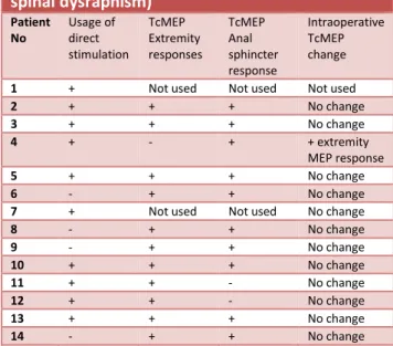
Intraoperative Changes in Neuromonitoring TCMEP responses were followed throughout the operations. In group 1, we could not use TCMEP in 2 patients because of neuromuscular blocker usage. We took TcMEP responses from extremity muscles in 11 of 12 TcMEP monitoring (92 %), in 1 patient (3 month age) we could not elicit TcMEP at the start of the operation (8%). However, extremity response (tibial muscle CMAP) was obtained just after untethering of spinal cord. We took anal sphincter TcMEP responses from 10 of 12 TcMEP monitoring (83%). No change was observed during the surgeries in group 1 (Table 3).
Table 3. Intraoperative monitoring modalities and responses of patients in Group 1 (occult spinal dysraphism) Patient No Usage of direct stimulation TcMEP Extremity responses TcMEP Anal sphincter response Intraoperative TcMEP change
1 + Not used Not used Not used
2 + + + No change 3 + + + No change 4 + - + + extremity MEP response 5 + + + No change 6 - + + No change
7 + Not used Not used No change
8 - + + No change 9 - + + No change 10 + + + No change 11 + + - No change 12 + + - No change 13 + + + No change 14 - + + No change
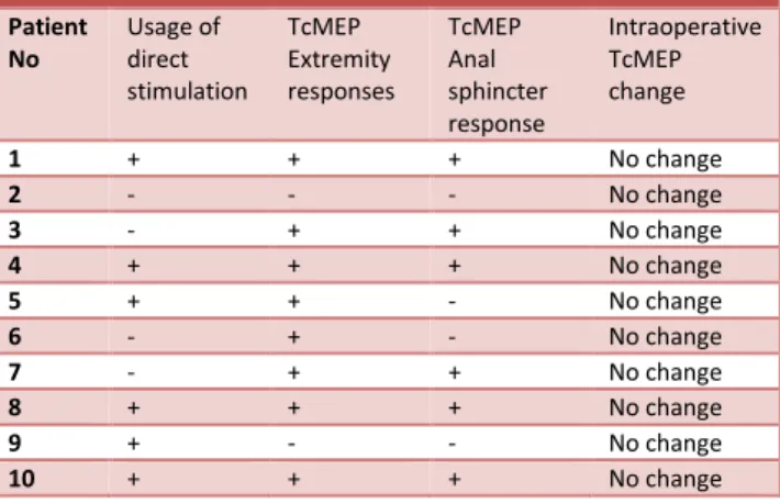
In group 2, TcMEP responses from lower extremity muscles were elicited in 8 of 10 patients (80%), in 2 patients (4 month and 2 month age) we could not take a response from lower extremity and also anal sphincter too. Anal sphincter TcMEP responses were elicited in 6 of 10 patients (60%). Two of those patients had lower extremity TcMEP responses while no response from anal sphincter (Table 4). No loss of intraoperative TcMEP response was seen in group 2 patients.
Table 4. Intraoperative monitoring modalities and responses of patients in Group 2 (TCS secondary to MMC) Patient No Usage of direct stimulation TcMEP Extremity responses TcMEP Anal sphincter response Intraoperative TcMEP change 1 + + + No change 2 - - - No change 3 - + + No change 4 + + + No change 5 + + - No change 6 - + - No change 7 - + + No change 8 + + + No change 9 + - - No change 10 + + + No change
Surgeons were warned in one operation from group 1, according to repeated responses in free-run EMG. In 10 of 14 operations in group 1, and 6 of 10 patients in group 2, direct stimulation with monopolar probe was used. Filum was stimulated if it was planned to dissected, no response were taken with maximum 5 mA current intensity. In 2 cases, we directly stimulated unidentifiable tissue and took responses from lower extremity muscles and surgical plan was changed accordingly.
Postoperative clinical changes
The motor functional evaluations were done 24 hours after the operations by same neurosurgeon. Urodynamic studies were performed minimum 3 months - maximum 12 months after surgery. When we look at the results of postoperative motor functions, there is no worsened or new postoperative motor deficits in both group of patients. In only one case from group 2, a better motor function was observed in distal muscles in 24th hours after the operation (Table 2). When urodynamic results were evaluated in 1 year period, we found a difference between two groups. In group 1, we could obtain postoperative urodynamic results in 9 of 14 patients. Seven of 9 were better compared to preoperative results (78%). In group 2, only 7 of 10 patients got results of postoperative urodynamic studies yet. Three of 7 results were better compared to preoperative studies (43%).
DISCUSSION
In previous studies, it was shown that preoperative motor deficits correlates with lower TcMEP monitorability for spinal cord surgeries (9,10). In our results, anal sphincter TcMEP responses and extremity TcMEP responses in occult spinal dysraphism patients were obtained in 83 % and 92 % respectively. Our results of TcMEP monitorability rates are similar with Sala and friends (11). They found the monitorability rates 97 % for limb muscles and 74 % for anal sphincter in children who underwent complex spinal cord untethering. Furthermore, the youngest patient with TcMEP response was 4-month old. In our results, the youngest patient with monitorable TcMEP was 2 month-old.
In TCS patients secondary to MMC, TcMEP responses were obtained in lower rates (60 % anal sphincter, 80 % extremity muscles). The lower monitorability rate of TcMEP in TCS patients secondary to MMC could be related to the more severe preoperative motor deficits. However, one of the two patients who had no extremity TcMEP responses in group 2 (patient number 2 and 9) had normal motor functions and the other had severe weakness in lower extremity (number 2). On the other hand, we could obtain extremity TcMEP responses from patients with more severe motor deficits in group 2 (Table 2, Table 4). Accordingly, those results are not enough to make a conclusion about the correlation between extremity TcMEP and lower extremity motor deficits in TCS patients secondary to MMC. The other possible factor interfere to monitorability of TcMEP in those patients may be the young ages of children. The three patients with no TcMEP responses were at 2 (in group 1), 3 and 4 month of ages (in group 2). Immaturity of central nervous system could be another reason of unresponsiveness of motor cortex (12).
The patient in group 1 who had no extremity TcMEP response, had monitorable anal sphincter responses. There was irregular on/off responses on tibial muscle at the start of the operation. After untethering the spinal cord the response become more stable and monitorable in the end of the
196 surgery. She had normal motor functions preoperatively, so there was no postoperative clinical confirmation of intraoperative change. The intraoperative improvement of TcMEP in tethered cord was anecdotally reported on case basis. It can be related to sudden changes in impaired oxidative metabolism in conus (11).
The correlations between anal sphincter TcMEP responses and preoperative clinical findings were also examined. In 2 patients from group 1, we could not elicit anal sphincter TcMEP response. Those patients had normal preoperative motor functions and extremity TcMEP responses were present. The corkscrew electrodes were at C3-C4 sites which induces more muscle twitch than other electrode montages (12). Since the transcranial stimulation induced a strong body twitch in those operations we could not increase the stimulation intensity to get an anal sphincter response. Therefore, the reason of absent anal sphincter TcMEP response may be due to inadequate intensity of transcranial stimulation. Since the electrode montage of C3-C4 limited us, (after the preparation of present paper) we continue TcMEP monitoring with C1-C2 electrode montages in our institution. In group 2, 4 patients had no TcMEP responses from anal sphincter. However, 2 of 4 patients had also no response from extremity muscles. The other 2 patients had hypoactive bladder in urodynamic studies preoperatively. Hypoactive bladder indicates an injury at lower motor neuron at sacral level- conus or sacral roots which also innervate external anal sphincter muscles. It can be deduced that hypoactive bladder is in correlation with absent anal sphincter TcMEP response in group 2.
Kumar and colleagues were used intraoperative mapping with direct stimulation at sacral level in patients with conus and cauda lesions including spinal dysraphism (14). He emphasized that mapping was helpful for dissection especially who have undergone surgery previously. Our results support same conclusions. In our operations, we did not dissect filum terminal without stimulation in a maximum 5 mA intensity. No muscle responses were obtained with filum stimulation and no postoperative worsened functions were found in patients. In 2 operations, we elicited muscle responses with direct stimulation of undefinable
tissue in surgical area and manipulations were changed according to responses. Direct stimulation helped surgery in a critical direction in those patients.
There are few studies focused on neuromonitoring in TCS syndrome secondary to MMC. Pouratian and colleagues reported the results of electrophysiologically guided untethering of secondary tethered spinal cord 38 patients who had undergone perinatal myelomeningocele repair (4). They use free-run EMG and direct stimulation of tissues to identify functional neural elements during untethering procedures. We use free-run EMG and direct stimulation during the closure of MMC lesions routinely. The reason to not use TcMEP in MMC closure is the safety questions of transcranial electrical stimulations in newborns. In secondary TCS operations as in the present study, we also add TcMEP technique to follow the integrity of spinal cord. Multimodality may provide a wide possibility of monitoring both roots and spinal cord simultaneously.
There are studies about TCS operations with intraoperative neuromonitoring which evaluated postoperative urological functions. The percentages of postoperative improvement in bladder functions were between 14-46 %. In most of them, only urological symptoms were evaluated, patients were adults or the pathologies were mixed (lipoma, myelomeningocele, tight filum etc.) (1,6,15). In very young children it is not possible to recognize bladder dysfunction due to tethering cord. Urodynamic studies are helpful for an early recognition of TCS and prevents a delay in surgery and irreversible injury. Hoving and colleagues used video urodynamics for evaluations (1). They found stable urological functions postoperatively in high risk group of TCS patients (lipomyelomeningocele or/and split cord). However, the time of the postoperative urodynamics did not mentioned which is suggested to perform at least 3 months after operations generally. In follow-up periods they found an improvement in urologic functions in the 4 of 40 patients (10 %). Pouratian and colleagues were evaluated bladder with cystometrogram and found a 20 % urological improvement postoperatively in TCS patients secondary to MMC (4). Untethering of spinal cord with intraoperative monitoring both in
asymptomatic occult spinal dysraphism and TCS secondary to MMC, may contribute to improvement of impaired urodynamic results by helping to preserve root functions. Our postoperative urodynamic change rates are high in both group according to literature. However, this can be resulted from the small number of patients in the groups which make difficult to draw a conclusion about postoperative urodynamic changes. Further studies need with large number of patients to investigate the postoperative changes in bladder functions after TCS operations with IONM.
Conclusion
The use of intraoperative monitoring and mapping techniques together is feasible and efficient during TCS operations (2,3,4,5,6). In our results, there is no new or worsened neurological or urological functions. Untethering of spinal cord can improve urodynamic results in both occult spinal dysraphism and MMC patients. Besides, we may not elicit anal sphincter TcMEP response, if patient has hypoactive bladder. Continuous information about spinal cord integrity with TcMEP gives a reassurance to surgeon about their strategy and mapping with direct stimulation make easier to find and preserve functional tissue especially while doing maneuvers in distorted neuroanatomy. Even the most of the patients with MMC had neurological deficits, it is feasible to use TcMEP and direct stimulation in those operations. Moreover, to preserve the residual functions in lower extremities during the operations is essential for those children to maintain their life quality.
Disclosure
The study received no specific grant or funding.
Conflict of Interest
All authors stated no conflict of Interest.
REFERENCES
1. Hoving EW, Haitsma E, Oude Ophuis CM, Journée HL. The value of intraoperative neurophysiological monitoring in tethered cord surgery. Childs Nerv Syst ;27:1445–52, 2011
2. Husain AM, Shah D. Prognostic value of neurophysiologic intraoperative monitoring in tethered cord syndrome surgery. J Clin
Neurophysiol 26:244–7, 2009
3. Kothbauer KF, Novak K: Intraoperative monitoring for tethered cord surgery: an update. Neurosurg Focus 16:E8, 2004
4. Pouratian N, Elias WJ, Jane JA Jr, Phillips LH 2nd, Jane JA Sr.: Electrophysiologically guided untethering of secondary tethered spinal cord syndrome. Neurosurg Focus: 29 (1):E3, 2010
5. Sala F, Squintani G, Tramontano V, Arcaro C, Faccioli F, Mazza C: Intraoperative neurophysiology in tethered cord surgey: techniques and results. Childs Nerv Syst 29: 1611–24, 2013
6. Von Koch CS, Quinones-Hinojosa A, Gulati M, Lyon R, Peacock WJ, Yingling CD: Clinical outcome in children undergoing tethered cord release utilizing intraoperative neurophysiological monitoring. Pediatr Neurosurg 37:81–6, 2002
7. Copp AJ, Adzick NS, Chitty LS, Fletcher JM, Holmbeck GM, Shaw GM: “Spina Bifida”Nature Reviews Disease Primers Published online:30 April 2015 doi: 10.1038/nrdp.2015.7
8. Balasubramaniam C, Laurent JP, McCluggage C, Oshman D, Cheek WR: Tethered-cord syndrome after repair of meningomyelocele. Childs Nerv Syst 6:208–11, 1990
9. Kothbauer K, Deletis V, Epstein FJ: Motor-evoked potential monitoring for intramedullary spinal cord tumor surgery: correlation of clinical and neurophysiological data in a series of 100 consecutive procedures. Neurosurg Focus 4: Article 1, 1998
10. Sala F, Palandri G, Basso E, Lanteri P, Deletis V, Faccioli F, et al: Motor evoked potential monitoring improves outcome after surgery for intramedullary spinal cord tumors: a historical control study. Neurosurgery 58:1129–43, 2006
11. Sala F, Tramontano V, Squintani G, Arcaro C, Tot E, Pinna G, Meglio M.: Neurophysiology of complex spinal cord untethering. J Clin Neurophysiol. 31(4):326-36, 2014
12. Szelényi A, Kothbauer KF, Deletis V.: Transcranial electric stimulation for intraoperative motor evoked potential monitoring: Stimulation parameters and electrode montages. Clin Neurophysiol. 118(7):1586-95, 2007
13. Riviello JJ, Kull L, Troup C, Holmes GL: Cortical stimulation in children: techniques and precautions. Tech Neurosurg 7:12–18, 2001
14. Kumar GS, Rajshekhar V, Babu KS. Intraoperative mapping of sacral nervous
198 system (S2-4). Br J Neurosurg:20:396–402, 2006
15. Quiñones-Hinojosa A, Gadkary CA, Gulati M, von Koch CS, Lyon R, Weinstein PR, Yingling CD: Neurophysiological monitoring for safe surgical tethered cord syndrome release in adults. Surg Neurol 62:127–33, 2004