ORIGINAL ARTICLE Volume: 30, Issue: 3, July 2019 pp: 199-205
Özkan ÖZGER1 Necati KAPLAN2
1Istinye University Faculty of Medicine, Canakkale Anadolu Hospital,
Department of Neurosurgery, Canakkale, Turkey.
2Rumeli University, Corlu Reyap Hospital, Neurosurgery Clinic, Tekirdag, Turkey.
ORCID Numbers:
Özkan ÖZGER: 0000-0001-7257-8379 Necati KAPLAN: 0000-0001-5672-0566
Address: Özkan ÖZGER,
Çanakkale Anadolu Hastanesi, Barbaros Mahallesi, Troya Caddesi, No:10, Çanakkale, Turkey. Phone: +90 4445058 / 2965 GSM: +90 507 7906639 E-mail: [email protected] Received: 11th January, 2019. Accepted: 27th April, 2019.
CLINICAL RESULTS OF PERCUTANEOUS
VERTEBROPLASTY IN THORACOLUMBAR
(T6-L5) VERTEBRAL COMPRESSION FRACTURES:
RETROSPECTIVE STUDY OF 111 PATIENTS
WITH 140 FRACTURED SEGMENTS
ABSTRACT
Object: Vertebroplasty was first applied by Harve Deramond to a patient with vertebral hemangioma in 1984. In recent years, the increase in the number of osteoporosis, trauma and tumor cases has increased the incidence of vertebral compression fractures (VCFs). Nowadays, percutaneous vertebroplasty (PVP) has been a widely used treatment for painful acute VCFs. It is a minimally invasive technique. In this procedure, polymethylmethacrylate (PMMA) is injected into the vertebral corpus. There are PMMA’s ability to increase stability at fracture site, thermal necrosis effect and chemotoxic effect on intra-osseous pain receptors. In this study, the safety and efficacy of PVP in patients with VCF were evaluated.
Methods: The patients who underwent PVP under sedoanalgesia or general anesthesia for single or multi-level thoracolumbar vertebrae fracture were reviewed retrospectively between January 2012 and March 2018. The study included 111 patients with VCF. 140 vertebral levels were treated with PVP. These VCFs were evaluated in 3 groups as osteoporotic, traumatic and pathological. We used the Oswestry Disability Index (ODI) for functional disability and the Visual Analog Scale (VAS) for pain severity. Our patients were followed up for 12 month after PVP.
Results: Patients mean age was 73,04 ± 7,17 years (91-56 years) and 18 (16,22 %) were male and 93 (83,78 %) were female. The most commonly affected vertebrae were T12 and L1 vertebrae corpus. Following PVP, VAS and ODI values decreased significantly in the last 12 months compared to preoperative levels (p<.001). Cement leakage was occurred in six patients (5.40 %).
Conclusions: PVP is an advantageous method. Because the procedure is fast and easy, a biopsy can be taken during the procedure, patients can soon stand up and be discharged; its complications are much less than open surgery. In addition to general anesthesia, it can be performed with sedoanalgesia. It is a reliable and effective technique for the treatment of pain due to osteoporotic and traumatic VCFs or metastatic lesions.
Keywords: Percutaneous vertebroplasty, Polymethylmethacrylate, Vertebral compression fractures
Level of Evidence: Retrospective clinical study, Level III.
INTRODUCTION
PVP was administered by Galibert and Deramond in patients with vertebral hemangiomas in 1984 (8). It is a minimally invasive procedure involving injection of bone cement (mostly PMMA) to the vertebral corpus fracture to improve pain and stability of the fracture (13). Minimally invasive PVP provides significant relief of pain and provides early postoperative ambulation. Therefore, it has been widely preferred as treatment in elderly
patients with osteoporotic VCF (23). Most PVP procedures are performed to relieve pain in patients with severe osteoporosis and those with stable fractures attached to one or more vertebral bodies. In addition, vertebroplasty is recommended for patients suffering from posttraumatic symptoms associated with vertebral fractures, patients with large angioma located within the vertebral body, increased risk of compression fractures, and patients with pain associated with İD
vertebral body metastatic disease (4). Osteoporotic fractures have become the main indication for vertebroplasty in many centers (10). We aim to analyse the clinical results following PVP for single or multi-level segment thoracolumbar VCFs. MATERIAL AND METHODS
Ethics approval and patient consents
Our study was a retrospective clinical study performed according to the principles of the World Medical Association Declaration of Helsinki, ‘Ethical Principles for Medical Research Involving Human Subjects’ (revised in 2013). Informed consent form was obtained from all patients. Patients studied
We retrospectively analyzed the files of 111 patients (93 females, 18 males) who were hospitalized for VCFs at the Neurosurgery Clinic between May 2015 - December 2018 and who had undergone PVP.
Study design
Patients had to have one or more vertebral compression fractures, severe pain in the lumbar or thoracic region, limited activity, and resistance to medical treatment. A thoracolumbar brace was provided to all patients before and after surgery. Acute (first 2 weeks) or sub-acute (2-8 weeks) VCFs were included in our study. Patients with anterior vertebral compression ratio greater than 85 %, motor and sensory loss, incontinence and unstable vertebral fractures were excluded from the study.
Imaging, VAS and ODI values
Preoperative and postoperative VAS measurements were used to evaluate the severity of the pain. Patients were scored between 0 and 10 points according to VAS requirements The most painless score was 0 points and the most painful score was 10 points.
The functional disability of patients were assessed using the ODI scale. There were 10 questions in the ODI survey. 6 stylish, scored from 0 to 5. The best score was 0 points and the worst score was 5 points. The total score was multiplied by two and calculated as a percentage. The maximum and minimum scores were 100 and 0, respectively. As the total score increases, the level of disability increases.
The patients were followed up in outpatient clinics and by phone interviews for 12 months after the surgery. VAS and
ODI evaluations of the patients were done preoperatively, and again at 1. month, 6. month, and 12. month. Vertebral MRI, X-ray or computed tomography (CT) was taken to patients. Surgical technique: Percutaneous vertebroplasty These procedures were performed in the operating room under sedo anesthesia (95 patients, 85,59%) or general anesthesia (16 patients, 14,41%). All patients were placed in the prone position and C-Arm scopy (X ray) was positioned appropriately. An antibioprophylaxis (intravenous 1 gm cephazolin sodium) was performed systemically before the procedure. Fractured vertebra was identified in both anteroposterior and lateral view images. C- Arm was positioned for an anteroposterior view to visualize the pedicles of the affected vertebra. 11-gauge Jamshidi biopsy needles were inserted through the upper external edge of the pedicle ring in AP imaging and the transpedicular approach was inserted percutaneously into the fracture vertebral body. Kirschner wires were used to place a cannula into the posterior half of the vertebral body. Bone biopsies were taken from the vertebral corpus from some of these patients. PMMA was injected through the pedicle into the vertebrae. Post-PVP PMMA leakage was evaluated by postoperative vertebrae radiography or vertebral CT. Figure 1a shows preoperative T12 compression fracture in the sagittal MRI sections. Figure 1b shows post-operative 1st month PMMA in the vertebra CT. Figure 1c shows post-operative 12th month T12 compression fracture in the sagittal MRI sections. These images belong to a 72-year-old female patient. Data of analysis
In this study, VAS and ODI values were examined measured before PVP and at the first, sixth, twelfth months after PVP. Statistical and visual analysis was used to analyze the data. Prior to the analysis of the data, the kurtosis and skewness values were examined to see if the data set met the assumption of normality. In all data the kurtosis values are in the range of -8686 and .092 and the skewness values are in the range of -.294 to .133. These findings show that the data show normal distribution. Since the data met the normality assumption, one of the parametric tests, Variance Analysis for Repeated Measurements (Repeated Measures ANOVA), was used to compare the change observed in pre- and post-intervention measurements. SPSS Statistic 22 package program was used to analyze the data and the significance value was analyzed as p <.05.
Figure-1. (a) T1-weighted and fat suppression sagittal magnetic resonance images reveal a fresh compression fracture at the T12 level (preoperative), (b) PMMA appearance in the T12 vertebral body in the thoracic CT axial section (post-operative 1st month), (c) T1-weighted and fat suppression sagittal magnetic resonance images reveal a fresh compression fracture at the T12 level (post-operative 12th month).
RESULTS
A total of 111 patients with traumatic, osteoporotic and pathological VCF and 140 vertebral levels were included in the study. The mean age was 73,04 ± 7,17 years (91-56 years), 18 (16,22%) were male and 93 (83,78 %) were female. The mean age of the males was 72.33 ± 8.43 (87-56) years, and the mean age of the females was 73.18 ± 6.95 (91-56) years. 68.47 % (n = 76) of the patients were treated for osteoporotic, 30.63 % (n = 34) traumatic and 0.90 % (n = 1) due to pathological compression fracture. 79.28 % (n = 88) had single level, 17.12 % (n = 19) had two levels, 3.60 % (n = 4) had three levels of VCF.
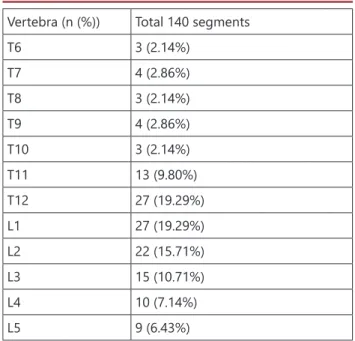
Most of the VCF was 59,29 % (n = 83) at the lumbar level. 40,71 % (n = 57) were at the thoracic level. The most affected level was T12 vertebra (n = 27, 19.29 %) and L1 vertebra (n = 27, 19.29 %). The mean preoperative anterior vertebral height loss rate was calculated as 26,61±14,57 %. The mean volume of PMMA injected to one vertebral level was 4.11±0.73 ml. In 6 patients (5,40 %) there was cement leakage. No neurological complications were associated with cement leakage. Postoperative hospital stay was calculated as 15.08 ± 9.50 hours (0.63 ± 0.40 days). Pathology was obtained from 61 patients. Only one patient (1.6 %) had multiple myeloma. All patients were mobilized in the first 4 hours postoperatively (Table-1,2).
Table-1. Demographic and clinical characteristics of study population
Study Population Patients (n=111)
Age (years, mean ±SD) 73,04±7,17
Gender (n,%) Male 18 (16,22%)Female 93 (83,78%)
Polymethyl methacrylate (PMMA) volume (ml, mean ±SD) 4.11 ± 0.73
Incidental metastatic tumours (n,%)
Note: Bone biopsy samples were taken from 61 patients
Multiple myeloma 1 (%1,6)
Postoperative hospital stay (hours, mean ± SD) 15.08 ± 9.50
Etiology of the VCF (n (%))
Osteoporotic 76 (68.47%)
Neoplastic 1 (0.90%)
Post-traumatic 34 (30.63%)
Table-2. The numbers (n) and percentages (%) of T6-L5 VCFs.
Vertebra (n (%)) Total 140 segments
T6 3 (2.14%) T7 4 (2.86%) T8 3 (2.14%) T9 4 (2.86%) T10 3 (2.14%) T11 13 (9.80%) T12 27 (19.29%) L1 27 (19.29%) L2 22 (15.71%) L3 15 (10.71%) L4 10 (7.14%) L5 9 (6.43%)
Table-3 shows the pre-test and post-test mean and standard deviation (SD) values for VAS and ODI, while Figure 2 shows the graph of these values.
The mean VAS of the patients before the intervention was 8.12 ± 1.11 and after the intervention was 2.19 ± 1.20 at the 1st month , 1.81 ± .98 at the 6th month, 1.41 ± .89 at the 12th month. After the surgery in the VAS values of the patients, a decrease was observed in all three measurements. According to the results of repeated measures analysis of variance, this decrease in VAS values are statistically significant, F =
1634.425, p <.001 (Table 4). According to post-hoc tests to determine the difference between the measurements, the difference observed between all measurements is statistically significant.
The mean ODI of the patients before the intervention was 73.72±10.93 and after the intervention was 21.15±12.11 at the 1st month , 18.00±10.58 at the 6th month, 14.52±8.97 at the 12th month. After the surgery in the ODI values of the patients, a decrease was observed in all three measurements. According to the results of repeated measures analysis of variance, this decrease in ODI values are statistically significant, F=1391.971, p<.001 (Table 5). According to post-hoc tests to determine the difference between the measurements, the difference observed between all measurements is statistically significant.
Figure-2. Pre-test and post-test (1st, 6th, 12th month) mean of VAS and ODI values
Table-3. Pre-test and post-test mean and standard deviation values of VAS and ODI values
n Pre-test
Post-test
1st month 6th monthPost-test 12th monthPost-test
Mean SD Mean SD Mean SD Mean SD
VAS 111 8.12 1.11 2.19 1.20 1.81 .98 1.41 .89
ODI 111 73.72 10.93 21.15 12.11 18.00 10.58 14.52 8.97
Table 4. ANOVA results of pre-test and post-test scores of VAS values
Source of variance Sum of Squares (SS) Sd Mean Squares (MS) F p
Subjects within 3583.500 333
Measurement 3357.532 3 1119.177 1634.425 .000
Table-5. ANOVA results for the pre-test and post-test scores of the ODI values
Source of variance Sum of Squares (SS) Sd Mean Squares (MS) F p
Subjects within 282709.000 333
Measurement 262004.207 3 87334.736 1391.971 .000
Error 20704.793 330 62.742
DISCUSSION
PVP is an accepted treatment modality for osteoporotic, malignant, and traumatic spinal fractures. The effectiveness of this technique has been demonstrated in numerous studies (7,9,11-12). These fractures cause severe disability and reduce the quality of life (19). Hence, bringing the patient back to functional status at the earliest is very important to prevent further osteoporosis. To achieve that, pain needs to be managed at the earliest. Vertebroplasty is one of the minimally invasive procedures to achieve such pain relief and stability where less viscous bone cement is injected into the vertebral body (1,3,6).
In 2016, Leali, Paolo Tranquilli et al. reported that the pain and disability caused by acute osteoporotic VCF appear to be treated with more efficacy through the PVP than with the conservative therapy alone (14).
The advantage of vertebroplasty in malignant spine disease is the less invasive nature compared to open spinal surgery and the apparent rapid pain relief compared to radiotherapy and other conventional treatment options. In the present study, the patients with painful spine metastasis were successfully treated without serious complications. One of the proposed mechanisms is the balancing of fractures. Other factors are vascular, chemical and thermal. Pain decreases with exothermic reaction of bone cement and compression of small nerve endings (5).
The absolute contraindications of vertebroplasty are irreversible coagulopathy, allergy to PMMA and the presence of infection in the body. PVP should not be applied to asymptomatic VCFs which may heal with conservative treatment. Disruption of the posterior vertebral corpus wall, tumor invasion to the spinal canal and collapse to less than one third of the vertebral body is a relative contraindication. These VCFs are difficult to treat. The risk of complications is higher during the surgical procedure (10). Complications of PVP include pulmonary embolism, cardiac perforation, fractures of adjacent vertebrae and infections. In addition to these complications, there may be bone cement extravasation into the spinal canal, paravertebral and intervertebral areas or venous systems (2). No significant complication was observed
in our study. Only 6 patients had cement leakage. These results show that PVP is usually a safe surgery for patients with VCF.
PVP and PKP (percutaneous kyphoplasty) have been compared in literature many times. In a recent study, PVP was found to be more advantageous than PKP in terms of operative time. However, PKP is more advantageous in terms of correcting kyphotic angle and restoration of vertebral height. There is no significant change in VAS and ODI values between PVP and PKP. Similarly, there is no difference in cement leakage rates (22). However, in a meta-analysis study published in 2016, it was reported that PVP caused more cement leakage than PKP. The cost of PKP was also higher. In terms of cost, PVP is more advantageous. In this meta-analysis study, there was no difference between PKP and PVP groups in terms the rate of adjacent and new vertebral fractures (15). In our study, VAS and ODI values were found to be quite significant at 1-year follow-up. Values in our study many kyphoplasty in the literature were not worse than VAS and ODI values.
As in the study of Takahara et al., The most common vertebral fractures is T12 or L1 (thoracolumbar junction) levels (20). In our series, T12 and L1 were the most commonly affected with 54 levels (38.57 %).
In a study conducted by Morsi et al., the mean duration of hospital stay for PVP and PKP was 22.4 hours and 24.5 hours respectively(16). In our study, this rate was
approximately 15 hours. This shows us that after the PVP, the duration of hospital stay and cost decreases as the experience increased.
Xu et al., calculated the average injected PMMA volume 4.3 ml In their study (21). Saracen A and Kotwica Z, injected maximum 0.5 ml PMMA even into the vertebral plane (18). In our case series, a mean volume of 4.11ml PMMA was injected per vertebra.
In an article published in 2010, 75 patients underwent routine bone biopsy during the PKP procedure. A high rate (11 patients) had pathology. These pathologies were metastatic lesions (7 patients), myeloma (3 patients) and leukemia (1 patient) (17). However, only one patient had multiple myeloma in our patient series (61 patients).
CONCLUSION
PVP provides stability in patients with osteoporotic, traumatic and pathological vertebral compression fractures. It is a safe surgical option with minimal complications and it prevents spinal deformity by reducing the collapse of the vertebral corpus. PVP can be performed quickly and should be preferred especially in elderly patients with secondary diseases. Routine bone biopsy during the surgical procedure may be significant in terms of incidental tumor detection. The low complication rate in our study may be related to the volume of injected PMMA (approximately 4ml). More work is needed in the future for the proof of all these claims. We consider PVP as a reliable and effective technique for the treatment of pain associated with osteoporotic, traumatic and pathological VCFs.
REFERENCES
1. Afzal S., Dhar S., Vasavada N.B., Akbar S. Percutaneous vertebroplasty for osteoporotic fractures. Pain
Physician 2007; 10: 559–563.
2. Al-Nakshabandi NA. Percutaneous vertebroplasty complications. Ann Saudi Med 2011; 31(3): 294–297. 3. Alexandru D, So W. Evaluation and management of vertebral
compression fractures. Perm J 2012; 16(4): 46–51. 4. Constantin C, Albulescu DM, Diţă DR, Georgescu CV,
Deaconu AC. Vertebral body clinico-morphological features following percutaneous vertebroplasty versus the
conservatory approach. Rom J Morphol Embryol 2018; 59(1): 159-164.
5. Cotten A, Boutry N, Cortet B, Assaker R, Demondion X, Leblond D, Chastanet P, Duquesnoy B, Deramond H. Percutaneous vertebroplasty: state of the art. Radiographics 1998; 18(2): 311–320.
6. Denaro V, Longo UG, Maffulli N, Denaro L. Vertebroplasty and kyphoplasty. Clin Cases Miner Bone Metab 2009; 6(2): 125–130.
7. Do HM, Jensen ME, Marx WF, Kallmes DF. Percutaneous vertebroplasty in vertebral osteonecrosis (Kummell’s spondylitis). Neurosurg Focus 1999; 7: e2.
8. Galibert P, Deramond H, Rosat P, Le Gars D. Preliminary note on the treatment of vertebral angioma by percutaneous acrylic vertebroplasty. Neurochirurgie 1987; 33(2): 166–168. 9. Grados F, Depriester C, Cayrolle G, Hardy N, Deramond
H, Fardellone P. Long-term observations of vertebral osteoporotic fractures treated by percutaneous vertebroplasty. Rheumatology (Oxford) 2000; 39(12): 1410– 1414.
10. Hargunani R, Le Corroller T, Khashoggi K, Murphy KJ, Munk PL. Percutaneous vertebral augmentation: the status of vertebroplasty and current controversies. Semin Musculoskelet Radiol 2011; 15(2): 117–124.
11. Hemama M, El Fatemi N, Gana R. Percutaneous vertebroplasty in Moroccan patients with vertebral compression fractures. Pan Afr Med J. 2017; 26: 225. doi:10.11604/pamj.2017.26.225.9872.
12. Klazen CA, Lohle PN, de Vries J, Jansen FH, Tielbeek AV, Blonk MC, Venmans A, van Rooij WJJ, Schoemaker MC, Juttmann JR, Lo TH, Verhaar HJJ, van der Graaf Y, van Everdingen KJ, Muller AF, Elgersma OEH, Di Halkema DR, Fransen H, Janssens X, Buskens E, Mali WPT. Vertebroplasty versus conservative treatment in acute osteoporotic vertebral compression fractures (Vertos II): an open-label randomised trial. Lancet 2010; 376(9746): 1085–1092.
13. Lapras C, Mottolese C, Deruty R, Lapras C, Remond J, Duquesnel J. [Percutaneous injection of methyl-metacrylate in osteoporosis and severe vertebral osteolysis (Galibert’s technic). Ann Chir 1989; 43: 371–376.
14. Leali PT, Solla F, Maestretti G, Balsano M, Doria C. Safety and efficacy of vertebroplasty in the treatment of osteoporotic vertebral compression fractures: a prospective multicenter international randomized controlled study. Clin Cases Miner Bone Metab. 2017; 13(3): 234–236.
15. Liang L, Chen X, Jiang W, Li X, Chen J, Wu L, Zhu Y. Balloon kyphoplasty or percutaneous vertebroplasty for osteoporotic vertebral compression fracture? An updated systematic review and meta-analysis. Ann Saudi Med. 2016; 36(3): 165– 174. doi:10.5144/0256-4947.2016.165.
16. Morsi AM, Abdelrahman AA, Khattab MF. Vertebroplasty versus kyphoplasty: a comparative study of safety and cost-effectiveness and tips to improve outcomes of vertebroplasty. Egyptian Orthop J 2017; 52(2): 144-149.
17. Pneumaticos SG, Chatziioannou SN, Savvidou C, Pilichou A, Rontogianni D, Korres DS. Routine needle biopsy during vertebral augmentation procedures. Is it necessary?. Eur
Spine J 2010; 19(11): 1894–1898.
doi:10.1007/s00586-010-1388-8.
18. Saracen A, Kotwica Z. Complications of percutaneous vertebroplasty: An analysis of 1100 procedures performed in 616 patients. Medicine (Baltimore) 2016; 95(24): e3850. doi:10.1097/MD.0000000000003850.
19. Suzuki N, Ogikubo O, Hansson T. Previous vertebral compression fractures add to the deterioration of the disability and quality of life after an acute compression fracture. Eur Spine J 2010; 19: 567–574.
20. Takahara K, Kamimura M, Moriya H, Ashizawa R, Koike T, Hidai Y, Ikegami S, Nakamura Y, Kato H. Risk factors of adjacent vertebral collapse after percutaneous vertebroplasty for osteoporotic vertebral fracture in postmenopausal women. BMC Musculoskelet Disord. 2016; 17: 12. doi:10.1186/ s12891-016-0887-0.
21. Xu BS, Hu YC, Yang Q, Xia Q, Ma XL, Ji N. Long-term results and radiographic findings of percutanous vertebroplasties with polymethylmethacrylate for vertebral osteoporotic fractures. Chin Med J (Engl) 2012; 125(16): 2832-2836.
22. Wang F, Wang LF, Miao DC, Dong Z, Shen Y. Which one is more effective for the treatment of very severe osteoporotic vertebral compression fractures: PVP or PKP? J Pain Res 2018; 11: 2625–2631. doi:10.2147/JPR.S179022.
23. Wang X, Kou JM, Yue Y, Weng XS, Qiu ZY, Zhang XF. Clinical outcome comparison of polymethylmethacrylate bone cement with and without mineralized collagen modification for osteoporotic vertebral compression fractures. Medicine (Baltimore) 2018; 97(37): e12204.