Original Investigation
Published Online: 26.11.2018Corresponding author: Hanifi UCPUNAR
[email protected]Ahmet SEVENCAN
1, Abdulhamit MISIR
1, Hanifi UCPUNAR
2, Mehmet Bulent BALIOGLU
3, Volkan GUR
1,
Suayip AKINCI
41Sanliurfa Training and Research Hospital, Department of Orthopaedics and Traumatology, Sanliurfa, Turkey 2Erzincan University Faculty Medicine, Department of Orthopedics and Traumatology, Erzincan, Turkey 3Istinye University, Department of Orthopaedics and Traumatology, Istanbul, Turkey
4Baltalimani Bone and Joint Diseases Training and Research Hospital, Department of Orthopaedics and Traumatology, Istanbul, Turkey
The Incidence and Interrelationship of Concomitant Anomalies
in Congenital Scoliosis
ABSTRACT
Maternal diabetes mellitus, hypoxia, vitamin deficiencies, and anticonvulsant medication use have been associated with CS (9). Development of the spinal cord and organs of mesoder-mal origin is closely related to development of the vertebral column (1). As a result, CS is often associated with anomalies of the spinal cord, cardiovascular system, and genitourinary system (12).
Previously, the incidences of intraspinal and other organ system pathologies associated with CS were reported
█
INTRODUCTION
C
ongenital scoliosis (CS) is characterized by a spinal curvature secondary to congenital vertebral malforma-tions. Deformity of the spine secondary to failure of nor-mal vertebral development at 4–6 weeks of gestation is an accepted criterion (16). A multifactorial etiology of scoliosis is accepted (2,10,15). The genetic factors associated with CS remain largely unknown (8). However, environmental factors are proven to have a strong relationship with CS (1,8,9,14,23).AIM: To report the incidence and interrelationship of concomitant anomalies in congenital scoliosis (CS) patients.
MATERIAL and METHODS: Whole-spine computed tomography and magnetic resonance imaging (MRI) examination, echocardi-ography, and renal ultrasonography (USG) evaluations of 231 patients with CS were reviewed. Additionally, intraspinal pathologies and structural cardiac and renal anomalies were recorded.
RESULTS: The incidence of intraspinal pathology was 53.7%. Echocardiography was performed in 140 of 231 patients, and congenital heart disease was detected in 38 patients. Renal USG was performed in 133 of 231 patients, and a renal disease was detected in 37 patients. In 133 patients, spinal MRI, echocardiography, and renal USG were performed. In 22 of 67 (32.8%) patients with an intraspinal anomaly, an additional cardiac anomaly was detected. In 27 of 67 (40.3%) patients with an intraspinal anomaly, an additional renal anomaly was detected. In 47.3% of patients with a cardiac anomaly, an additional renal anomaly was detected. In 15 of 133 patients (11.2%) intraspinal, cardiac, and renal anomalies were identified.
CONCLUSION: Surgeons should evaluate additional anomalies in CS if patients report having a congenital anomaly. Because cardiac and renal anomalies increase intra- and postoperative complication risks, a careful and comprehensive preoperative evaluation is needed.
KEYWORDS: Congenital scoliosis, Intraspinal, Cardiac, Renal, Anomaly
Ahmet SEVENCAN : 0000-0002-6698-0406 Abdulhamit MISIR : 0000-0002-5270-1429 Hanifi UCPUNAR : 0000-0001-8394-0708
Mehmet Bulent BALIOGLU : 0000-0003-2702-5796 Volkan GUR : 0000-0003-4736-6555 Suayip AKINCI : 0000-0002-0422-5886
(1,3,16,19,20,22). The aim of this study was to report the inci-dences of a larger series and to analyze the interrelationship between associated spinal cord, cardiovascular, and urogeni-tal anomalies.
█
MATERIAL and METHODS
Between 2009 and 2016, a total of 231 patients with CS were diagnosed in Baltalimani Bone and Joint Diseases Training and Research Hospital. Their medical records were retrospectively reviewed. There were 137 (59%) females and 94 (41%) males. The mean age at the time of diagnosis was 12.6 ± 7.1 years (range: 1 month–17 years). Patients with a history of spinal trauma, tumor, infection, syndromic patients, and meningocele were excluded. Genetic consultation notes were used in the distinction between syndromic and non-syndromic patients.
A whole-spine computed tomography scan with three-dimensional reconstructions of all 231 patients was evaluated. Congenital scoliosis was classified according to vertebral malformation as failure of formation, failure of segmentation, and mixed groups (24). Magnetic resonance imaging (MRI) of the whole spine, including the brainstem, was evaluated by a neuroradiologist for spinal anomalies. Intraspinal anomalies were classified as syringomyelia, diastematomyelia, tethered cord, or intradural mass. Caudal displacement of cerebellar tonsils of more than 5 mm or below the level of the foramen magnum was considered as an Arnold Chiari malformation. A pediatric cardiologist examined 140 patients for cardiac anomalies by echocardiography. An experienced radiologist evaluated 133 of 231 patients for urogenital anomalies with ultrasonography (USG). In 133 patients, echocardiography and renal USG was performed. Because this is a retrospective study, we could not recruit external consultation notes and reports of imaging studies. A pediatric cardiologist and urogenital radiologist have been available at our institution since 2011.
Statistical Analysis
Mean, standard deviation, lowest and highest frequency, and ratio were used to describe the data. The chi-square test was used to evaluate the relationships between categorical variables. A p value<0.05 was considered to indicate a statistically significant result. SPSS IBM Statistics 22 (IBM, Armonk, New York, USA) was used for all statistical analyses.
█
RESULTS
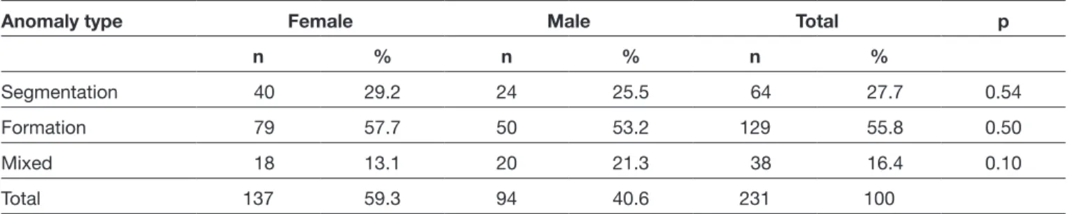
A total of 231 patients with CS were evaluated in this study. Failure of formation was seen in 129 patients (55.8%). A failure of segmentation was seen in 64 patients (27.7%). A mixed defect was seen in 38 patients (16.5%). The sex distributions by anomaly type and a comparison of their frequencies are presented in Table I. There was no statistically significant difference between genders or CS types.
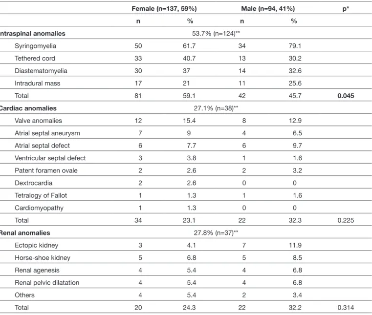
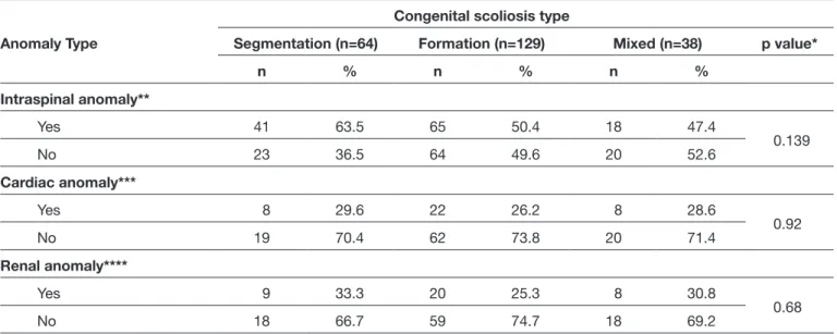
All patients underwent MRI evaluation for a potential intraspinal pathology. An intraspinal pathology was detected in 124 patients (53.7%). The most common intraspinal anomaly among CS patients was syringomyelia followed by the tethered cord, diastematomyelia, and intradural mass (Table II). An intraspinal pathology was found in 45.7% of male patients and 59.1% of female patients. The distributions of intraspinal anomalies in male and female patients are shown in Table II. The incidence of intraspinal pathology was significantly higher in female patients (p=0.045). The distribution of intraspinal anomalies was similar to that of vertebral anomalies (p=0.14) (Table III).
An echocardiography was performed in 140 (60.6%) of 231 patients. Congenital heart disease was detected on echocardiography in 38 of 140 patients (27.1%). The most common cardiac pathologies were valve-related pathologies (mitral valve prolapse, aortic valve insufficiency, bicuspid aortic valve, aortic valve stenosis, pulmonary valve insufficiency), followed by atrial septal defect, patent foramen ovale, dextrocardia, and cardiomyopathy (Table II). Congenital heart disease was found in 32.3% of male patients and 23.1% of female patients. There was no statistically significant difference between male and females in the incidence of cardiac pathology (p=0.225). According to scoliosis type, cardiac pathology was found in 29.6% of patients with failure of segmentation, 26.2% of patients with failure of formation, and 28.4% of patients with a mixed type. According to vertebral anomalies, the distribution of cardiac anomaly was similar (p=0.92) (Table III).
A renal USG was performed in 133 of 231 patients. Congenital renal disease was found in 37 of 133 (27.8%) patients. The most common renal pathology was ectopic kidney and horseshoe kidney, followed by renal agenesis, renal pelvic dilatation, and other urogenital diseases such as cystic kidney, hydronephrosis, and ectasia. Congenital renal disease was found in 32.2% of male patients and 24.3% of female patients
Table I: Distribution of Vertebral Anomaly Types in Female and Male Patients
Anomaly type Female Male Total p
n % n % n %
Segmentation 40 29.2 24 25.5 64 27.7 0.54
Formation 79 57.7 50 53.2 129 55.8 0.50
Mixed 18 13.1 20 21.3 38 16.4 0.10
Total 137 59.3 94 40.6 231 100
(Table II). There was no significant difference between males and females in the incidence of renal pathology (p=0.92). According to scoliosis type, urogenital pathology was found in 33.3% of patients with failure of segmentation, 25.3% of patients with failure of formation, and 30.8% patients with the mixed type. The distribution of renal anomalies was similar to that of vertebral anomalies (p=0.68) (Table III). The Arnold Chiari malformation was detected in 19 of 231 patients (8.2%). There was no statistically significant difference between gender groups (p=0.357).
Figure 1 shows the relationship between intraspinal, cardiac, and renal anomalies detected in patients with CS who were evaluated with CT-scan, MRI, echocardiography, and renal
USG. In 22 of 67 (32.8%) patients with an intraspinal anomaly, an additional cardiac anomaly was detected. In 27 of 67 (40.3%) patients with an intraspinal anomaly, an additional renal anomaly was detected. In 18 of 38 (47.3%) patients with a cardiac anomaly, an additional renal anomaly was detected. In 15 of 133 patients (11.2%), intraspinal, cardiac, and renal anomalies were identified.
█
DISCUSSION
Spinal deformity in CS is the visible part of the iceberg. Intraspinal anomalies and other organ defects are frequent (16). In our study, we evaluated the incidences and interrelationships of intraspinal, cardiac, and renal anomalies. The incidence
Table II: Distribution of Intraspinal, Cardiac, and Renal Anomaly Types in Female and Male Patients
Female (n=137, 59%) Male (n=94, 41%) p* n % n % Intraspinal anomalies 53.7% (n=124)** Syringomyelia 50 61.7 34 79.1 Tethered cord 33 40.7 13 30.2 Diastematomyelia 30 37 14 32.6 Intradural mass 17 21 11 25.6 Total 81 59.1 42 45.7 0.045 Cardiac anomalies 27.1% (n=38)** Valve anomalies 12 15.4 8 12.9
Atrial septal aneurysm 7 9 4 6.5
Atrial septal defect 6 7.7 6 9.7
Ventricular septal defect 3 3.8 1 1.6
Patent foramen ovale 2 2.6 2 3.2
Dextrocardia 2 2.6 0 0 Tetralogy of Fallot 1 1.3 1 1.6 Cardiomyopathy 1 1.3 0 0 Total 34 23.1 22 32.3 0.225 Renal anomalies 27.8% (n=37)** Ectopic kidney 3 4.1 7 11.9 Horse-shoe kidney 5 6.8 5 8.5 Renal agenesis 4 5.4 4 6.8
Renal pelvic dilatation 4 5.4 4 6.8
Others 4 5.4 2 3.4
Total 20 24.3 22 32.2 0.314
*p values indicate the comparisons of the distrubution of intraspinal, cardiac, and renal anomaly frequency between the genders.
**An intraspinal pathology was detected 53.7% (n=124) in 231 patients.
Congenital heart disease was detected 27.1% (n=38) on echocardiography in 140 patients. A congenital renal disease was found 27.8% (n=37) in 133 patients.
Previously, it was suggested that thorax abnormalities might cause a change in the heart shape and, subsequently, may lead to mitral valve prolapse in idiopathic scoliosis (7). The results of our study and the study of Liu et al. (16) support this assumption in CS patients.
The incidence of cardiac anomalies was found to be similar in patients with formation, segmentation, and mixed defects.The incidence of urogenital anomalies in our study was in line with those reported in the literature, which vary from 21% to 34% (1,4,11,13,17). Also, the incidence of urogenital anomalies was similar in patients with formation, segmentation, and mixed defects. According to our results, 57.9% of patients with cardiac anomaly had an additional intraspinal anomaly. This is in contradiction with Liu et al. (16), who previously reported of intraspinal anomalies was found to be 53.7%, which was
higher than previous studies (1,4-6,18,21). Syringomyelia (67.7%) was the most common type of intraspinal anomaly in this study, which was different from the results of previous studies (1,4-6,19,21,22).
The incidence of intraspinal anomaly was similar in patients with formation, segmentation, and mixed defects. Our results are in contradiction with Basu et al. (1), and Shen et al. (23). The incidence of cardiac anomalies was found to be 27.1% (38 of 140), which was in line with the study of Basu et al. (1). This incidence was higher than reported in most of the previous studies (3,5,22). Valve-related pathologies (14.3%) (MVP being the most common) were found to be the most common type of cardiac anomaly. Liu et al. reported similar results (16).
Table III: Distribution of Intra-Spinal, Cardiac, and Renal anomalies according to Congenital Scoliosis Types Anomaly Type
Congenital scoliosis type
Segmentation (n=64) Formation (n=129) Mixed (n=38) p value*
n % n % n % Intraspinal anomaly** Yes 41 63.5 65 50.4 18 47.4 0.139 No 23 36.5 64 49.6 20 52.6 Cardiac anomaly*** Yes 8 29.6 22 26.2 8 28.6 0.92 No 19 70.4 62 73.8 20 71.4 Renal anomaly**** Yes 9 33.3 20 25.3 8 30.8 0.68 No 18 66.7 59 74.7 18 69.2
*p values indicates the comparisons of the frequencies of the intraspinal, cardiac, and renal anomaly between the congenital scoliosis types.
n: numbers.
Figure 1: Interrelationships of the anomalies in congenital scoliosis. A whole-spine computed tomography scan with three-dimensional reconstructions, magnetic resonance imaging, echocardiography, and renal ultrasonography evaluation was performed to identify potential intraspinal, cardiac, and renal anomaly in 133 of 231 patients.
7. Dhuper S, Ehlers KH, Fatica NS, Myridakis DJ, Klein AA, Friedman DM, Levine DB: Incidence and risk factors for mitral valve prolapse in severe adolescent idiopathic scoliosis. Pediatr Cardiol 18(6):425–428, 1997
8. Giampietro PF: Genetic aspects of congenital and idiopathic scoliosis. Scientifica 2012:152365, 2012
9. Giampietro PF, Blank RD, Raggio CL, Merchant S, Jacobsen FS, Faciszewski T, Shukla SK, Greenlee AR, Reynolds C, Schowalter DB: Congenital and idiopathic scoliosis: Clinical and genetic aspects. Clin Med Res 1(2):125–136, 2003 10. Giampietro PF, Raggio CL, Blank RD, McCarty C, Broeckel
U, Pickart MA: Clinical, genetic and environmental factors associated with congenital vertebral malformations. Mol Syndromology 4(1-2):94–105, 2013
11. Guerrero G, Saieh C, Dockendorf I, Diaz V: Genitourinary abnormalities in children with congenital scoliosis. Rev Chil Paediatr 60(5):281–283, 1989
12. Hedequist D, Emans J: Congenital scoliosis. A review and update. J Pediatr Orthop 27:106–116, 2007
13. Hensinger R, Lang JE, MacEwen GD: Klippel-Feil syndrome: A constellation of associated abnormalities. J Bone Joint Surg Am 56:1246–1250, 1974
14. Hensinger RN: Congenital scoliosis: Etiology and associations. Spine 34(17):1745–1750, 2009
15. Li Z, Yu X, Shen J: Environmental aspects of congenital scoliosis. Environ Sci Pollut Res Int 22(8):5751–5755, 2015 16. Liu YT, Guo LL, Tian Z, Zhu WL, Yu B, Zhang SY, Qiu GX:
A retrospective study of congenital scoliosis and associated cardiac and intraspinal abnormities in a Chinese population. Eur Spine J 20(12):2111–2114, 2011
17. MacEwen GD, Winter RB, Hardy JH: Evaluation of kidney abnormalities in congenital scoliosis. J Bone Joint Surg Am 54:1451–1455, 1972
18. McMaster MJ: Occult intraspinal anomalies and congenital scoliosis. J Bone Joint Surg Am 66:588–601, 1984
19. Prahinski JR, Polly DW Jr, McHale KA, Ellenbogen RG: Occult intraspinal anomalies in congenital scoliosis. J Pediatr Orthop 20(1):59–63, 2000
20. Rai AS, Taylor TKF, Smith GHH, Cumming RG, Plunkett-Cole M: Congenital abnormalities of the urogenital tract in association with congenital vertebral malformations. J Bone Joint Surg Br 84(6):891–895, 2002
21. Rajasekaran S, Kamath V, Kiran R, Shetty AP: Intraspinal anomalies in scoliosis: An MRI analysis of 177 consecutive scoliosis patients. Indian J Orthop 44(1):57–63, 2010
22. Sear JW: Kidney dysfunction in the postoperative period. Br J Anesth 95(1):20-32, 2005
23. Shen J, Wang Z, Liu J, Xue X, Qiu G: Abnormalities associated with congenital scoliosis. A retrospective study of 226 Chinese surgical cases. Spine (Phila Pa 1976) 38(10):814–818, 2013 24. Winter RB: Congenital spinal deformity. In: Lonstein JE (ed)
Moe’s Textbook of Scoliosis and Other Spinal Deformities, 3rd ed. Philadelphia: W.B. Saunders Co., 1995:257–294
rare presentation of both cardiac and intraspinal anomalies in the same patient with CS.
Our study described high incidences of additional anomalies in patients with cardiac, renal, and intraspinal anomalies.These have not been described previously. In this study, intraspinal anomalies and congenital heart disease were found to be associated with congenital renal anomalies. More than half of the patients with the Arnold Chiari malformation had an additional intraspinal anomaly. The incidence of all intraspinal, cardiac, and renal anomalies was found to be 11.2% (15 of 133 patients). This has not been described previously. High incidences of all three anomalies in the Turkish population may be associated with genetic or environmental factors. Detailed investigations on this topic are needed.
Cardiac and renal anomalies bring intra- and postoperative risks. Excessive correction in the presence of advanced thoracic deformities and cardiac pathologies may cause sudden cardiopulmonary decompensation (7). Also, intra- and postoperative metabolic disorders may develop in patients with congenital renal anomalies (22).
There are some limitations of this study. First, this is a retrospective study. Second, not all patients underwent renal and cardiac examination; therefore, bias may have occurred in the statistical analysis. The lack of genetic analysis and detailed patient histories are additional limitations.
█
CONCLUSION
For patients reporting a congenital anomaly, surgeons should be alert to evaluate additional anomalies in CS. Because cardiac and renal anomalies increase intra- and postoperative complication risks, a careful and comprehensive preoperative evaluation is needed.
█
REFERENCES
1. Basu PS, Elsebaie H, Noordeen MH: Congenital spinal deformity: A comprehensive assessment at presentation. Spine (Phila Pa 1976) 27(1):2255–2259, 2012
2. Batra S, Ahuja S: Congenital scoliosis: Management and future directions. Acta Orthop Belg 74(2):147–160, 2008 3. Beals RK, Robbins JR, Rolfe B: Anomalies associated with
vertebral malformations. Spine(Phila Pa 1976) 18(10):1329– 1332,1993
4. Bernard TN Jr, Burke SW, Johnston CE III, Roberts JM: Con-genital spine deformities: A review of 47 cases. Orthopaedics 8(6):777–783, 1985
5. Bollini G, Launay F, Docquier PL, Viehweger E, Jouve JL: Congenital abnormalities associated with hemivertebrae in relation to hemivertebrae location. J Pediatr Orthop B 19(1):90–94, 2010
6. Bradford DS, Heithoff KB, Cohen M: Intraspinal abnormalities and congenital spine deformities: A radiographic and MRI study. J Paediatr Orthop 11:36–41, 1991