Ibrahim Devrim GURSOY
1, Sureyya BARUN
1, Saban Remzi ERDEM
2, Ulya KESKIN
2, Murat KIZILTAS
1,
Pergin ATILLA
3, Sevda MUFTUOGLU
3, Deniz YUCE
4, Firat NARIN
5, Mert ERTUNC
6, Yildirim SARA
6,
Hande CANPINAR
71Gazi University, School of Medicine, Department of Pharmacology, Ankara, Turkey 2Baskent University, School of Medicine, Department of Pharmacology, Ankara, Turkey
3Hacettepe University, School of Medicine, Department of Histology and Embryology, Ankara, Turkey 4Hacettepe University, School of Medicine, Department of Preventive Oncology, Ankara, Turkey 5Hacettepe University, Institute of Neurological Sciences and Psychiatry, Ankara, Turkey 6Hacettepe University, School of Medicine, Department of Pharmacology, Ankara, Turkey 7Hacettepe University, School of Medicine, Department of Basic Oncology, Ankara, Turkey
This study has been presented at the 29th International Epilepsy Congress between 28th August and 1st September 2011 at Rome, Italy
Investigation of the Possible Protective Effects of Ketamine
and Dantrolene on the Hippocampal Apoptosis and Spatial
Learning in Rats Exposed to Repeated Electroconvulsive
Seizures as a Model of Status Epilepticus
ABSTRACT
AIM: To evaluate the possible neuroprotective effects of ketamine and dantrolene on the hippocampal apoptosis and spatial learning in rats exposed to repeated electroconvulsive seizures (ECS) as a model of status epilepticus (SE).
MATERIAL and METHODS: Twenty-four rats were assigned to 4 groups. 1st Group was Sham. 2nd Group was ECS: ECS was induced by ear electrodes via electrical stimulation. The same ECS protocol was applied to the 3th and 4th Groups which received ketamine (40 mg/kg s.c.) or dantrolene (5 mg/kg i.p.) 1 h before each ECS, respectively. Following 30 days of recovery, the cognitive status of the animals was evaluated via Morris Water Maze (MWM). The same experimental protocol was repeated 14 days afterward to evaluate the retention of the memory. Hippocampal apoptosis was examined in corresponding experimental groups.
RESULTS: All the animals in four groups learned the task with no significant difference between groups in MWM. The ECS+ketamine group showed memory impairment 14 days afterward. ECS+dantrolene group was not different from controls. ECS caused long term apoptotic processes in dentate gyrus (DG) and non-apoptotic neuronal injury in CA1 and CA2.
CONCLUSION: Dantrolene and ketamine inhibited apoptosis and showed neuroprotective effects. Although ketamine and dantrolene inhibited ECS-induced apoptosis and non-apoptotic injury, they did not produce similar effects on memory retention. It will be warranted to evaluate cognitive dysfunction by taking into consideration the other factors in addition to apoptosis and neurodegenerative changes.
KEYWORDS: Status epilepticus, Electroconvulsive seizure, Ketamine, Dantrolene, Cognition, Rats Corresponding author: Sureyya BARUN
[email protected]Firat NARIN : 0000-0002-5985-4460 Mert ERTUNC : 0000-0002-1323-3722 Yildirim SARA : 0000-0003-1079-0782 Hande CANPINAR : 0000-0002-8973-6289 Ibrahim Devrim GURSOY : 0000-0001-8735-321X
Sureyya BARUN : 0000-0003-3726-8177 Remzi ERDEM : 0000-0002-7537-2170 Ulya KESKIN : 0000-0003-2563-4993 Murat KIZILTAS : 0000-0002-0207-4711 Pergin ATILLA : 0000-0001-5132-0002 Sevda MUFTUOGLU : 0000-0003-0664-0204 Deniz YUCE : 0000-0003-0725-5447 Received: 26.08.2019 Accepted: 23.03.2020 Published Online: 24.07.2020
Original Investigation
DOI: 10.5137/1019-5149.JTN.27023-19.3█
INTRODUCTION
S
ince memory-relevant structures are frequently involved during status epilepticus (SE), learning and memory functions may be impaired. Cognitive dysfunction has been shown in most patients to be associated with neuronal loss in hippocampus. SE induced by various agents including kainic acid, pilocarpine or intracranial electrical stimulation in animals also lead to neuronal loss in hippocampus associated with cognitive impairment (6,26,36,41,70). However, it is unknown whether cognitive impairment results from the loss of memory processing neurons or increased activity-induced plasticity that inhibits normal memory formation (4,27). Morris Water Maze (MWM) test is most widely used paradigms for testing visual-spatial learning and memory in rats (8). Hippocampal lesions are associated with impairments in spatial learning and memory (1,57).Various studies show that apoptotic pathways are activated due to repeated seizure activity (29). It has been clarified that prolonged seizures lead to apoptotic DNA fragmentation and DNA laddering in rat brains (17,61). Loss of Ca2+ homeostasis
produces apoptosis by promoting mitochondria-mediated caspase activation (58).
Glutamate overflow is playing a pivotal role in epileptic seizure, status epilepticus or seizure induced neuronal damage. Glutamate-induced NMDA receptor activation increases Ca2+
influx and leads to cell necrosis and apoptosis (46). Ketamine is a non-competitive glutamate NMDA receptor antagonist and although controversial studies, it has been shown to have anti-apoptotic and neuroprotective properties (12,33,34,64). Calcium, in particular, intracellular Ca2+ plays a crucial role in
the development of neuronal injury (19). Significant Ca2+ efflux
from the endoplasmic reticulum (ER) induces the expression of stress molecules and protein misfolding, triggering apoptosis (35,80). Dantrolene, which is a specific ryanodine receptor (RYR) antagonist, may protect neurons from disruptions in Ca2+-homeostasis by inhibiting Ca2+ release from the ER (62).
Therefore, it has recently been used to diminish cell death resulting from ischemia, spinal cord injury and epileptic seizures (47,60,75).
Electroconvulsive seizure (ECS) is used as a model to induce SE in experimental animals (18). In previous studies, repeated ECS treatments impaired the spatial learning and gave rise to a number of morphological and functional changes in the hippocampus of rats (23,43,67). Although ECS does not lead to remarkable neuronal loss in hippocampus, apoptotic changes have not been investigated in this model in correlation with cognitive deficits (23,65).
In this study, the aim was to investigate the possible neuroprotective effects of ketamine and dantrolene on hippocampal apoptosis and cognitive functions in rats exposed to repeated ECS applications. Spatial learning and memory were tested via MWM.
Clarifying the underlying mechanisms in repeated SE-induced cognitive impairment will facilitate our understanding of the pathophysiology. Thus, it might be possible that new
treatment strategies could be developed against SE-induced brain injury.
█
MATERIAL and METHODS
Experimental Design
This study was performed according to the Helsinki Declaration about animal studies. The experiments were conducted in accordance with Guide for the Care and Use of Laboratory Animals Eighth Edition (NIH publications). Gazi University Scientific Research Committee granted the study with project number 01/2010-70. Baskent University Ethical Committee approved the protocol on 08.19.2010 with approval number DA 10/18.
A total of 48 Wistar rat, which were 2 months of age were included in 8 groups in the study, as 6 rats in each group. The rats were exposed to standard conditions of 20-22 °C of temperature, and 12 hours of light-darkness cycle. Food and water were not restricted. Twenty-four rats were randomly assigned to the following 4 study groups in order to evaluate spatial learning and memory through the MWM test:
1. Control Group (Control): Ear electrodes were applied for 30 seconds without stimulation.
2. ECS Group: Stimulation was applied to rats by ear electrodes as 20 Volts and for 2 seconds, which supplied by May STPT 05 Research Stimulator (maximum energy 150 Volts) equipment. ECS treatment was repeated every other day for 5 times, and after 2 hours of the last application, ECS stimulus was administered and the total number of ECSs was completed to 6 cycles. This protocol was adapted from a previous study (65). The rats were kept for 1 month for recovery.
3. Ketamine+ECS (Ketamine) Group: Ketamine was applied to rats 1 hour prior to each application of ECS at sub-anesthetic doses (40 mg/kg s.c.).
4. Dantrolene+ECS (Dantrolene) Group: Dantrolene was applied to rats 1 hour prior to each application of ECS (10 mg/kg i.p.).
Chemicals were not administered before the 6th ECS treatment.
Following 30 days of recovery, spatial learning and memory were tested via MWM.
MWM Protocol
Animals were evaluated for cognitive functions and memory by the MWM test at Baskent University Animal Laboratory. A black and cylindrical tank was used for the experiments. The tank was filled with water of 50 cm in-depth, and the temperature was stabilized to 22±1°C. It was also surrounded by a screen and isolated from the environmental stimulants. The water tank was divided into four quadrants by fictitious lines. The escape platform was placed in the same quadrant at a depth of 2.5 cm from the surface. A video camera was placed on the top of the tank and connected to a computer that runs an image processing software (53).
All rats were trained two times a day for 4 days, and evaluated during the remaining 5 days (between 1st and 5th days). At the
trainings, rats were placed facing to the wall of the tank, and the starting point was randomized for four quadrants. Rats were allowed for exploring the tank for finding the escape platform, and the ones that could not find the platform were assisted. The rats were kept on the platform for 15 seconds for forming the memory. On the 19th day, MWM experiments
were repeated to test the retention of the memory.
The parameters evaluated at the MWM experiments were as follows:
1. Escape Latency Time (ELT)(time to finding of escape platform, seconds)
2. Mean ELT (arithmetic mean of ELT values for north, south, east and west)
3. Swimming speed (cm/sec)
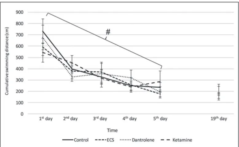
4. Cumulative swimming distance (cm)
Apoptosis Group
Twenty-four rats in the apoptosis group were assigned to 4 groups including 6 rats in each group (Control, ECS, ECS+Ketamine, ECS+Dantrolene). These rats were scarified under thiopental anesthesia (20 mg/kg) at the end of the 30th
day without subjecting to MWM experiments, and the TUNEL method was used for evaluation of the morphological and temporal characteristics of apoptosis in the hippocampus.
Histological Evaluation and Determination of Apoptosis
Rats were sacrificed by decapitation after thiopental sodium administration (20 mg/kg, Pental Sodyum, IE Ulagay, Istanbul, Turkey). Hippocampi were removed and alternatively sampled from the left and right sites.
The hippocampi samples of control, ECS, ECS+dantrolene and ECS+ketamine groups were fixed in 10% buffered formalin for 72 hours (h), processed according to routine light microscopy method and embedded in paraffin. The serial sections of 5 mm were stained with hematoxylin-eosin for histological evaluations, and TUNEL for determining the apoptosis. All samples were evaluated by Leica DM6000B light microscope, and the images were transferred to electronic media by DC 500 Leica digital camera.
The transverse sections of 5 mm were obtained by VSLM1 “vibroslice vibrating slicer” device in oxygenated dissection solution at freezing temperature (low Ca2+-high Mg2+ artificial
cerebrospinal fluid; ACSF), and incubated at 34 °C for 30 minutes. The sections were kept in ACSF containing 124 mM NaCl, 5 mM KCl, 12 mM NaH2PO4, 26 mM NaHCO3, 10 mM
d-glucose, 2 mM CaCl2, 1 mM MgCl2, and 95% O2 and 5%
CO2 for at least 60 minutes at room temperature.
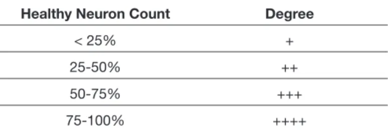
The healthy and contracted neurons with basophilic cytoplasm were counted in CA1, CA2 and dentate gyrus regions at 40x magnification and 5 fields, by 2 histologists blinded to the study groups, and were evaluated according to the criteria showed in Table I.
Table I: Histological Neuronal Evaluation Criteria Healthy Neuron Count Degree
< 25% +
25-50% ++
50-75% +++
75-100% ++++
TUNEL (+) cells were counted at 40x magnification in all samples stained with TUNEL method, and regional apoptotic indexes were calculated [TUNEL (+) cell count/100 cells].
Statistical Analyses
The analyses were performed with SPSS 18.0 (IBM Inc., USA) software. Qualitative data were presented with frequencies, and quantitative data were presented with means and standard deviations. Statistical comparisons were performed by using repeated- measures ANOVA for the dependent groups, and by using one-way ANOVA for the independent groups. The statistical significance level of Type-I error was 5% in the study.
█
RESULTS
The Results of MWM Experiments
Mean ELT
All the animals in 4 experimental groups learned the platform finding task. The change percent of mean ELT values was calculated for 5 days. The 2nd, 3rd, 4th and 5th days values
were significantly lower than 1st day values. There were no
significant differences between the mean ELT of four study groups for each day for 5 days. For determining the retention in each group, mean ELT values were calculated for the 19th day
and compared with the 5th day mean ELT values. The analyses
revealed that the ECS+ketamine application inhibited the progression of memory retention in rats on the 19th day. And,
ECS+dantrolene application was found to have no negative effects on spatial learning and memory retention (Figure 1).
Cumulative swimming distance
The cumulative swimming distances were gradually decreased in all groups. But, the differences between groups were not statistically significant. These results are in accordance with the mean ELT values (Figure 2).
Swimming speed
The swimming speed was not significantly differed between groups during 5 days. Ketamine significantly decreased the swimming speed on the 19th day (Figure 3).
Histological Evaluation and Determination of Apoptosis
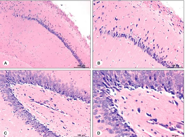
Control group, Hematoxylin-eosin staining
Mild edema, which was more significant in the neuropil, was observed below piamater in all samples of the electroshock-free control group. Also, edema was observed in the basal
margin of granular neuron groups in dentate gyrus adjacent to the CA4 region, and around vessels in hippocampus.
There were basophilic and condensed neurons in hippocampus, which are more significant in some fields. Some neurons were contracted and basophilic viewed between healthy neurons in CA1 and CA3 regions. In the dentate gyrus region, most of the granular neurons were normal, but some dense neurons with basophilic cytoplasm were significant (Figure 4A-D).
Control group, TUNEL staining
In one sample in the control group, TUNEL (+) neurons were determined in a particular field of the CA1 region. None of the remaining samples revealed TUNEL (+) neurons in the CA1 and CA2 regions. There were no TUNEL (+) neurons in dentat gyrus regions of the samples (Figure 5A-D).
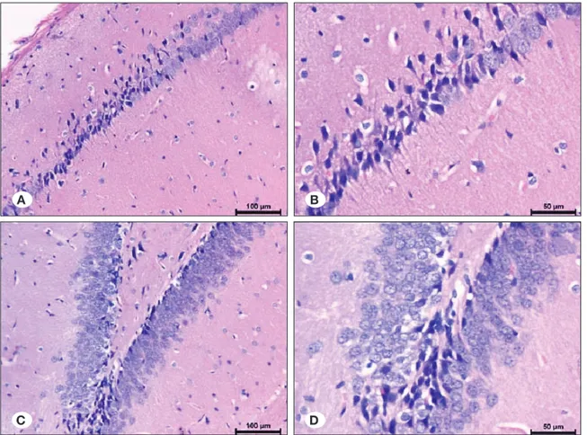
ECS group, Hematoxylin-eosin staining
Mild edema was observed in neuropil, particularly significant below piamater. Edema was also observed around the pyknotic neurons with basophilic condensed cytoplasm in the CA1-4 regions and the vessels of Ammon’s horn. There was mild stasis in the vessels.
The pyknotic and basophilic cytoplasm neurons were more than normal structured pyramidal neurons in localized fields in the CA1 and CA2 regions. The numbers of pyknotic and basophilic cytoplasm neurons were lower in the CA1 region, and increased through CA2. In the dentate gyrus, there were pyknotic and basophilic neurons especially in the terminal regions, along with the granular normal neurons (Figure 6A-D).
ECS group, TUNEL staining
Few numbers of TUNEL (+) neurons were determined between the pyknotic neurons and healthy neurons in CA1 and CA2 regions. TUNEL (+) cells were more in some localized fields through CA2. There were more TUNEL (+) neurons than the CA1 and CA2 regions, between healthy and pyknotic neurons in terminal parts of the dentate gyrus (Figure 7A-D).
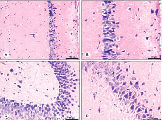
ECS+ketamine group, Hematoxylin-eosin staining
There was mild edema in the neuropil, particularly significant below piamater, similar to control, ECS and dantrolene groups. There was also mild edema around some vessels. There was no stasis in most of the vessels.
Most of the neurons in the CA1 and CA2 regions were healthy, and there were limited number of neurons with basophilic cytoplasm between the normal neurons. These were not degenerated neurons. Most of the granular neurons in the dentate gyrus region were also healthy. There were very few numbers of neurons with basophilic cytoplasm between them. Apart from the dantrolene group, there were no infiltrative neurons (Figure 8A-D).
ECS+ketamine group, TUNEL staining
There were rare TUNEL (+) neurons in the CA1 region in all samples, but one. And, in that sample there were more TUNEL (+) neurons in a localized field in the CA1 region. There were no TUNEL (+) neurons in the dentate gyrus region (Figure 9A-D).
Figure 1: Learning-memory curves at Morris water-maze. +: Ketamin group between day 5 and day 19; p<0.005; #: temporal change in escape latencies in all groups; p<0.005; **: ketamine and other groups (19th day); p<0.01.
Figure 2: Mean swimming distances in Morris water-maze. #: Temporal change in cumulative swimming distance in all groups; p<0.005.
Figure 3: Mean swimming speed in Morris water-maze.
**: Ketamin and other groups (day 19); p<0.005; #: Dantrolene day 5 and day 19; p<0.05; ++: Ketamine day 5 and day 19; p<0.005.
Figure 5: Distribution of apoptotic neurons in study groups. Figure 4: Distribution of healthy neurons in study groups.
A B
C D
A B
Figure 7: Distribution of TUNEL positive neurons in ECS group (Tunel method; x400, x200, x100, x200). Figure 6: Distribution of pyknotic and healthy neurons in ECS group (H&E; x100, x200, x200, x400).
A B
C D
A B
Figure 9: Distribution of neurons in ECS and Ketamine groups (TUNEL method, x100, x200, x400, x400). Figure 8: Distribution of neurons in ECS and Ketamine groups (H&E; x100, x200, x200, x400).
A B
C D
A B
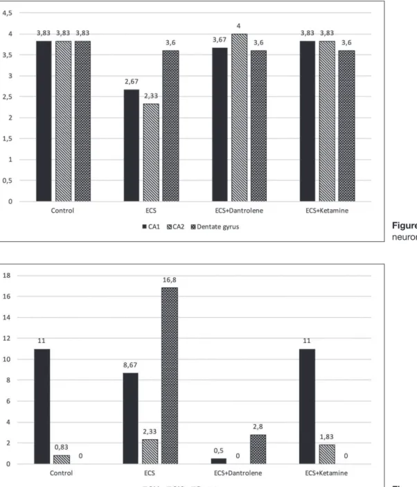
between the CA1, CA2, and dentate gyrus regions in each group, a statistically significant difference was observed only in the ECS group. Several healthy neurons were higher in the dentate gyrus (p<0.05) (Table II, Figure 12).
Distribution of healthy neurons between study groups
There were statistically significant differences for distributions of normal neurons in CA1 and CA2 regions of control, ECS, ECS+dantrolene and ECS+ketamine groups (p=0.005, p=0.001, respectively). Further analyses revealed that these differences were related to the ECS group. The distribution of healthy neurons was significantly lower than the other groups. There was no statistically significant difference in the distribution of healthy neurons in the dentate gyrus region between study groups (p>0.05) (Table II, Figure 12).
Distribution of apoptotic neurons in each study group
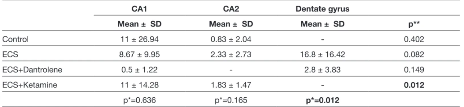
There was a statistically significant difference in the number of apoptotic cells between CA1, CA2, and dentate gyrus regions, only in the ECS+ketamine group (p=0.012). Further analyses revealed that this difference was related to the dentate gyrus region. The number of apoptotic cells was significantly higher in the dentate gyrus region of the ECS group. The antiapoptotic effect of ketamine was found to be higher than dantrolene (Table III, Figure 13).
Distribution of apoptotic neurons between study groups
There was a statistically significant difference for several apoptotic cells in the dentate gyrus region between control,
ECS+dantrolene group, Hematoxylin-eosin staining
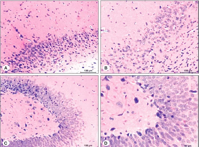
There were clefts due to edema in the neuropil, particularly below pia-mater, such as control and ECS groups. But, the edema in neuropil was relatively milder and localized, when compared to the ECS group. There was also edema around vessels like other groups. Apart from the ECS group, edema was not observed in the field through the CA4 region below the granular neurons in the dentate gyrus region, excluding only one sample. Also, apart from the ECS group, there was no edema around the pyknotic neurons.
Several of condensed neurons with pyknotic basophilic cytoplasms in the CA1 and CA3 regions of Ammon’s horn were higher than the control group, but similar to numbers of pyknotic neurons in the ECS group. Infiltrative cells were observed in two samples. And, in the dentate gyrus region, few numbers of pyknotic neurons with basophilic cytoplasm were observed between granular neurons (Figure 10A-D).
ECS+dantrolene group, TUNEL staining
In two samples, some TUNEL (+) neurons were observed in basal fields of the dentate gyrus region. There were no TUNEL (+) neurons in the the CA1 and CA2 regions, excluding one sample. Few numbers of TUNEL (+) cells in some field of dentate gyrus region were observed in two samples (Figure 11A-D).
Distribution of healthy neurons in each study group
When the distribution of healthy neurons was compared
Table II: Distribution of Healthy Neurons in CA1, CA2 and Dentate Gyrus Regions
CA1 CA2 Dentate gyrus
Mean ± SD Mean ± SD Mean ± SD p**
Control 3.83 ± 0.41 3.83 ± 0.41 3.83 ± 0.41
-ECS 2.67 ± 0.52 2.33 ± 0.52 3.6 ± 0.55 0.004
ECS+Dantrolene 3.67 ± 0.52 4 ± 0 3.6 ± 0.55 0.410
ECS+Ketamine 3.83 ± 0.41 3.83 ± 0.41 3.6 ± 0.55 0.410
p*=0.001 p*<0.001 p*=0.826
*: One-way ANOVA, **: Repeated measures ANOVA.
Table III: Distribution of Apoptotic Cells in CA1, CA2, and Dentate Gyrus Regions
CA1 CA2 Dentate gyrus
Mean ± SD Mean ± SD Mean ± SD p**
Control 11 ± 26.94 0.83 ± 2.04 - 0.402
ECS 8.67 ± 9.95 2.33 ± 2.73 16.8 ± 16.42 0.082
ECS+Dantrolene 0.5 ± 1.22 - 2.8 ± 3.83 0.149
ECS+Ketamine 11 ± 14.28 1.83 ± 1.47 - 0.012
p*=0.636 p*=0.165 p*=0.012
Figure 11: Distributon of neurons in ECS and Dantrolene groups. (TUNEL method; x200, x400, x200, x400). Figure 10: Distribution of pyknotic and healthy neurons in ECS and Dantrolene groups (H&E; x100, x200, x200, x400).
A B
C D
A B
effects (14,16,69). However, it is difficult to analyze its actions in clinical studies since clinical evidences are scarcer and more difficult since it is used in polytherapy situations (56). In this study, although spatial learning was not affected, ketamine administration before each ECS application impaired the development of memory-retention. In consistent with this study, previous studies show that ketamine impairs cognition and memory (52,56). However, the exact mechanisms are not clear.
Acute ketamine administration gives rise to impairment in spatial learning (50). A hypothesis was put forward that the hypofunction of NMDA receptors are responsible for the defi-cits in cognitive functions (10,66). In addition, acute ketamine treatment leads to a prominent increase in dopamine level ECS, ECS+dantrolene and ECS+ketamine groups (p=0.012).
Further analyses revealed that this difference was related to the ECS group (Table III, Figure 13).
█
DISCUSSION
Epilepsy is associated with co-morbidities including learning and memory impairment (20,21). Although cognitive impair-ment is the most debilitating comorbidity, current treatimpair-ment strategies do not contribute to improvement in the quality of life (49).
Ketamine is a non-competitive NMDA receptor antagonist and acts by binding to phencyclidine site (3,59). It has been shown that ketamine exhibits antiepileptic and neuroprotective
Figure 12: Distribution of healthy neurons in study groups.
Figure 13: Distribution of apoptotic neurons in study groups.
(47,60,75). Apoptosis may also be triggered by the Ca2+
released from ER (72). It has been shown that dantrolene may produce neuroprotective effects by inhibition of apoptosis and neuronal cell death through blockade of Ca2+ release from ER
(11,30,40,79).
In this study, dantrolene produced no harmful effect on spatial learning and memory retention. In addition, dantrolene abolished ECS-induced tonic-clonic muscle contractions. Although, dantrolene was also tested in status epilepticus in human and conflicting results were obtained (31,74), furher studies are required to take the advantage of this agent. Thus, dantrolene appears to be worthy of testing in excitotoxic conditions as a neuroprotective agent.
ECS treatment led to apoptosis in dentate gyrus and non-apoptotic neuronal injury in CA1 and CA1 regions in this study. Both ketamine and dantrolene inhibited apoptosis in dentate gyrus. Additionally, they showed neuroprotective effcets in CA1 and CA2. ECS-induced neurodegenerative changes are consistent with other studies performed with different experimental epilepsy models (43,61,70). Neuronal loss or neurodegenerative alterations in hippocampus are associated with cognitive dysfunctions (9,68,73). However, the functions of all hippocampal regions have not been identified precisely (39). Nevertheless, it is known that synchronization of the neurons in dentate gyrus plays a critical role in memory retention and carries the memory from the entorhinal cortex to CA3 (8,76). In the present study, it was observed that ECS induces long term apoptotic processes. However, the antiapoptotic properties of ketamine and dantrolene did not lead to the parallel effects on memory retention. Therefore, it is not possible to explain the hippocampus-based cognitive dysfunctions by apoptotic or non apoptotic neuronal loss mechanisms. It will be warranted to evaluate the physiopathology by taking into consideration the aforementioned factors in addition to neuronal loss or neurodegenerative changes.
█
CONCLUSION
In the present study, ketamine led to a decline in swimming rate which is associated with the memory-retention deficits in animals. This result may be due to major depression or motor deficit. In favour of the major depression, repeated use of ketamine has been shown to reduce the function of the prefrontal dopaminergic system and compensatory upregulation of presynaptic D1 receptors (55). Since there is no report about the ketamine-related motor deficit in literature, a decline in swimming rate seems to result from major depression. However, it needs furher investigation.
█
REFERENCES
1. An L, Zhang T: Vitamins C and E reverse melamine-induced deficits in spatial cognition and hippocampal synaptic plasticity in rats. Neurotoxicology 44:132-139, 2014
2. Andrade C, Thyagarajan S, Singh NM, Vinod PS, Sanjay Kumar Rao N, Chandra JS: Celecoxib as an in vivo probe of cyclooxygenase-2 mechanisms underlying retrograde amnesia in an animal model of ECT. J Neural Transm 115: 1063-1070, 2008
and activation of D1/D5 receptor activation inhibits long-term potentiation (LTP) (15,38,42,48). Another effect of ketamine is to produce disinhibition on glutamatergic neurons, resulting in an increase in the release of glutamate (5,42,48,83). Increased glutamate activates the post-synaptic non-NMDA receptors that trigger metabotropic glutamate receptors (mGluR)-de-pendent long-term depression (LTD) (22,82). It seems that these results account for the ketamine-induced cognitive dysfunction in acute administration. However, it is not pos-sible to explain ketamine-induced memory retention on the long-term basis observed in the present study based upon the hypofunction of NMDA receptors and the inhibitory effect of dopamine on LTP via D1/D5 receptors.
Since there are controversial reports on the effects of ketamine on cognitive functions on a long run, it is not clear whether it specifically affects the consolidation stage of the memory (51). It is known that LTD plays a critical role in both the formation and retention of memory as well as LTP (7,54). Impairment in hippocampus-dependent memory retention occurs due to LTD formation in the synapses between Schaffer collaterals and CA1 region. AMPA receptor internalization is absolutely necessary for the formation of LTD (25,81). In consistent with the present study, it has been shown that ketamine causes memory-retention deficit through induction of LTD in Schaffer-CA1 synapses (15). Taken these together, impairment in memory-retention on a long-term basis in this study is most likely due to LTD formation resulting from NMDA receptor internalization in Schaffer-CA1 synapses. In favour of this study, although ketamine-induced LTD may last by 24 h, impairment in memory retention may last for weeks (45). Protein synthesis and gene expressions may also take place in the mechanisms of ketamine (44).
Additionally, ketamine impairs the consolidation stage of the long-term memory, at least in part by preventing a learning-induced increase in BDNF levels in hippocampus (24,28,37). Accordingly, it can impair the cell morphology in immature GABAergic neurons, leading to interaction with the build-up of neural networks in the brain (77,78). Therefore, it is feasible to speculate that activity-induced plasticity may also be another explanation for the ketamine-induced impairment in memory retention in this study.
The most important result obtained from the present study is that ketamine impaired the memory-retention in an excitotoxic condition due to the ECS treatments (2). However, in a previous study, when it was administered alone for 6 months (30 mg/ kg), cognitive dysfunction was not observed (71). Although acute ketamine administration shows the neuroprotective effect (12,64), our results suggest that ketamine use in excitotoxic conditions may be harmful on a long-term basis. Further studies are required to clarify the exact mechanisms. Calcium released from intracellular stores has a role in neuronal injury (19). Dantrolene is an RYR antagonist and approved for the treatment of malign hyperthermia, malign hyperpyrexia and neuroleptic malign syndrom (13,32,63). In various studies, dantrolene exhibits neuroprotection againts spinal cord ischemia, stroke and epilepsy induced neuronal injuries via inhibition of Ca2+-induced Ca2+ release from ER
20. Friedman A, Behrens CJ, Heinemann U: Cholinergic dysfunction in temporal lobe epilepsy. Epilepsia 48:126-130, 2007
21. Gaitatzis A, Carroll K, Majeed A, Sander W: Epidemiology of the comorbidity of epilepsy in the general population. Epilepsia 45:1613-1622, 2004
22. Gean PW, Chang FC, Yi PL, Lin JH, Tsai JJ: Activation of metabotropic glutamate receptors in conjuction with postsynaptic depolarization triggers a long-term depression of the N-methyl-D-aspartate receptor-mediated synaptic potential in the rat hippocampus. J Biomed Sci 2:166-173, 1995
23. Gombos Z, Spiller A, Cottrell GA, Racine RJ, Burnham WM: Mossy fiber sprouting induced by repeated electroconvulsive shock seizures. Brain Res 844:28-33, 1999
24. Goulart BK, De Lima MNM, De Farias CB, Reolon GK, Almeida VR, Quevedo J, Kapczinski F, Schröder N, Roesler R: Ketamine impairs recognition memory consolidation and prevents learning-induced increase in hippocampal brain-derived neurotrophic factor levels. Neuroscience 167:969-973, 2010
25. Han J, Kesner P, Metna-Laurent M, Duan T, Xu L, Georges F, Koehl M, Abrous DN, Mendizabal-Zubiaga J, Grandes P, Liu Q, Bai G, Wang W, Xiong L, Ren W, Marsicano G, Zhang X: Acute cannabinoids impair working memory through astroglial CB1 receptor modulation of hippocampal LTD. Cell 148:1039-1050, 2012
26. Hannesson DK, Howland J, Pollock M, Mohapel P, Wallace AE, Corcoran ME: Dorsal hippocampal kindling produces a selective and enduring disruption of hippocampally mediated behaviour. J Neurosci 21:4443-4450, 2001
27. Hauser WA: Status epilepticus: Frequency, etiology, and neurological sequelae. Adv Neurol 34:3-14, 1983
28. Heldt SA, Stanek L, Chhatwal JP, Ressler KJ: Hippocampus-specific deletion of BDNF in adult mice impairs spatial memory and extinction of aversive memories. Mol Psychiatry 12:656-670, 2007
29. Henshall DC, Simon RP: Epilepsy and apoptosis pathways. J Cereb Blood Flow Metab 25:1557-1572, 2005
30. Hernandez-Fonseca K, Massieu L: Disruption of endoplasmic reticulum stores is involved in neuronal death, induced by glycolysis inhibition in cultured hippocampal neurons. J Neurosci Res 82:196-205, 2005
31. Hiasa A, Sasaki R, Takeuchi T, Tomimoto H: A case of status epilepticus with a possible therapeutic effect by dantrolene sodium. Rinsho Shinkeigaku 51:777-780, 2011
32. Horni G, Meirik K, Lund MB: Neuroleptic malign syndrome in a patient treated with olanzapine. Tidsskr Nor Laegeforen 123: 2867-2869, 2003
33. Jevtovic-Todorovic V, Benshoff N, Olney JW: Ketamine potentiates cerebrocortical damage induced by the common anaesthetic agent nitrous oxide in adult rats. Br J Pharmacol 130:1692-1698, 2000
34. Jevtovic–Todorovic V, Wozniak DF, Benshoff ND, Olney JW: A comparative evaluation of the neurotoxic properties of ketamine and nitrous oxide. Brain Res 895:264-267, 2001 3. Anis NA, Berry SC, Burton NR, Lodge D: The dissociative
anesthetics, etamine and phencyclidine, selectively reduce excitation of central mammalian neurons by N-methyl-aspartate. Br J Pharmacol 79:565-575, 1983
4. Armstrong DD: The neuropathology of temporal lobe epilepsy. J Neuropathol Exp Neurol 52:433-444, 1993
5. Behrens MM, Ali SS, Dao DN, Lucero J, Shekhtman G, Quick KL, Dugan LL: Ketamine induced loss of phenotype of fast spiking interneurons is mediated by NADPH-oxidase. Science 318:1645-1647, 2007
6. Ben-Ari Y, Tremblay E, Otterson OP: Injections of kainic acid into the amygdaloid complex of the rat: An electrographical, clinical and histological study in relation to the pathology of epilepsy. Neuroscience 5:515-528, 1980
7. Bliss TV, Collingridge GL: A synaptic model of memory: Long term potentiation in the hippocampus. Nature 361:31-39, 1993
8. Bragin A, Jando G, Nadasdy Z, van Landeghem M, Buzsaki G: Dentate EEG spikes and associated interneuronal population bursts in the hippocampal hilar region of the rat. J Neurophysiol 73:1691-1705, 1995
9. Byeon JH, Kim GH, Kim JY, Sun W, Kim H, Eun BL: Cognitive dysfunction and hippocampal damage induced by hypoxic-ischemic brain injury and prolonged febrile convulsions in immature rats. J Korean Neurosurg Soc 58:22-29, 2015 10. Castner SA, Williams GV: Tuning the engine of cognition: A
focus on NMDA/D1 receptor interactions in prefrontal cortex. Brain Cogn 63:94-122, 2007
11. Chen X, Wu J, Lvovskaya S, Herndon E, Supnet C, Bezprozvanny I: Dantrolene is neuroprotective in Huntington’s disease transgenic mouse model. Mol Neurodegener 6:81, 2011
12. Choi SJ, Kim MH, Lim SW, Gwak MS: Effect of ketamine on apoptosis by energy deprivation in astroglioma cells using flow cytometry system. J Korean Med Sci 20:113-120, 2005 13. Correia AC, Silva PC, da Silva BA: Malignant hyperthermia:
Clinical and molecular aspects. Rev Bras Anestesiol 62:820-837, 2012
14. Dorandeu F, Dhote F, Barbier L, Baccus B, Testylier G: Treatment of status epilepticus with ketamine, are we there yet? CNS Neurosci Ther 19:411-427, 2013
15. Duan TT, Tan JW, Yuan Q, Cao J, Zhou QX, Xu L: Acute ketamine induces hippocampal synaptic depression and spatial memory impairment through dopamine D1/D5 receptors. Psychopharmacology 228:451-461, 2013
16. Esaian D, Joset D, Lazarovits C, Dugan PC, Fridman D: Ketamine continuous infusion for refractory status epilepticus in a patient with anticonvulsant hypersensitivity syndrome. Ann Pharmacother 47:1569-1576, 2013
17. Filipowski RK, Jetman M, Kaminska B, Kaczmarek L: DNA fragmantation in rat after intraperitoneal administration of kainate. Neuroreport 5:1538-1540, 1994
18. Fisher RS: Animal models of the epilepsies. Brain Res Rev 14:245-278, 1989
19. Frandsen A, Schousboe A: Dantrolene prevents glutamate cytoxicity and Ca2+ release from intracellular stores in cultured cerebral cortical neurons. J Neurochem 56:1075-1078, 1991
50. Moosavi M, Yadollahi Khales G, Rastegar K, Zarifkar A: The effect of sub-anesthetic and anesthetic ketamine on water maze memory acquision consolidation and retrieval. Eur J Pharmacol 677:107-110, 2012
51. Morgan CJ, Curran HV: Acute and chronic effects of ketamine upon memory: A review. Psychopharmacology 188:408-424, 2006
52. Morgan CJ, Mofeez A, Brandner B, Bromley L, Curran HV: Acute effects effects of ketamine on memory systems and psychotic symptoms in healthy volunteers. Neuropsychopharmacology 29:208-218, 2004
53. Morris R: Developments of water maze procedure for studying spatial learning in the rat. J Neurosci Methods 11:47-60, 1984 54. Morris RG: NMDA receptors and memory encoding.
Neuropharmacology 74:32-40, 2013
55. Narendran R, Frankle WG, Keefe R, Gil R, Martinez D, Slifstein M, Kegeles LS, Talbot PS, Huang Y, Hwang DR, Khenissi L, Cooper TB, Laruelle M, Abi-Dargham A: Altered prefrontal dopaminergic function in chronic recreational ketamine users. Am J Psychiatry 162:2352-2359, 2005
56. Newcomer JW, Farber NB, Jevtovic-Todorovic V, Selke G, Melson AK, Hershey T, Craft S, Olney JW: Ketamine-induced NMDA receptor hypofunction as a model of memory impairment and psychosis. Neuropsychopharmacology 20:106-118, 1999
57. O’Reilly KC, Alarcon JM, Ferbinteanu J: Relative contributions of CA3 and medial entorhinal cortex to memory in rats. Front Behav Neurosci 8:292, 2014
58. Orrenius S, Zhivotovsky B, Nicotera P: Regulation of cell death: The calcium-apoptosis link. Nature Rev Mol Cell Biol 4:552-565, 2003
59. Orser BA, Pennefather PS, MacDonald JF: Multiple mechanisms of ketamine blockade of N-methyl-Daspartate receptors. Anesthesiology 86:903-917, 1997
60. Pelletier MR, Wadia JS, Mills LR, Carlen PL: Seizure-induced cell death produced by repeated tetanic stimulation in vitro: Possible role of endoplasmic reticulum calcium stores. J Neurophysiol 81:3054-3064, 1999
61. Pollard H, Charriaut-Marlangue C, Cantagrel S, Represa A, Robain O, Moreau J, Ben-Ari Y: Kainate-induced apoptotic cell death in hippocampal neurons. Neuroscience 63:7-18, 1994
62. Popescu BO, Oprica M, Sajin M, Stanciu CL, Bajenaru O, Predescu A, Vidulescu C, Propescu LM: Dantrolene protects neurons against kainic acid induced apoptosis in vitro and in vivo. J Cell Mol Med 6:555-569, 2002
63. Reulbach U, Dütsch C, Biermann T, Sperling W, Thuerauf N, Kornhuber J, Bleich S: Managing an effective treatment for neuroleptic malign syndrome. Crit Care 11:R4, 2007
64. Rothman SM, Thurston JH, Hauhart RE, Clark GD, Solomon JS: Ketamine protects hippocampal neurons from anoxia in-vitro. Neuroscience 21:673-678, 1987
65. Rowley HL, Marsden CA, Martin KF: Generalised seizure-induced changes in rat hippocampal glutamate but not GABA release are potentiated by repeated seizures. Neurosci Lett 234:143-146, 1997
35. Kaufman RJ: Stress signaling from the lumen of the endoplasmic reticulum: Coordination of gene transcriptional and translational controls. Genes Dev 13:1211-1233, 1999 36. Kelsey JE, Sanderson KL, Frye CA: Perforant path stimulation
in rats produces seizures, loss of hippocampal neurons, and a deficit in spatial mapping which are reduced by prior MK-801. Behav Brain Res 107:59-69, 2000
37. Knapman A, Heinzmann JM, Hellweg R, Holsboer F, Landgraf R, Touma C: Increased stress reactivity is associated with cognitive deficits and decreased hippocampal brain-derived neurotrophic factor in a Mouse model of affective disorders. J Psychiatr Res 44:566-575, 2010
38. Lindefors N, Barati S, O’Connor WT: Differential effects of single and repeated ketamine administration on dopamine, serotonin and GABA transmission in rat medial prefrontal cortex. Brain Res 759:205-212, 1997
39. Lisman J: Formation of the non-functional and functional pools of granule cells in the dentate gyrus: role of neurogenesis, LTP and LTD. J Physiol 589:1905-1909, 2011
40. Liu J, Tang TS, Tu H, Nelson O, Herndon E, Huynh DP, Pulst SM, Bezprozvanny I: Deranged calcium signaling and neurodegeneration in spinocerbellar ataxia type 2. J Neurosci 29:9148-9162, 2009
41. Lopes da Silva FH, Gorter JA, Wadman WJ: Kindling of the hippocampus induces spatial memory deficits in the rat. Neurosci Lett 63:115-120, 1986
42. Lorrain DS, Baccei CS, Bristow LJ, Anderson JJ, Varney MA: Effects of ketamine and N-methyl-D-aspartate on glutamate and dopamine release in the rat prefrontal cortex: Modulation by a group II selective metabotropic glutamate receptor agonist LY379268. Neuroscience 117:697-706, 2003
43. Lukoyanov NV, Sa MJ, Madeira MD, Paula-Barbosa MM: Selective loss of hilar neurons and impairment of initial learning in rats after repeated administration of electroconvulsive shock seizures. Exp Brain Res 154:192-200, 2004
44. Lyons MR, West AE: Mechanisms of specifity in neuronal activity-regulated gene transcription. Prog Neurobiol 94:259-295, 2011
45. Manahan-Vaughan D, von Haebler D, Winter C, Juckel G, Heinemann U: A single application of MK 801 causes acute symptoms of acute psychosis, deficits in spatial memory, and impairment of synaptic plasticity in rats. Hippocampus 18: 125-134, 2008
46. Meldrum BS: Glutamate as a neurotransmitter in the brain: Review of physiology and pathology. J Nutr 130:1007S-1015S, 2000
47. Mody I, MacDonald JF: NMDA receptor-dependent excitotox-icity: The role of intracellular Ca2+ release. Trends in Pharma-cological Sciences 16:356-359, 1995
48. Moghaddam B, Adams B, Verma A, Daly D: Activation of glutamatergic neurotransmission by ketamine: A novel step in the pathway from NMDA receptor blockade to dopaminergic and cognitive disruptions associated with the prefrontal cortex. J Neurosci 17:2921-2927, 1997
49. Mojs E, Gajevska E, Glowacka MD, Samborski W: The prevalance of cognitive and emotional disturbances in epilepsy and its consequences for therapy. Ann Acad Med Stetin 53:82-87, 2007
74. Takashima S: Dantrolene sodium may not be effective in the treatment of status epilepticus. Rinsho Shinkeigaku 52:262, 2012
75. Thorell WE, Leibrock LG, Agrawal SK: Role of RyRs and IP3 receptors after traumatic injury to spinal cord white matter. J Neurotrauma 19:335-342, 2002
76. Vida I, Frotscher M: A hippocampal interneuron associated with the mossy fiber system. Proceed Nat Acad Sci 97:1275-1280, 2000
77. Vutskits L, Gascon E, Potter G, Tassonyi E, Kiss JZ: Low concentrations of ketamine initiate dendritic atrophy of differentiated GABAergic neurons in culture. Toxicology 234:216-226, 2007
78. Vutskits L, Gascon E, Tassonyi E, Kiss JZ: Effects of ketamine on dendritic arbor development and survival of immature GABAergic neurons in vitro. Toxicol Sci 91:540-549, 2006 79. Wei H, Perry DC: Dantrolene is cytoprotective in two models
of neuronal cell death. J Neurochem 67:2390-2398, 1996 80. Welihinda AA, Tirasophon W, Kaufman RJ: The cellular
response to protein misfolding in the endoplasmic reticulum. Gene Expression 7:293-300, 1999
81. Wong TP, Howland JG, Robillard JM, Ge Y, Yu W, Titterness AK, Brebner K, Liu L, Weinberg J, Christie BR, Philips AG, Wang YT: Hippocampal long-term depression mediates acute stress induced spatial memory retriaval impairment. Proc Natl Acad Sci 104:1141-1146, 2007
82. Yi PL, Chang FC, Tsai JJ, Hung CR, Gean PW: The involvement of metabotropic glutamate receptors in long-term depression of N-methyl-D-aspartate receptor-mediated synaptic potential in the rat hippocampus. Neurosci Lett 185:207-210, 1995 83. Zhang Y, Behrens MM, Lisman JE: Prolonged exposure to
NMDAR antagonist supresses inhibitory synaptic transmission in prefrontal cortex. J Neurophysiol 100:959-965, 2008 66. Rujescu D, Bender A, Keck M, Hartmann AM, Ohl F, Raeder H:
A pharmacological model for psychosis based on N-methyl-D-aspartate receptor hypofunction: Molecular, cellular and behavioral abnormalities. Biological Psychiatry 59:721-729, 2006
67. Scott BW, Wojtowicz JM, Burnham WM: Neurogenesis in the dentate gyrus of the rat following electroconvulsive shock seizures. Exp Neurol 165:231-236, 2000
68. Scoville WB, Milner B: Loss of recent memory after bilateral hippocampal lesions. J Neuropsychiatry Clin Neurosci 12:103-113, 2000
69. Shu L, Li T, Han S, Ji F, Pan C, Zhang B, Li J: Inhibition of neuron-specific CREB dephosphorylation is involved in propofol and ketamine-induced neuroprotection against cerebral ischemic injuries of mice. Neurochem Res 37:49-58, 2012
70. Sloviter RS: Dexreased hippocampal inhibition and a selective loss of interneurons in experimental epilepsy. Science 235:73-76, 1987
71. Sun L, Lam WP, Wong YW, Lam LH, Tang HC, Wai MS, Mak YT, Pan F, Yew DT: Permanent deficits in brain functions caused by long-term ketamine treatment in mice. Hum Exp Toxicol 30:1287-1296, 2011
72. Szegezdi E, Duffy A, O’Mahoney ME, Logue SE, Mylotte LA, O’Brien T, Samali A: ER stress contributes to ischemia-induced cardiomyocyte apoptosis. Biochem Biophys Res Commun 349:1406-1411, 2006
73. Takada SH, Haemmerle CA, Motta-Teixeira LC, Machado-Nils AV, Lee VY, Takase LF, Cruz-Rizzolo RJ, Kihara AH, Xavier GF, Watanabe IS, Nogueira MI: Neonatal anoxia in rats: hippocampal cellular and subcellular changes related to cell death and spatial memory. Neuroscience 284:247-259, 2015