Arachnoid Cyst With Spontaneous Subdural Hematoma and
Intracystic Hemorrhage in a Child
Çocukta Araknoid Kist Zemininde Gelișen Spontan Subdural Hematom ve İntrakistik Hemoraji
Gökmen Kahiloğulları, Ümit Eroğlu, Melih Bozkurt, Ağahan Ünlü
1Ankara University Faculty of Medicine, Neurosurgery AD
Araknoid kistler, embriyolojik dönemde, araknoid membranda gelișimsel anomaliler sonucunda ortaya çıkan tümöral olmayan sıvı birikimleridir. Bu çalıșmada, cerrahi olarak tedavi edilen araknoid kist zemininde spontan gelișen intrakistik hemoraji ve subdural hematom olgusu sunuldu. Hasta bașağrısı, vertigo, kusma ve somnolans șikâyetleriyle bașvurdu. Travma öyküsü yoktu. Kompüterize tomografide aynı tarafta intrakistik hemoraji ve subdural hematom tespit edildi. Hasta opere edildi ve șikayetleri operasyon sonrası düzeldi. Bu olgu literatürde bildirilen yirmi ikinci, travma öyküsü olmaksızın, araknoid kist zemininde gelișen intrakistik hemoraji ve subdural hematom olgusudur.
Anahtar Sözcükler : Araknoid kist, spontan intrakistik hemoraji, subdural hematom
Arachnoid cysts are non-tumorus fluid collections due to abnormality in the embryonic development of the arachnoid membrane. In this study, a surgically treated arachnoid cyst that presented with intracystic hemorrhage and subdural hematoma developed spontaneously is presented. The patient was admitted to the clinic with headache, vertigo, vomiting and somnolence. There was no history of trauma. Computerized tomography revealed intracystic hemorrhage and subdural hematoma on the same side. The patient underwent surgical intervention and improved post-operatively. This is the twenty-second case of spontaneous intracystic hemorrhage and subdural hematoma which developed on the basis of arachnoid cyst in the literature.
Key Words: Arachnoid cyst, spontaneous intracystic hemorrhage, subdural hematoma
Chronic subdural hematoma (SDH) has generally been assumed to occur several weeks following a head injury particularly in elderly (1). Arachnoid cysts are non-tumorous fluid collections which develop as a result of abnormal embryonic brain development during the early weeks. The most common location of the arachnoid cyst is the temporal fossa. Approximately, 1% of non-traumatic intracranial mass lesions are arachnoid cysts. Usually, these cysts are asymptomatic. Enlargement or hemorrhage of the cyst may cause symptoms (2-6). The potentially serious effects of such hematomas suggest that participants in professional sports with a high incidence of head injuries should be alerted to this
possible risk (7). Nevertheless, arachnoid cysts with chronic SDH in the absence of head trauma are rare; only 37 such cases have been reported in the literature. In twenty-one of the reported cases, as in ours, there was an ipsilateral subdural hematoma associated with the arachnoid cyst. We report a case of an arachnoid cyst in which subdural hematoma and intracystic hemorrhage developed spontaneously.
CASE REPORT
A previously asymptomatic 6-year-old boy was examined in the emergency service with complaints of headache, vertigo, vomiting, and somnolence for six days. His neurological examination did not reveal any abnormality. A
Ankara Üniversitesi Tıp Fakültesi Mecmuası 2013, 66 (2)
DOI: 10.1501/Tıpfak_000000848
CERRAHİ TIP BİLİMLERİ/ SURGICAL SCIENCES
Olgu Sunumu/Case Reports
Received: May 07,2012 Accepted: Feb 27,2014 Corresponding Author
Uz. Dr. Gökmen Kahiloğulları Phone: 0312 5082933 E-mail: [email protected]
Ankara Üniversitesi Tıp Fakültesi Mecmuası 2013, 66 (2)
Arachnoid Cyst With Spontaneous Subdural Hematoma and Intracystic Hemorrhage in a Child 92
Figure 1: CT scan shows SDH in left
frontotemporoparietal region.
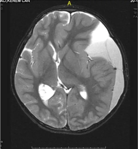
computed tomography (CT) scan revealed a hypodense mass in the left fronto-temporoparietal region (Figure 1). Magnetic resonance imaging (MRI) examination presented a chronic left cerebral subdural hematoma along with an intracystic hematoma in the left middle cranial fossa in addition to a midline shift to the right side (Figure 2).
Examination for coagulopathy was negative. A left fronto-temporoparietal craniotomy was performed, and the chronic SDH was perceived to be separated from the hematoma within the arachnoid cyst by the membrane of the arachnoid cyst and the capsule of the chronic SDH. Intracystic blood clots were removed and the membrane was widely fenestrated. The patient tolerated the procedure well and recovered completely. A postoperative CT scan confirmed the absence of the subdural hematoma and intracystic hematoma (Figure 3)
DISCUSSION
The natural history of arachnoid cysts is still not well defined (8,9). Arachnoid cysts arise from embryonic developmental anomalies involving duplication or
splitting of the arachnoid membrane. Arachnoid cysts represent 1% of all non-traumatic intracranial masses (10, 11). The most common site for this etiology is at the sylvian fissure (49%) followed by the cerebellopontine angle (11%), supracollicular (10%), vermian (9%), sellar, suprasellar (9%), interhemispheric (5%), cerebral convexity (4%), and clival (3%) regions (12-14). The first case of an arachnoid cyst with intracystic bleeding was described by Davidoff and Dyke in 1938. Tearing of the outer wall of the arachnoid cyst is associated with subdural or intracystic hemorrhage inflicted by a rupture of bridging veins, unsupported blood vessels around the cyst wall, and leptomeningeal vessels located at the base of the cyst (15-17). The
arachnoid cyst may enlarge overtime as a result of secretion of fluid from the cyst walls (18-20). The ensuing increased pressure may rupture and may manifest as a hematoma if there is also an accompanying vascular disruption. The most successful and comprehensive treatment for a patient with an arachnoid cyst and intracystic and/or SDH is surgery. Membranectomy and cyst communication to the basal cisterns must be executed.
CONCLUSION
Rarely, spontaneous rupture of arachnoid cysts can result in intra cystic hemorrhage, SDH or subdural hygroma. Surgery is a helpful and successful treatment for the symptomatic patients with an arachnoid cyst combined with intracystic and subdural hemorrhage.
Figure 2: MRI shows left chronic
subdural hematoma and intracystic hematoma including left middle cr.
Figure 3: Postoperative CT scan
showed the absence of the subdural hematoma and intracystic hematoma.
Journal Of Ankara University Faculty of Medicine 2013, 66 (2)
Gökmen Kahiloğullarıı, Ümit Eroğlu, Melih Bozkurt, Ağahan Ünlü 93 REFERENCES
1. Markwalder TM. Chronic subdural
hematomas: a review. J Neurosurg 1981;54:637-645.
2. Arai H, Sato K, Wachi A, Okuda O,
Takeda N. Arachnoid cysts of the middle cranial fossa: experience with 77 patients who were treated with cystoperitoneal shunting. Neurosurg 1996;39:1108-1112.
3. Galassi E, Piazza G, Gaist G, Frank F.
Arachnoid cysts of the middle cranial fossa: a clinical and radiological study of 25 cases treated surgically. Surg Neurol 1980;14:211-219.
4. Iaconetta G, Esposito M, Maiuri F,
Cappabianca P. Arachnoid cyst with intracystic hemorrhage and subdural hematoma: case report and literature review. Neurol Sci 2006;26:451-455.
5. Parsch CS, Krauss J, Hofmann E,
Meixensberger J, Roosen K. Arachnoid cysts associated with subdural hematomas and hygromas: analysis of 16 cases, long-term follow-up, and review of the literature. Neurosurg 1997;40:483-490.
6. Rengachary SS, Watanabe I, Brackett
CE. Pathogenesis of intracranial arachnoid cysts. Surg Neurol 1978;9:139-144.
7. Mori K, Yamamoto T, Horinaka N,
Maeda M. Arachnoid cyst is a risk factor for chronic subdural hematoma in juveniles: twelve cases of chronic
subdural hematoma associated with arachnoid cyst. J Neurotrauma 2002;19:1017-1027.
8. Sato K, Shimoji T, Yaguchi K, et al.
Middle fossa arachnoid cyst: clinical, neuroradiological, and surgical features. Childs Brain 1983;10:301-316.
9. van der Meche FG, Braakman R.
Arachnoid cysts in the middle cranial fossa: cause and treatment of progressive and non-progressive symptoms. J Neurol Neurosurg Psychiatry 1983;46:1102-1107.
10. Aoki N, Sakai T. Intraoperative
subdural hematoma in a patient with arachnoid cyst in the middle cranial fossa. Childs Nerv Syst 1990;6:44-46.
11. Huang D, Abe T, Kojima K, et al.
Intracystic hemorrhage of the middle fossa arachnoid cyst and subdural hematoma caused by ruptured middle cerebral artery aneurysm. Am J Neuroradiol 1999;20:1284-1286.
12. Bilginer B, Onal MB, Oguz KK,
Akalan N. Arachnoid cyst associated with subdural hematoma: report of three cases and review of the literature. Childs Nerv Syst 2009;25:119-124.
13. LaCour F, Trevor R, Carey M.
Arachnoid cyst and associated subdural hematoma. Observations on conventional roentgenographic and computerized tomographic diagnosis. Arch Neurol 1978;35:84-89.
14. Olsen NK, Madsen HH. Arachnoid
cyst with complicating intracystic and subdural hemorrhage. Rontgenblatter 1990;43:166-168.
15. Albuquerque FC, Giannotta SL.
Arachnoid cyst rupture producing subdural hygroma and intracranial hypertension: case report. Neurosurg 1997;41:951-955.
16. Yilmaz C, Cetinalp E, Caner H,
Altinors N. Disappearance of arachnoid cyst after rupturing into subdural space. Acta Neurochir (Wien) 2007;149:731-733
17. Wester K. Gender distribution and
sidedness of middle fossa arachnoid cysts: a review of cases diagnosed with computed imaging. Neurosurg 1992;31:940-944.
18. Eustace S, Toland J, Stack J. CT and
MRI of arachnoid cyst with complicating intracystic and subdural haemorrhage. J Comput Assist Tomogr 1992;16:995-997.
19. Page AC, Mohan D, Paxton RM.
Arachnoid cysts of the middle fossa predispose to subdural hematoma formation fact or fiction? Acta Neurochir Suppl (Wien) 1988;42:210-215.
20. Rakier A, Feinsod M. Gradual
resolution of an arachnoid cyst after spontaneous rupture into the subdural space. Case report. J Neurosurg 1995;83:1085-1086.
Ankara Üniversitesi Tıp Fakültesi Mecmuası 2013, 66 (2)
Arachnoid Cyst With Spontaneous Subdural Hematoma and Intracystic Hemorrhage in a Child 94