ORIGINAL RESEARCH
Comparison of Transoral Power-Assisted Endoscopic
Adenoidectomy to Curettage Adenoidectomy
Özmen Öztürk · Şenol Polat
To view enhanced content go to www.advancesintherapy.com Received: June 6, 2012 / Published online: July 31, 2012 © Springer Healthcare 2012
ABSTRACT
Introduction: The aim of this prospective and
randomized study was to compare patients who underwent curettage adenoidectomy (CA) or transoral power-assisted endoscopic adenoidectomy (PAEA) performed as isolated procedures by a single surgeon over a period of 7 years. The success of an adenoidectomy has been evaluated by assessing the amount of reduction in the adenoid size and the symptomatic improvement at the sixth postoperative month.
Methods: Patients’ nasal airway obstruction was
graded by the parents of the patient on a visual
analog scale (VAS), with a range from 0 to 10, with grade 10 representing total obstruction. The ratio of the choanal opening obstructed by an adenoid mass was measured and expressed in percentages as representative of the adenoid size. Preoperative and 6-month postoperative adenoid sizes were compared.
Results: Fifty-three patients (CA group
27 patients/PAEA group 26 patients) completed the study. In the CA group, VAS score improved from the preoperative score of 8.63 ± 0.88 to the 6-month score of 2.22 ± 1.01 (P < 0.0001); and in the PAEA group, the preoperative VAS score of 8.69 ± 0.84 improved to 2.08 ± 1.05 in the same period (P < 0.0001). No statistical significance was found when VAS score improvements were compared (P = 0.4569). The average ratio of choanal opening obstructed by an adenoid mass improved from the preoperative ratio of 89.41% ± 6.48% to the 6-month ratio of 7.85% ± 2.28% in the CA group; and the preoperative ratio of 90.19% ± 6.95% in the PAEA group improved to 3.65% ± 1.38% after 6 months. The reduction of adenoid size was significantly superior in PAEA than CA (P < 0.0001). The operative time in PAEA was significantly shorter than that in CA (P < 0.0001).
C o n c l u s i o n : A l t h o u g h s y m p t o m a t i c
improvement at the 6-month follow-up is Ö. Öztürk (*)
Istanbul Medipol University, School of Medicine, Department of Otorhinolaryngology
Haydarpasa – Harem Yolu, Kosuyolu, Kadikoy, 34718, Istanbul, Turkey
e-mail: [email protected] Ş. Polat
Acibadem University, School of Medicine, Department of Otorhinolaryngology, Istanbul, Turkey
Enhanced content for Advances in Therapy articles is available on the journal web site: www.advancesintherapy.com
choanal and laterally based adenoidal tissue without leaving a residue [2, 6, 7]. The ensuing completeness is expected to provide an improvement in the elimination of symptoms and an advantage in decreasing the risk of a recurrence [2, 7]. Furthermore, a finer control of adenoid resection under direct vision decreases the risk of damaging neighboring structures [6–8].
In this prospective study, a comparison of patients who underwent conventional CA or transoral power-assisted endoscopic adenoidectomy (PAEA) by a single surgeon over a period of 7 years is presented to confirm the clinical impression that PAEA is superior to CA by demonstrating the level of success in operative time, symptomatic improvement, and parents’ satisfaction.
MATERIALS AND METHODS
A prospective and randomized clinical study was conducted in the Acibadem Healthcare Group, Acibadem Kozyatagi Hospital, Otorhinolaryngology Department, between January 2004 and December 2010, to compare PAEA with CA to determine the difference in operative time and complications, assess the success of operative techniques in eliminating the adenoid mass, and confirm symptomatic improvement at the sixth postoperative month.
Inclusion Criteria
The study was limited to patients younger than 16 years. Patients with the presence of nasal airway obstruction with sleep disordered breathing (classified as ranging from persistent snoring for more than 3 months to obstructive sleep apnea), otitis media with effusion or recurrent otitis media, and chronic or recurrent rhinosinusitis were included [9, 10].
statistically indifferent, PAEA has been shown to be superior to CA with its superior performance in providing a near-total elimination of the adenoid mass in a shorter operating time.
Keywords: Adenoid; Adenoidectomy;
Complications; Curettage; Endoscopic surgical procedure
INTRODUCTION
Adenoidectomy, a frequently performed surgical procedure in the pediatric group, is indicated mainly when adenoid hypertrophy is the definite disorder in nasal airway obstruction, with sleep disordered breathing, otitis media with effusion, recurrent otitis media, and chronic and/or recurrent rhinosinusitis [1–5]. The patients usually present with rhinorrhea, chronic mouth breathing, excessive snoring and apneic episodes, enuresis, daytime somnolence, and neurocognitive and learning problems. Adenoidectomy provides a symptomatic recovery with a strong impact on improving the quality of life and health status of the pediatric population. Among the adenoidectomy techniques performed by using conventional surgical instruments (e.g., a variety of forceps, adenotomes, adenoid curettes, and electrocautery), curettage adenoidectomy (CA) still remains the most widely preferred surgical procedure [1–3, 6, 7].
With an improvement in microdebrider technology, power-assisted adenoidectomy (PAA) has evolved as a distinct technique for performing a more complete surgery via transnasal and/or transoral routes [3, 6]. In the transoral PAA approach, a microdebrider with a bent or a bendable blade is advanced to the nasopharynx under direct vision. Performing PAA under the assistance of an endoscopic view enables precision in removing intranasal,
Surgical Methods
Initial Steps Common to Both Surgical Techniques
An adenoidectomy was performed under general anesthesia with orotracheal intubation. For documentation and measurement of the adenoid size, the operation began with a digital video recording of the endonasal view of the adenoid pad and the choanal opening by using a 2.7 mm, 0° Hopkins® II telescope (Karl Storz-Endoskope, Karl Storz GmbH & Co. KG). After recording an acceptable image of the adenoid pad and the choanal opening, the child was placed in the Rose position, with a roll under the shoulders and a head ring enhancing neck extension. A left-curved Boyle-Davis mouth gag suspended on a Draffin bipod was used to retract the tongue and lower jaw. The patient was covered with sterile drapes, and the palate was palpated to exclude a submucosal clefting.
After these common steps, either CA or PAEA was used for performing adenoidectomy. The recording of operation time started when a microdebrider or a curette first touched the adenoid tissue, and stopped when the hemostasis was announced to be complete by the surgeon. The length of the procedure was recorded in minutes and seconds.
PAEA Technique
The distal and proximal ends of two soft rubber catheters were passed through the mouth, and crossed externally with a clamp to retract the soft palate and increase nasopharyngeal exposure. The powered microdebrider XPS® 3000 (Medtronic Xomed, Inc., Jacksonville, FL, USA) was used for PAEA. The tip of the microdebrider (RAdenoid®, pediatric blade 4 mm; Medtronic Xomed, Inc.) guarding an inner rotating blade had a cutting window for resection of adenoid tissue.
Exclusion Criteria
The exclusion criteria were the presence of documented allergic rhinitis, the co-existence of craniofacial abnormalities, and a history of a previous adenotonsillectomy or sino-nasal surgery. Adenoidectomies were performed as isolated procedures by a single surgeon (first investigator) and all children who underwent adenoidectomy in conjunction with other procedures (i.e., tonsillectomy, myringotomy, ventilation tube insertion, turbinate cauterization, and radiofrequency ablation of the tonsils) were excluded from the study.
Preoperative Assessment
During the assessment stage, the severity of the patient’s nasal airway obstruction was graded by the parents on a visual analog scale (VAS) ranging from 0 to 10, with grade 0 representing completely normal nasal breathing, and grade 10 representing total nasal airway obstruction.
During a routine otolaryngologic examination, the presence of adenoid tissue was determined by using a flexible nasopharyngoscope (Rhino-Fiberscope, with 2.5 mm diameter; Karl Storz-Endoskope, Karl Storz GmbH & Co. KG, Tuttlingen, Germany) in the clinic. If the ratio of the cross-sectional area of the adenoid mass at the choanal plane to the area of the choanal opening was determined to be larger than approximately 75%, the adenoid size was accepted as obstructive, and an adenoidectomy was indicated.
Random selection of the patients was performed before the commencement of the study. Numbers allocated in turn to either CA (odd numbers) or PAEA (even numbers) were equally sequenced on an Excell worksheet. Patients were listed and enumerated on the worksheet in order of their application date to the clinic.
Under an endoscopic view of a 4 mm, 70° Hopkins II telescope, the microdebrider was inserted transorally into the nasopharyngeal hollow, and the foot switch was depressed to adjust the blade at a speed of 1,500 rpm in oscillating mode. The handpiece of the microdebrider was connected to a continuous suction and irrigation system, and the adenoid tissue sucked into the cutting window was resected by the rotating blade.
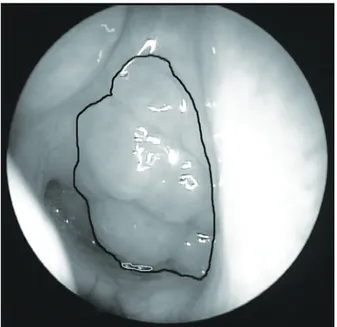
PAEA began from the choanal sill, and the resection was performed with a side-to-side sweeping motion of the microdebrider (Fig. 1), progressing posteriorly and inferiorly until the inferior border of the adenoid pad was reached. Care was taken to preserve the velopharyngeal function by leaving a rim of adenoid tissue just above the Passavant’s ridge. Furthermore, for the resection of peritubaric and laterally based adenoid tissue,
the shaver worked in the furrow between the adenoid pad and the lateral nasopharyngeal wall. Ample care was taken by keeping the tip of the microdebrider under continuous endoscopic view all through the operation to protect the nearby structures. A gentle resection was performed to keep the depth of resection on a level above the prevertebral fascia.
CA Technique
The adenoid mass was examined with digital palpation, and removed with a proper adenoid curette having a horizontal sharp edge for cutting through the adenoid base. Initially, a large adenoid curette was swept from the posterior border of the vomer to the inferior margin of the nasopharyngeal hollow with a slight side-to-side rocking motion. After removing the bulk of adenoid mass, the procedure was repeated with medium and smaller-sized curettes to remove choanal and peritubaric adenoid tissue. Removal was then confirmed by using an endoscopic examination.
End Steps Common to Both Surgical Techniques
For hemostasis, a tonsil pack was placed in the operative field for a period of approximately 60 seconds, and then removed. In all patients, any remaining bleeding point was controlled with bipolar electrocautery at a low power setting. After achieving hemostasis, the hardware was removed, and the child was left to the anesthetist. Intraoperative and immediate complications (e.g., hemorrhage, injury to a neighboring structure, laryngospasm, and prolonged recovery) were recorded.
Postoperative Follow-Up
All procedures were performed on an outpatient basis, and all patients followed the Fig. 1 The technique of transoral power-assisted
endoscopic adenoidectomy (PAEA). PAEA began high from the choana and the resection was performed with a “side to side” sweeping motion of the microdebrider (arrows), progressing downward until the inferior border of the adenoid pad was reached. For the resection of a laterally based adenoid tissue, the shaver worked in the depression between the adenoid pad and the lateral nasopharyngeal wall (black lines)
same postoperative instructions. A control examination was performed on day 7, and complications (e.g., dehydration with fever, postoperative hemorrhage, and atlanto-axial subluxation) were noted.
The patients were reviewed 6 months after the operation, and the parents were invited to repeat VAS grading to determine the degree of improvement in the postoperative nasal airway patency. Subsequently, a digital video recording of the endonasal view of the choanal opening was performed by using a flexible nasopharyngoscope to obtain a processable image of any adenoid residue.
Assessment of Data and Statistical Analysis
All of the preoperative assessments and surgery were performed by the first investigator. The first investigator had more experience with CA before this study, and started using microdebriders for performing PAEA 3 months before the start of the study. Postoperative follow-up evaluations were performed by the second investigator blinded to information about the selected adenoidectomy technique.
By comparing the preoperative and postoperative VAS scores, the degree of satisfaction of the parents associated with an improvement of nasal patency was noted. Subsequently, the percentage of choanal opening obstructed by an adenoid mass was used as a grading of nasal patency. By using the endonasal images obtained from the intraoperative and the 6-month postoperative digital video recordings, the ratio of the cross-sectional area of the adenoid mass at a plane passing vertically through the choana to the choanal opening area was measured to determine the adenoid size. The images were processed as a JPEG file, and after manually outlining the adenoidal and choanal limits by
using the lasso tool of the Adobe Photoshop CS5 version 12.0.4 extended software, the record measurement tool was used to measure the longitudinal cross-sectional area of the adenoid pad and the choanal opening in square pixels (Fig. 2). The ratio of the adenoid mass area (A) to the choanal opening area (C) (adenoid area/ choanal area; A/C) expressed in percentages was used as a reflection of the adenoid size and the severity of airway obstruction. Preoperative and 6-month postoperative A/C ratios were compared to accomplish an objective assessment of improvement in nasal patency and elimination of adenoid mass.
Fig. 2 The technique of measuring the ratio of choanal opening obstructed by an adenoid mass. The images were processed as a JPEG file, and after manually outlining the adenoidal mass (black lines) and choanal opening (white lines) limits by using “lasso tool” of the Adobe Photoshop CS5 version 12.0.4 extended software, the “record measurement tool” was used to measure the cross-sectional areas of the adenoid mass and the choanal opening in square pixels. Then the ratio of the “adenoid mass area” to the “choanal opening area” was expressed in percentages. In the figure, the adenoid mass was detected to obstruct 99.5% of the choanal opening (adenoid mass area 73 316 pixel2, choanal opening area 73 718 pixel2)
(mean 5 years 7 months; median 5 years) included in the current study. The two treatment groups were well matched for age (P = 0.7729) and gender (P = 0.7857) (Table 1).
Preoperative and 6-month postoperative mean VAS scores were evaluated in both groups (Figs. 4–6). In the CA group (n = 27), the preoperative VAS score of 8.63 ± 0.88 improved to the 6-month postoperative VAS score of 2.22 ± 1.01 (P < 0.0001) (Fig. 4). In the PAEA group (n = 26), an improvement was detected from the preoperative VAS score of 8.69 ± 0.84 to the 6-month postoperative VAS score of 2.08 ± 1.05 (P < 0.0001) (Fig. 5). Although symptoms resolved in both groups, no significant difference was found when the amount of All of the data obtained were coded and
entered in Prism 5 for Mac OS X version 5.0d. Data were expressed as mean ± SD. Appropriate statistical analysis with a two-tailed
t test was performed for data that followed a
Gaussian distribution. The Mann-Whitney
U nonparametric test was used for data that did
not follow a normal distribution. Fisher’s exact test was used for the comparison of gender. A significance level of P < 0.05 was chosen to define statistical significance.
Ethical Considerations
This report was approved by the ethics committee of Acibadem Health Group, Acibadem University, Faculty of Medicine, and was carried out in accordance with the Declaration of Helsinki. As a hospital policy, parents were informed preoperatively about the determined adenoidectomy technique. Informed consent has been received from the parents for inclusion of their children in this study.
RESULTS
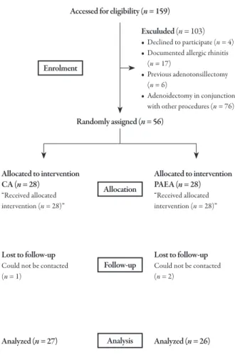
A group of 56 children, randomly assigned into two matched groups, underwent either PAEA (n = 28) or CA (n = 28) over a period of 7 years, between January 2004 and December 2010 (Fig. 3). Patients were contacted by telephone and appointments were scheduled for their 6-month follow-up visits. Fifty-three patients (27 patients in the CA group and 26 patients in the PAEA group) returned for the 6-month control examination and completed the study (Fig. 3). Three patients who failed to return for the follow-up examination could not be contacted.
There were 29 male (54.7%) and 24 female (45.3%) patients, ranging in age from from 1 year 8 months to 15 years 6 months
Accessed for eligibility (n = 159)
Allocation Randomly assigned (n = 56) Allocated to intervention CA (n = 28) “Received allocated intervention (n = 28)” Allocated to intervention PAEA (n = 28) “Received allocated intervention (n = 28)” Exculuded (n = 103) • Declined to participate (n = 4) • Documented allergic rhinitis
(n = 17)
• Previous adenotonsillectomy (n = 6)
• Adenoidectomy in conjunction with other procedures (n = 76)
Follow-up Lost to follow-up
Could not be contacted (n = 1)
Lost to follow-up
Could not be contacted (n = 2)
Analyzed (n = 26)
Analyzed (n = 27) Analysis Enrolment
Fig. 3 Flow-chart of the study. CA curettage
adenoidectomy, PAEA power-assisted endoscopic
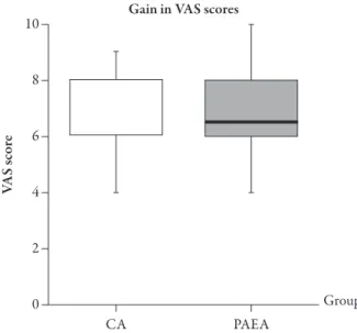
VAS score improvements were compared between the groups (P = 0.4569) (Fig. 6).
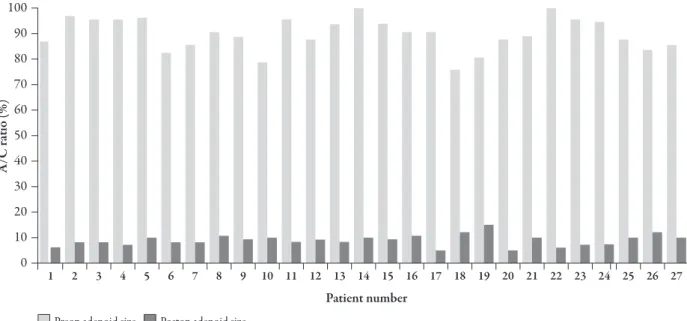
Preoperative and 6-month postoperative A/C ratios (a reflection of adenoid size) were compared in both groups (Figs. 7–9). The average ratio of choanal opening obstructed by an adenoid mass in the CA group improved from the preoperative ratio of 89.41% ± 6.48% to the
6-month postoperative ratio of 7.85% ± 2.28% (P < 0.0001) (Fig. 7); and the preoperative ratio of 90.19% ± 6.95% in the PAEA group improved to the postoperative ratio of 3.65% ± 1.38% after 6 months (P < 0.0001) (Fig. 8). There was a significant difference in the amount of A/C ratio reduction (a reflection of adenoid mass elimination) when both groups were compared (P < 0.0001) (Fig. 9).
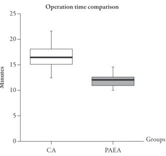
Although the duration of surgery showed wide variation in both groups, the operative time of PAEA was significantly shorter than that of CA (P < 0.0001) (Fig. 10). While the operative time varied from 10 minutes to 14 minutes 30 seconds in the PAEA group (mean 11 minutes 49 seconds; median 12 minutes), it was found to be between 12 minutes 30 seconds and 1 minutes 30 seconds (mean 16 minutes 31 seconds, median 16 minutes 30 seconds) in the CA group.
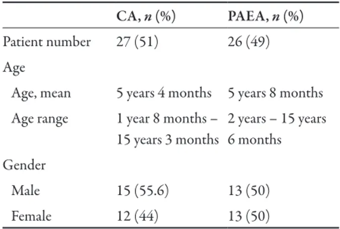
Various minor complications were detected in both groups. One patient in the CA group experienced an early postadenoidectomy Table 1 Patient demographics
CA, n (%) PAEA, n (%)
Patient number 27 (51) 26 (49) Age
Age, mean 5 years 4 months 5 years 8 months Age range 1 year 8 months –
15 years 3 months 2 years – 15 years 6 months Gender Male 15 (55.6) 13 (50) Female 12 (44) 13 (50)
CA curettage adenoidectomy, PAEA power-assisted
endoscopic adenoidectomy 10 8 6 4 2 0 VA S score 1
Preop VAS score Postop VAS score
2 3 4 5 6 7 8 9 10 11 12 13 14 15 16 17 18 19 20 21 22 23 24 25 26 27 Patient number
Fig. 4 The preoperative and the 6-month postoperative visual analog scale scores in curettage adenoidectomy patients (P < 0.0001). CA curettage adenoidectomy, VAS visual analog scale
Another patient from the PAEA group experienced a late bleed, on the eighth postoperative day. The slight bleeding was coming from the left lateral nasopharnygeal wall, inferior to torus tubarius, and stopped without any intervention under close follow-up. The third patient in the CA group had postoperative myalgia and limitation of neck movement for a period of 1 week. The investigation with imaging studies and necessary consultations to exclude a probable cervical subluxation were inconclusive, and myalgia resolved completely after 2 weeks. In addition, when both groups were viewed together, four patients (7.5%) had laryngospasm and prolonged recovery from anesthesia.
Neither surgical type resulted in a major complication or permanent damage to a nasal or nasopharyngeal structure. Postoperatively, velopharyngeal closure was adequate in both groups and speech quality was determined to be normal in all patients. No recurrences were detected during the follow-up period of 6 months. bleed during the recovery period. Bleeding
was immediately treated by the surgeon before the patient had left the operating theatre.
10 8 6 4 2 0 VA S score 1
Preop VAS score Postop VAS score
2 3 4 5 6 7 8 9 10 11 12 13 14 15 16 17 18 19 20 21 22 23 24 25 26 Patient number
Fig. 5 The preoperative and the 6-month postoperative visual analog scale scores in power-assisted endoscopic adenoidectomy patients (P < 0.0001). PAEA power-assisted endoscopic adenoidectomy, VAS visual analog scale
Fig. 6 Box plots comparing the gains in visual analog scale scores of curettage adenoidectomy and power-assisted endoscopic adenoidectomy groups (P = 0.4569). CA curettage adenoidectomy, PAEA power-assisted
endoscopic adenoidectomy, VAS visual analog scale
8 6 4 2 0 10 CA PAEA VA S score Groups Gain in VAS scores
100 90 80 70 60 50 40 30 20 10 0 A/C rat io (%)
Preop adenoid size Postop adenoid size
1 2 3 4 5 6 7 8 9 10 11 12 13 14 15 16 17 18 19 20 21 22 23 24 25 26 27 Patient number
Fig. 7 The preoperative and the 6-month postoperative adenoid mass area/choanal opening area ratios reflecting adenoid sizes in the curettage adenoidectomy group (P < 0.0001). A/C adenoid mass area/choanal opening area, CA curettage
adenoidectomy 100 90 80 70 60 50 40 30 20 10 0 A/C rat io %
Preop adenoid size Postop adenoid size
1 2 3 4 5 6 7 8 9 10 11 12 13 14 15 16 17 18 19 20 21 22 23 24 25 26 Patient number
Fig. 8 The preoperative and the 6-month postoperative adenoid mass area/choanal opening area ratios reflecting adenoid sizes in power-assisted endoscopic adenoidectomy patients (P < 0.0001). A/C adenoid mass area/choanal opening area, PAEA power-assisted endoscopic adenoidectomy
DISCUSSION
Although removal of a hypertrophied adenoid mass with conventional CA frequently elicits a significant improvement in symptoms, performing adenoidectomy without leaving an adenoid remnant is difficult to achieve with a blind surgical technique [2, 7, 11, 12]. Studies evaluating the success of a blind adenoidectomy prove that the removal of adenoid tissue using a traditional instrument was often incomplete [2, 6, 7, 11–13]. An endoscopic control examination of the nasopharynx performed at the end of a conventional adenoidectomy was reported to detect residual adenoid tissue at a high rate, particularly in the vicinity of the posterior nasal cavity, choana and peritubaric area [2, 13]. Any residue has the potential to hypertrophy, which may subject the patient to the recurrence
of symptoms, and a revision surgery may eventually be necessitated [1, 7, 12]. To reduce the incidence of regrowth, a control examination under endoscopic vision has to be routinely planned to detect and remove any remnant at the end of a blindly performed conventional adenoidectomy [2].
PAEA is a current alternative for decreasing the risk of recurrence and consequent revision surgery by providing completeness in adenoidectomy in a shorter operating time [1, 7, 8]. Extension of adenoid tissue into the choana and nasal cavity makes the approach demanding for a conventional method, and in PAEA, the curved blade that properly fits into the nasopharynx optimizes precision in the removal of adenoid tissue under the assistance of an endoscopic visualization providing excellent illumination and focus [2]. With this technique, resection of any residual adenoid tissue around the choana, posterior part of the nasal passage and torus tubarius is well executed, and the Fig. 9 Box plots comparing the gains in adenoid mass
area/choanal area ratios, reflecting the amount of
reduction in adenoid sizes, in the curettage adenoidectomy and power-assisted endoscopic adenoidectomy groups (P < 0.0001). A/C adenoid mass area/choanal opening
area, CA curettage adenoidectomy, PAEA power-assisted
endoscopic adenoidectomy
Fig. 10 Box plots of the operating time in the curettage adenoidectomy and power-assisted endoscopic adenoidectomy groups (P < 0.0001). CA curettage adenoidectomy, PAEA
power-assisted endoscopic adenoidectomy 90 80 70 60 50 40 30 20 10 0 100 CA PAEA A/C ra tio (%) Groups Gain in A/C ratios (reduction of adenoid sizes)
20 15 10 5 0 25 CA PAEA Minute s Groups Operation time comparison
displayed during the VAS grading for the evaluation of symptom improvement. The comparison of preoperative and 6-month postoperative VAS scores revealing the parental satisfaction level showed that both surgical methods were effective in providing a comparable amount of symptom improvement after 6 months. The difference between the VAS scoring of the adenoidectomy groups did not reach statistical significance, because it is known that an adenoid mass becomes symptomatic when more than approximately 50% of the postnasal space has been obstructed [12, 14]. In the current study, the choanal obstruction in both surgical groups was found to be less than the above-mentioned limit of critical choanal obstruction (7.85% ± 2.28% in CA and 3.65% ± 1.38% in PAEA at the 6-month postoperative examination), and thus the benefit provided by reduction in adenoid size was not sufficient to cause a statistical difference in symptomatic grading.
As a hospital policy, it was impossible to blind the parents to the selected adenoidectomy technique, and this information caused a possible bias in favor of PAEA during the subjective VAS grading of the 6-month postoperative follow-up reassessment. This is because parents had a probable tendency to be satisfied by a newer technique using a better technology.
Although the technical advantage provided by PAEA for the near-total elimination of adenoid mass did not cause a significant difference in the symptomatic outcome when compared to CA, any adenoid remnant left after CA can be a nidus for the progressive hypertrophy of residual adenoid tissue, which increases the risk of causing symptom recurrence or repeat surgery [1, 2, 7]. With its superior effectiveness in reducing the risk of an adenoid regrowth, PAEA is considered to ensure stability in the permanent well-being state of nasal airway patency. While a control examination at the sixth postoperative month risk of collateral injury to the neighboring
nasopharyngeal structures and pharyngeal muscles is minimized [2, 8]. A more complete resection results in a better chance of resolving any disease process related to the presence of adenoid tissue.
In the current study, when CA and PAEA were compared, the improvement in choanal patency after adenoid tissue elimination and a reduction in adenoid size reached statistical significance in favor of PAEA (P < 0.0001). The precision of the microdebrider and the direct vision provided by an endoscope afforded a superior technical advantage for the near-total elimination of an adenoid mass. While the average ratio of choanal opening obstructed by an adenoid mass in the CA group improved from the preoperative ratio of 89.41% ± 6.48% to the 6-month postoperative ratio of 7.85% ± 2.28%; in the PAEA group, the preoperative ratio of 90.19% ± 6.95% improved to the postoperative ratio of 3.65% ± 1.38% after 6 months (P < 0.0001). Determining the definite size of an adenoid pad with transnasal endoscopy has limitations. Small variations in the placement of an endoscope may change the angle of panaromic view and distort the appearance of the adenoid pad and choana. A markedly deformed view may affect the correct measurement of adenoid and choanal areas measured in square pixels. Also, while a rigid endoscope was used for the preoperative recordings, a flexible endoscope was chosen for the postoperative evaluations. So, in order to decrease the variability, the A/C ratio expressed in percentages has been taken into consideration. As both measurements are equally affected by the endoscopic aberration and the type of endoscope used, comparing the A/C ratio was preferred as a reflection of the adenoid size and the severity of airway obstruction.
The superior result provided by PAEA for adenoid size reduction was not proportionally
detects the hypertrophy of a nonobstructing adenoid remnant, a long-term follow-up period will establish the value of surgical techniques in monitoring a symptomatic adenoid tissue regrowth. In the current study, no recurrence has been detected in both adenoidectomy groups after 6 months.
As adenoidectomy is one of the most common operations performed in the pediatric age group, procedure time gains importance. In PAEA, both the cutting and the hemostasis times are shortened. One retrospective study comparing PAA with CA showed that operative time was significantly shorter with a microdebrider than a curette (11 minutes vs. 19 minutes) [6]. A subsequently performed prospective randomized study comparing PAA with CA supported the previous finding that operative time was 20% faster with a microdebrider (616 seconds vs. 732 seconds) [7]. In the current study, the interval between the first touch of the instrument to the adenoid tissue and completion of adenoid resection was significantly shorter with PAEA than CA (P < 0.0001).
Generally, the ability of the microdebrider to provide a well-controlled dissection down to a lesser vascular plane of nasopharynx reduces the amount of blood loss and shortens the time for hemostasis [6–8]. In PAA, adenoid tissue sucked into the cutting window of the microdebrider is fragmented with each oscillation of the blade, leaving an excoriated surface that bleeds during the resection [6]. Bleeding during an endoscopic procedure is usually a challenging problem, but when the experience of the surgeon increases, an efficiently used irrigation-suction set up in PAEA secures a well-controlled resection and an unobstructed endoscopic view [6, 15]. Patients are usually children, and thus the operating area is restricted, and the surgeon has to manage time-consuming problems such as bleeding in a narrow surgical field [16]. The most bloodless
approach to perform PAEA is to start resection high from the choana and progress in an orderly fashion to the inferior border of the adenoid bed, with the cutting tip of the microdebrider in continuous view [6, 17].
In PAEA, although the time for hemostasis is noticeably shortened, additional time and effort are spent in supporting the effectiveness of a microdebrider [15]. The effect of a microdebrider depends on suction efficacy [15]. Any adenoid fragment clogging the suction channel reduces the efficiency of the shaver, and continuous irrigation of the operative field is essential [15]. Operating room staff are required to organize the microdebrider system, and time is spent during set up. An additional drawback of the shaver system is its limited availability and higher cost. For the surgeons, training is required before performing PAEA, but the learning curve is shortened if there is familiarity with the mirror visualized CA and/or endoscopic sinonasal surgery [17]. Although there is a trend towards using new technologies in surgery, otolaryngologists still continue to use traditional adenoidectomy techniques due to the above-mentioned disadvantages.
On the other hand, several disadvantages are unique to CA. While removal of an adenoid tissue with a sharp curette damages prevertebral muscles due to an uncontrolled and excessive resection, the multiple pass of a dull curette scraping the adenoid pad during surgery may provoke a bloody but an incomplete dissection [1, 6]. The risk of velopharyngeal insufficiency is also increased with an aggressive blind adenoidectomy [6, 18].
It has been shown that both techniques are safe in experienced hands [2, 8, 17], but complications have been reported after both techniques [6, 7]. Early bleeding in a CA patient in our study was probably due to the aggressive use of a dull curette. In a PAEA patient, a late bleed on the 8th day was due to a scab peeling off from a damaged peritubal mucosa. This complication
was caused by a previous traumatization of the lateral nasopharyngeal structure during the adenoidectomy. This patient was operated on when the surgeon was relatively inexperienced (fourth patient of the PAEA group).
The authors’ study population is relatively small, because only adenoidectomies performed as isolated procedures were included, and exclusion criteria were used to increase the precision in the present study.
CONCLUSION
Although the PAEA approach offers several advantages (i.e., shorter procedure time, an improved field of vision, effective management of bleeding with continuous suction, extreme accuracy in removing the adenoid tissue with an instrument having a better manageability, increased safety in avoiding unnecessary trauma to the surrounding structures, and better satisfaction of the surgeon) over CA, the clinical efficacy remains comparable from an aspect of symptom improvement and patient outcome. Although symptomatic improvement in both techniques is indifferent; at the 6-month follow-up, PAEA has been shown to be superior to the conventional CA technique for providing completeness in adenoidectomy in a shorter operating time. Performing PAEA gains importance in providing a permanent patency of the nasopharynx and decreasing the risk of recurrence.
ACKNOWLEDGMENTS
The authors are indebted to Hasan Catalca, PhD, from the School of Health Sciences, Istanbul Medipol University, School of Medicine for statistical supervision. Özmen Öztürk, MD, assistant professor, takes full responsibility for the integrity of the whole work. Both authors contributed to the design, conduct, analysis,
and/or interpretation of the study, and participated in the preparation, review, and approval of this article. Dr. Öztürk is the guarantor for this article, and takes responsibility for the integrity of the work as a whole.
Conflict of Interest. The authors have no
conflict of interest or financial ties to disclose.
REFERENCES
1. Al-Mazrou KA, Al-Qahtani A, Al-Fayez AI. Effectiveness of transnasal endoscopic powered adenoidectomy in patients with choanal adenoids. Int J Pediatr Otorhinolaryngol. 2009;73:1650–2. 2. Costantini F, Salamanca F, Amaina T, Zibordi F.
Videoendoscopic adenoidectomy with microdebrider. Acta Otorhinolaryngol Ital. 2008;28:26–9.
3. Mitchell RB. Adenoidectomy techniques for sleep disordered breathing. Operative Techniques in Otolaryngology. 2005;16:229–31.
4. Wang DY, Clement PA, Kaufman L, Derde MP. Chronic nasal obstruction in children. A fiberscopic study. Rhinology. 1995;33:4–6.
5. Ungkanont K, Damrongsak S. Effect of adenoidectomy in children with complex problems of rhinosinusitis and associated diseases. Int J Pediatr Otorhinolaryngol. 2004;68:447–51. 6. Koltai PJ, Kalathia AS, Stanislaw P, Heras HA.
Power-assisted adenoidectomy. Arch Otolaryngol Head Neck Surg. 1997;123:685–8.
7. Stanislaw P Jr, Koltai PJ, Feustel PJ. Comparison of power-assisted adenoidectomy vs adenoid curette adenoidectomy. Arch Otolaryngol Head Neck Surg. 2000;126:845–9.
8. Rodriguez K, Murray N, Guarisco JL. Power-assisted partial adenoidectomy. Laryngoscope. 2002;112:26–8. 9. American Thoracic Society. Cardiorespiratory sleep
studies in children. Establishment of normative data and polysomnographic predictors of morbidity. Am J Respir Crit Care Med. 1999;160:1381–7. 10. Rosenfeld RM, Culpepper L, Doyle KJ, et al.; American
Academy of Pediatrics Subcommittee on Otitis Media with Effusion; American Academy of Family Physicians; American Academy of Otolaryngology – Head and Neck Surgery. Clinical practice guideline: otitis media with effusion. Otolaryngol Head Neck Surg. 2004;130(5 Suppl.):S95–118.
11. Cannon CR, Replogle WH, Schenk MP. Endoscopic-assisted adenoidectomy. Otolaryngol Head Neck Surg. 1999;121:740–4.
12. Buchinsky FJ, Lowry MA, Isaacson G. Do adenoids regrow after excision? Otolaryngol Head Neck Surg. 2000;123:576–81.
13. Havas T, Lowinger D. Obstructive adenoid tissue: an indication for powered-shaver adenoidectomy. Arch Otolaryngol Head Neck Surg. 2002;128:789–91. 14. Kemaloglu YK, Goksu N, Inal E, Akyildiz N.
Radiographic evaluation of children with nasopharyngeal obstruction due to the adenoid. Ann Otol Rhinol Laryngol. 1999;108:67–72.
15. Vokurka J. Shaver (micro debridor) in otorhinolaryngology. International Congress Series. 2003;1240:1411–15.
16. Inancli HM, Enoz M. Odds and evens for endoscopic adenoidectomy. Acta Otorhinolaryngol Ital. 2009;29:51; author reply 51–2.
17. Heras HA, Koltai PJ. Safety of powered instrumentation for adenoidectomy. Int J Pediatr Otorhinolaryngol. 1998;10;44:149–53.
18. van Gelder L. Open nasal speech following adenoidectomy and tonsillectomy. J Commun Disord. 1974;7:263–67.