Acta Orthop Traumatol Turc 2013;47(5):376-378 doi:10.3944/AOTT.2013.3081
CASE REPORT
Correspondence: Ak›n U¤rafl, MD. ‹stanbul Medipol Hastanesi T›p Fakültesi, Ortopedi ve Travmatoloji Anabilim Dal›, 34273 Ba¤c›lar, ‹stanbul, Turkey. Tel: +90 212 – 460 77 77 e-mail: [email protected]
Submitted: November 5, 2012 Accepted: January 23, 2013 ©2013 Turkish Association of Orthopaedics and Traumatology
Available online at www.aott.org.tr doi:10.3944/AOTT.2013.3081 QR (Quick Response) Code:
Osteoblastoma is a benign local aggressive tumor mostly localized in the vertebra or long bones. Carpal location and recurrence are extremely rare. Treatment options include either curettage or wide en bloc resection which causes functional disability in the hand and wrist and should be reserved only for recurrence. We present a case of recurrent trapezoid osteoblastoma previously treated with curet-tage of the trapezoid bone and a case of primary triquetral osteoblastoma.
Key words: Curettage; osteoblastoma; trapezoid bone; triquetral bone.
Osteoblastoma is a benign primary bone tumor first described as “giant osteoid osteoma” by Dahlin and Johnson in 1954.[1]
Later, in 1956, Lichtenstein and Jaffe named this tumor “osteoblastoma” in two different arti-cles.[2]
It is an uncommon benign but locally aggressive tumor, most commonly located in the vertebral column or metaphysis of long bones. This tumor is very rare in the carpal bones.[2,3]
Trapezoid bone location is uncommon and has not been previously addressed in the literature. We report a case of a recurrent osteoblastoma in the trapezoid bone, previously treated with curettage of the trapezoid bone, as well as a case of primary triquetral osteoblastoma.
Case report
Case 1
A 12-year-old girl presented to our clinic with posttrau-matic left hand pain and swelling in February 2008. The
pain increased at night and had a good response to non-steroidal analgesic drugs. Radiographs, computerized tomography and magnetic resonance imaging (MRI) revealed findings resembling avascular necrosis of the trapezoid bone, periosteal reaction at the second metacarpal and generalized edema in the dorsal com-partment (Figs. 1a and b). Pathological evaluation of the sample of the lesion obtained by curettage was consistent with osteoid osteoma or osteoblastoma (Fig. 1c). Clinically, the lesion was accepted as osteoid osteoma. The patient was completely free of pain and swelling with full range of wrist motion at the 12th month follow-up. However, at the 18th month follow-up, the patient had recurrent pain and swelling in the left hand. An oste-olytic recurrent lesion of 23-mm diameter located in the trapezoid bone but expanding across the capitate bone was detected in the radiographs and MRI. Initial diagno-sis was osteoblastoma and the lesion was excised with curettage. No adjuvant treatment was applied.
Osteoblastoma of the trapezoid bone and
triquetral bone: report of two cases
‹brahim KAYA1, Burak BOYNUK2, Caner GÜNERBÜYÜK3, Ak›n U⁄RAfi4
1
Department of Orthopedics and Traumatology, Haseki Training and Research Hospital, ‹stanbul, Turkey;
2
Department of Orthopedics and Traumatology, Bak›rköy Ac›badem Hospital, ‹stanbul, Turkey;
3
Department of Orthopedics and Traumatology, 29 May›s Hospital, ‹stanbul, Turkey;
4
Kaya et al. Osteoblastoma of the trapezoid bone and triquetral bone 377
Pathological evaluation of the samples revealed osteoblastoma. The patient had complete pain relief and no swelling with sufficient grasp strength at the postop-erative 35th month. Osseous fusion of the car-pometacarpal and intercarpal bones was detected in fol-low-up radiographs (Fig. 1d).
Case 2
In June 2011, a 19-year-old male presented to our clin-ic with persistent right hand pain of a duration of two years. The patient took analgesic drugs consistently and the pain increased at night. Radiologic examination revealed a locally sclerotic lesion of 20-mm diameter in the triquetral bone (Figs. 2a, b and c). Curettage and grafting of the lesion was performed and pathological examination defined the lesion as osteoblastoma (Fig. 2d). The patient had dramatic relief of complaints after the operation and was free of pain with full range of wrist motion and full grasp strength at the 13th month follow-up. There was no sign of recurrence in radiolog-ic examination.
Discussion
Osteoblastoma, a benign but locally aggressive tumor, constitutes 1% of all bone tumors.[2-4] It is most
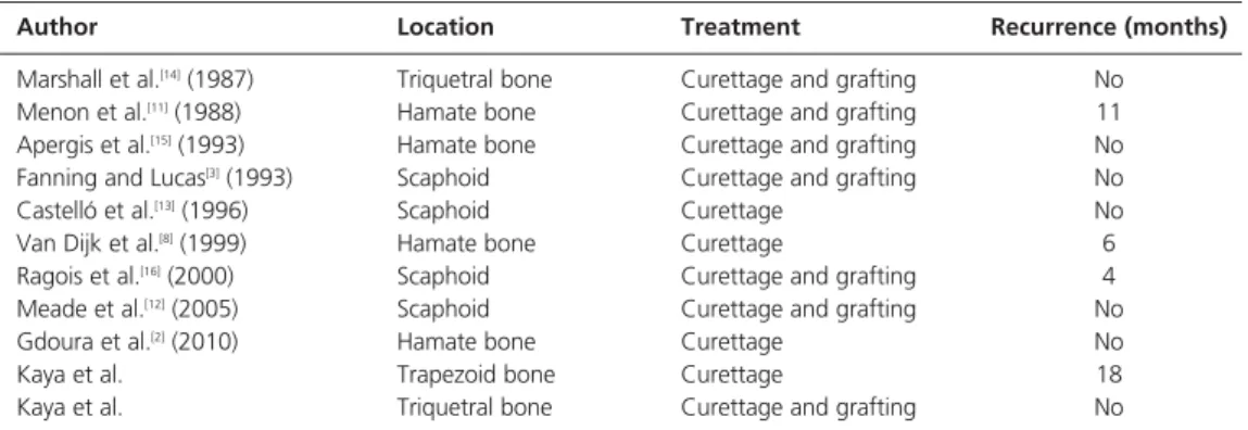
com-monly located in the vertebral column (40%), pelvis and long bones. Carpal location is extremely rare. In total, nine cases have been reported, with the navicular bone was affected in 5 cases, triquetral bone in 1 and hamate bone in 4 (Table 1). Trapezoidal bone location has not been previously addressed in the literature. The young population between 10 and 25 years of age and males are more often affected.[2]
Gdoura et al. reported that pain has no specific pat-tern and no correlation with rest or activity or time of day and that non-steroidal anti-inflammatory drugs do not relieve pain.[2]
However, non-steroidal anti-inflam-matory drugs provided good pain relief in our cases in which pain had circadian characteristics.
In the presence of cortical destruction, the osteoblastoma is separated from the adjacent tissue by a reactive bony shell. In our recurrent case, cortical disruption was noted. Giant cell bone tumor, aneurys-mal bone cyst, chondrosarcoma and osteosarcoma should be taken into consideration in the differential diagnosis in cases with cortical destruction.[2]
Dorfman and Weiss pointed out the importance of differential diagnosis between osteolytic aggressive osteoblastoma and well-differentiated osteosarcoma.[5]
Fig. 1. Views from Case 1 with osteoblastoma of the trapezoid bone. (a) Preoperative CT scan, (b) T1-weighted image of coronal MRI view, (c) pathology specimen (H&E ×400), and (d) 35th month follow-up anteroposterior X-ray of the patient. [Color figure can be viewed in the online issue, which is available at www.aott.org.tr]
(a) (b) (c) (d)
Fig. 2. Views from Case 2 with triquetral osteoblastoma. (a) Anteroposterior X-ray of the wrist, (b) T1-weighted and (c) T2-weighted images of coronal MRI view, and (d) pathology specimen with osteoblasts of different sizes, with abundant cytoplasm with regular nucleus and nucleoli are seen (H&E ×400). [Color figure can be viewed in the online issue, which is available at www.aott.org.tr]
378 Acta Orthop Traumatol Turc
Microscopically, the tumor consists of more vascu-larized connective tissue and has a greater diameter and has less reactional tissue surrounding the tumor than osteoid osteoma (greater than 2 centimeters) which has similar microscopic findings.[6,7]
Despite its benign character, early recurrence (10 to 19%) and malign transformation, even with pulmonary metasta-sis, can occur.[2,8-10]
In curettage of osteoblastoma, the bone window must be at least the same size as the tumor diameter as tumor cells left in the bone may cause recurrence. Curettage must be performed carefully using a burr motor. Bone grafting should be performed following curettage if nec-essary. Menon et al. treated a recurrent osteoblastoma in the hamate bone with curettage and grafting.[11]
Van Dijk et al. treated a recurrent osteoblastoma in hamate bone with en bloc resection and temporary interposition of the dead space with cement followed by vascularized iliac bone graft.[8] In general, wide
resection is the suggested treatment method but con-sidering the consequent functional and constitutional morbidity it should be reserved as the last choice.
In conclusion, carpal osteoblastoma is an extremely rare benign but locally aggressive tumor. Diagnosis is difficult due to ambiguous symptoms and radiographic findings. Treatment is curettage and graft augmenta-tion when necessary. In aggressive cases when the tumor progression is rapid, wide resection may be the preferred treatment. However, considering its conse-quent catastrophic results, this should be saved as the last treatment option. Well-differentiated osteosarco-ma must be considered in the differential diagnosis. Conflicts of Interest: No conflicts declared.
References
1. Dahlin DC, Johnson EW JR. Giant osteoid osteoma. J Bone Joint Surg Am 1954;36-A:559-72.
2. Gdoura F, Trigui M, Ellouze Z, Hamed YB, Ayadi K, Keskes H. Hamatum osteoblastoma. Orthop Traumatol Surg Res 2010;96:712-6.
3. Fanning JW, Lucas GL. Osteoblastoma of the scaphoid: a case report. J Hand Surg Am 1993;18:663-5.
4. Lucas DR, Unni KK, McLeod RA, O'Connor MI, Sim FH. Osteoblastoma: clinicopathologic study of 306 cases. Hum Pathol 1994;25:117-34.
5. Dorfman HD, Weiss SW. Borderline osteoblastic tumors: problems in the differential diagnosis of aggressive osteoblas-toma and low-grade osteosarcoma. Semin Diagn Pathol 1984; 1:215-34.
6. Xarchas KC, Leviet D. Osteoblastoma of the carpal scaphoid frequency and treatment. Acta Orthop Belg 2002;68:532-6. 7. Kenan S, Floman Y, Robin GC, Laufer A. Aggressive osteoblastoma. A case report and review of the literature. Clin Orthop Relat Res 1985;(195):294-8.
8. Van Dijk M, Winters HA, Wuisman PI. Recurrent osteoblastoma of the hamate bone. A two-stage reconstruc-tion with a free vascularized iliac crest flap. J Hand Surg Br 1999;24:501-5.
9. Jackson RP. Recurrent osteoblastoma: a review. Clin Orthop Relat Res 1978;(131):229-33.
10. Kunze E, Enderle A, Radig K, Schneider-Stock R. Aggressive osteoblastoma with focal malignant transforma-tion and development of pulmonary metastases. A case report with a review of literature. Gen Diagn Pathol 1996; 141:377-92.
11. Menon J, Rankin D, Jacobson C. Recurrent osteoblastoma of the carpal hamate. Orthopedics 1988;11:609-11. 12. Marshall JH, Sonsire JM, Nielsen PE, Nigogosyan G, Terzian
J. Digital angiography and osteoblastoma of the triquetrum. J Hand Surg Am 1987;12:256-8.
13. Apergis E, Tsamouri M, Theodoratos G, Maris I, Antoniou N. Osteoblastoma of the hamate bone: a case report. J Hand Surg Am 1993;18:137-40.
14. Castelló JR, Garro L, San Miguel P, Campo M. Osteoblastoma of the scaphoid – long-term results following curettage: a case report. J Hand Surg Am 1996;21:426-7.
15. Ragois P, Leclerc P, Hallonet D. Aggressive osteoblastoma of the carpal scaphoid bone. [Article in French] Rev Chir Orthop Reparatrice Appar Mot 2000;86:94-7.
16. Meade RA, Allende CA, Tsai TM. Osteoblastoma of the scaphoid: a case report. J Surg Orthop Adv 2005;14:125-8.
Author Location Treatment Recurrence (months)
Marshall et al.[14](1987) Triquetral bone Curettage and grafting No
Menon et al.[11](1988) Hamate bone Curettage and grafting 11
Apergis et al.[15](1993) Hamate bone Curettage and grafting No
Fanning and Lucas[3](1993) Scaphoid Curettage and grafting No
Castelló et al.[13](1996) Scaphoid Curettage No
Van Dijk et al.[8](1999) Hamate bone Curettage 6
Ragois et al.[16](2000) Scaphoid Curettage and grafting 4
Meade et al.[12](2005) Scaphoid Curettage and grafting No
Gdoura et al.[2](2010) Hamate bone Curettage No
Kaya et al. Trapezoid bone Curettage 18
Kaya et al. Triquetral bone Curettage and grafting No