DOI 10.1007/s00167-015-3872-5 KNEE
Comparison of quadriceps muscle volume after unilateral total
knee arthroplasty with and without tourniquet use
Olcay Guler1 · Mahir Mahirogullari1 · Mehmet Isyar1 · Ahmet Piskin2 · Sercan Yalcin1 · Serhat Mutlu3 · Bünyamin Sahin4
Received: 30 March 2015 / Accepted: 10 November 2015 / Published online: 21 November 2015 © European Society of Sports Traumatology, Knee Surgery, Arthroscopy (ESSKA) 2015
20 % of its volume in Group A postoperatively at 1 month (p < 0.001). A significant difference was found in all tissue measurements, except the connective and bone tissue vol-umes of the thigh between the operated and contralateral non-operated knees in Group A. No significant difference was identified between the operated and contralateral non-operated knees in Group B. The total WOMAC score was significantly higher, and the total KSS was significantly lower in Group A than in Group B during the postoperative follow-up period of 1–6 months (p < 0.001 for all) but not 12 months (n.s.).
Conclusion Tourniquet use in TKA decreases the thigh and quadriceps muscle volumes and postoperatively delays the recovery of knee function. Therefore, caution should be exercised for tourniquet use during TKA in daily clinical practice and using alternative methods for tourniquet appli-cation in preventing intraoperative blood loss.
Level of evidence III.
Keywords Total knee replacement · Total knee
arthroplasty · Tourniquet use · Quadriceps muscle · Atrophy · Quantitative analysis · Magnetic resonance imaging
Introduction
Intraoperative tourniquet use is a common method to decrease blood loss and obtain a bloodless surgical area during total knee arthroplasty (TKA), an operation associated with sig-nificant blood loss because of extensive soft tissue and bone damage [13, 23]. However, the benefits and risks of tourniquet use are contradictory. Although tourniquet use can shorten the operation time, allow a better visualization of the surgical area, control intraoperative blood loss, and increase the tibial cement
Abstract
Purpose Determination of the effect of tourniquet use in total knee arthroplasty (TKA) on thigh and quadriceps muscle volume using magnetic resonance imaging (MRI).
Methods A total of 148 knees of 74 patients (mean age 66.5 ± 4.8 years; female/male, 62/12) with bilateral pri-mary varus gonarthrosis underwent unilateral TKA with a tourniquet (Group A, n = 35) or without a tourniquet (Group B, n = 39). The total thigh volume and connec-tive, bone, and muscle tissue volumes were stereologi-cally measured on preoperative and postoperative MRI. The Knee Society Score (KSS) and Western Ontario and McMaster Universities Osteoarthritis Index (WOMAC) score were calculated to evaluate functional outcomes.
Results After TKA, the knees of patients in Group A exhibited a significant decrease in all tissue measure-ments, except bone tissue volume; however, the knees of patients in Group B exhibited no significant difference in tissue measurements. Although no difference was found between the operated and contralateral non-operated thighs (4076.9 and 4073.4 cm3, respectively) in Group B postop-eratively at 1 month (p > 0.05), the operated thighs had lost
* Olcay Guler
1 Orthopedics and Traumatology Department, Medical Faculty,
Medipol University, Atatürk Bulvarı. No:27 Unkapani, Fatih, 34083 Istanbul, Turkey
2 Orthopedics and Traumatology Department, Medical Faculty,
Ondokuz Mayıs University, Samsun, Turkey
3 Orthopedics and Traumatology Department, Kanuni Sultan
Suleyman Training Hospital, Istanbul, Turkey
4 Anatomy Department, Medical Faculty, Ondokuz Mayıs
mantle thickness, recent studies have shown that the overall blood loss does not change with tourniquet use because occult bleeding (also known as hidden blood loss) can continue for several hours after the operation [20, 23]. Furthermore, tourni-quet use is a well-known risk factor for thromboembolism after TKA [23, 29, 33]. Other potential complications of tourniquet use include postoperative thigh pain, wound pain because of reactive hyperperfusion after tourniquet deflation [23], vascu-lar injury [28], and nerve palsy [18]; furthermore, in rare cases, tourniquet use includes acute pulmonary oedema, cardiac arrest [6, 17], and thigh fat necrosis [24].
Most of the previous studies on the efficacy and safety of tourniquets have focused on blood loss and thrombo-embolism, whereas only a few have evaluated the effect of tourniquets on the physical function [14, 15]. Tourniquet use causes electromyographical changes in the quadriceps muscles and increases the plasma myoglobin levels [10,
22], suggesting that tourniquets can induce muscle damage. However, the effect of tourniquets on muscle volume and patients’ postoperative functionality has not been studied to date. Further studies on the effect of tourniquets on the physical function are currently being undertaken to clarify the benefits of tourniquet use [15].
In the present study, it was aimed to determine the effect of tourniquet use in TKA on thigh and quadriceps muscle volume using magnetic resonance imaging (MRI), which has not been reported to date. MRI was used to estimate muscle volume, a technique considered to be the gold standard [16]. The functional outcomes of patients who underwent TKA with and without tourniquet use were also compared. Our hypothesis was that tourniquet use during TKA has negative effects on the functional outcomes by decreasing thigh and quadriceps muscle volumes.
Materials and Methods
In total, 174 knees of 87 patients with stage 4 bilat-eral primary varus gonarthrosis according to the
Kellgren–Lawrence radiological classification scale for tibiofemoral joint degeneration [12] underwent unilateral TKA with or without tourniquet use from 2012 to 2014 and were included in this retrospective case–control study. Thirteen patients were excluded from this study because of the following reasons: muscle strength of less than 5/5 (1 patient), body mass index of >35 kg/m2 (1 patient), history of hip surgery (1 patient), contralateral control knee sur-gery in the previous year (9 patients), and contraindication for MRI (1 patient). Therefore, 148 knees of 74 patients were analysed.
The patients were divided into two groups: Group A comprised 35 patients who underwent TKA with tourni-quet use, and Group B comprised 39 patients who under-went TKA without tourniquet use (Table 1). The decision of tourniquet use was taken by the surgeon.
The study was approved by the institutional ethics committee of Medipol University, which operates under the national and international standards (no, 68; date, 12/02/2015), and conducted in accordance with the latest version of the Declaration of Helsinki.
Surgical technique
All operations were performed by two surgeons (O.G. and S.M.) with a 7-year experience each. The decision of tourniquet use during TKA was solely based on each sur-geon’s clinical and personal experiences. In practice, one of the two surgeons (O.G.) always used a tourniquet during arthroplasty surgery, whereas the other (S.M.) never used. Their decision was not affected by any patient-related fac-tor. The standard TKA to retain the posterior cruciate liga-ment was performed through parapatellar entrance with a fixed-bearing prosthesis using cement (Vanguard; Biomet, Warsaw, IN, USA) under spinal–epidural plus general anaesthesia. In Group A, after the leg had been exsangui-nated, a tourniquet was kept inflated throughout the opera-tion at twice the arterial blood pressure and released after suture application.
Table 1 Basic characteristics of each study group
TKA total knee arthroplasty, n.s. not significant TKA with tourniquet (Group A)
TKA without tourniquet (Group B) Total p value Number of patients 35 39 74 – Number of knees 70 78 148 – Age (years) 67.4 ± 4.3 (55–74) 65.8 ± 5.1 (58–78) 66.5 ± 4.8 (55–78) n.s. Sex (female/male) 30/5 32/7 62/12 n.s. Site of TKA Right 16 17 – Left 19 22 –
From the first day after surgery, the patients were allowed to walk using two walking sticks for a period of 1 month and using one stick thereafter. The standard post-operative rehabilitation program was applied to all patients. Continuous passive motion was performed in flexion post-operatively for 15 days, depending on the patients’ toler-ance level. Isometric quadriceps exercises were continued postoperatively for 3 months. The patients were evaluated postoperatively at 1, 3, 6, and 12 months.
MRI and evaluation
MRI was performed preoperatively and postoperatively at 1, 3, 6, and 12 months by two surgeons who performed the operations (O.G. and S.M.). To ensure the accuracy of image fusion, both surgeons performed the measurements on all axial MR images between the hip joint and femur supracon-dylar region. All imaging studies were conducted on a 1.5-Tesla MRI device (Sigma HDXT; General Electric, Chicago, IL, USA). T2-weighted MRI scans were obtained in the axial and sagittal planes covering the hip joint and supracondylar region of the femur, and the hip and knee joints were fully extended. The area was continuously scanned at an axial slice thickness of 8 mm with 2-mm spaces, a matrix of 320 × 224, and Nex = 2. Images were stored in a picture archiving and communication system (PACS; General Electric, Chicago, IL, US) and viewed on a RadiAnt DICOM Viewer for Windows (Medixant, Poznan, Poland). Serial images of the thigh were exported for volume analysis. For each thigh, another software package (ImageJ 1.45 s; NIH, Bethesda, MD, USA) was used to measure the sectional areas of the examined structures on MRI. Cavalieri’s principle of the stereological technique was used for this process. The sectional areas of total thigh, bone, soft tissue, and quadriceps muscles were individually meas-ured. The tissue to be measured was marked leaving no space in the outer surface margin. In accordance with the shape of tissue in the sectional area, the volume within the outer surface was measured by combining the marked points. The structures were multiplied by the section thickness, and the final volume was obtained in cm3 with one decimal point of accuracy. All
volume calculations were performed on MS Excel worksheets (2007; Microsoft, Redmond, WA, USA), where the results were automatically obtained by entering the section thickness and area of the structure into the worksheet.
Regions of interest were delineated in ImageJ to assess the total section surface areas of the thigh (Fig. 1), bone (Fig. 2), and soft tissue (fat, muscle, and connective tissue) (Fig. 3). The section surface area of the region of interest was automatically calculated using the software. These val-ues were then used for the estimation of the volume for all structures in cm3. For standardization, the volume of the region between the trochanter minor in the proximal femur and supracondylar aspect of the distal femur was calcu-lated. Furthermore, the muscle volumes of the vastus later-alis, vastus medilater-alis, rectus femoris, and vastus intermedius (Fig. 4) were stereologically estimated in cm3.
Study parameters
The total thigh volume; volumes of the connective, bone, and muscle tissues of both thighs; and muscle volumes of the vastus medialis, vastus lateralis, rectus femoris, and
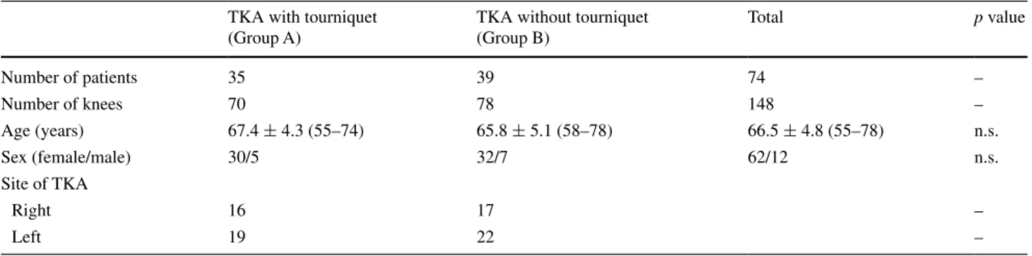
Fig. 1 Calculation of the total volume of the right thigh on single axial T2-weighted MRI. The yellow line shows the thigh circumference
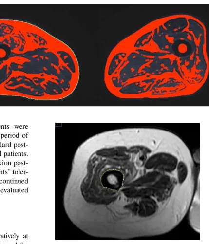
Fig. 2 Calculation of right femoral bone and muscle tissue volumes on single axial T2-weighted MRI
vastus intermedius were stereologically measured on pre-operative and postpre-operative MRI. Moreover, the preopera-tive and postoperapreopera-tive Knee Society Score (KSS) and West-ern Ontario and McMaster Universities Osteoarthritis Index (WOMAC) score [2], which are the routine scoring sys-tems used preoperatively and postoperatively in all patients who underwent TKA in our clinic, were used to evaluate the patients’ functional outcomes.
Statistical analysis
Study data were summarized using descriptive statistics, i.e. mean, standard deviation, and range for continuous variables and frequency and percentage for categorical variables. The change over time and the difference between the operated and contralateral knees were analysed using the preoperative and follow-up tissue volume and KSS/ WOMAC scores of both groups.
Repeated-measures analysis of variance was used to test the difference between more than two dependent and normally distributed variables. A paired-samples t test was applied for the post hoc analysis of significant results.
The difference between two and more than two depend-ent and non-normally distributed variables was analysed using the Wilcoxon signed-rank test and the Friedman test, respectively. The Wilcoxon signed-rank test was used as a post hoc test following the Friedman test. The inter-observer variability for interpreting the MR images was determined by comparing the 95 % confidence intervals of every parameter calculated by each of the two surgeons. The power of the study was calculated by post hoc power analysis on the total WOMAC score results at postopera-tive first month and found to be 98.3 % through an online power calculator program (http://clincal.com/).
The statistical level of significance was set as p ≤ 0.05. Analysis was performed using the MedCalc Statistical Software (version 12.7.7; MedCalc Software bvba, Ostend, Belgium).
Results
The study groups were considered to be similar in terms of age and sex (Table 1). The operated site was the left thigh in 19 of 35 patients from Group A and in 22 of 39 patients from Group B.
Tissue measurements
MRI tissue volume measurements for thighs on the oper-ated and contralateral non-operoper-ated sides are summarized in Tables 2 and 3 and Fig. 5. Although no difference was found between the operated and contralateral non-operated thigh volumes in Group B postoperatively at 1 month, the operated side lost 20 % of its volume (p < 0.001) in Group A postoperatively at 1 month. However, the muscle tissue
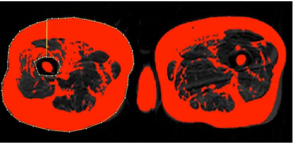
Fig. 3 Calculation of right thigh soft tissue (fat and con-nective tissue) volume on single axial T2-weighted MRI. The outer yellow line indicates the thigh circumference. The area between the two yellow lines indicates the non-bone soft tissue
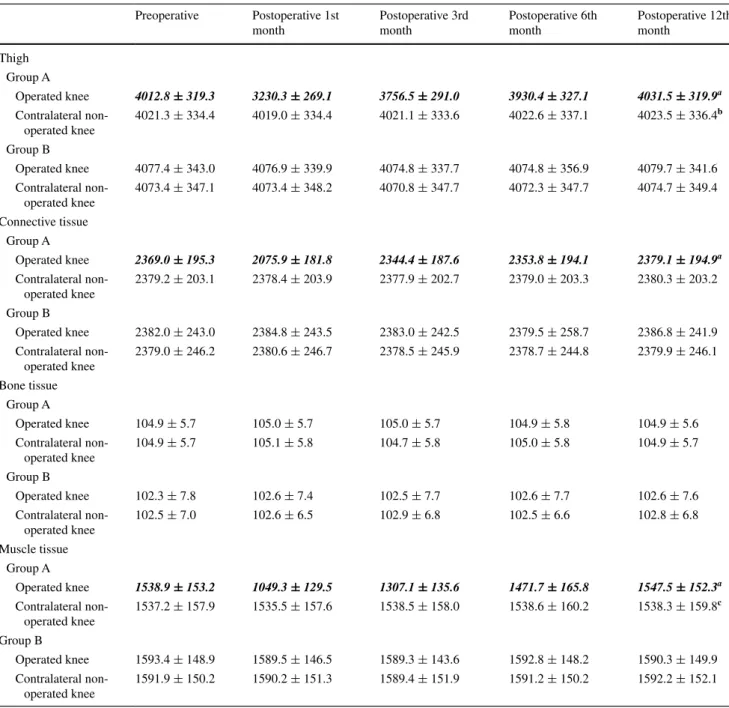
Fig. 4 Calculation of the muscle volume of the right vastus lateralis (VL), vastus medialis (VM), rectus femoris (RF), and vastus interme-dius on single axial T2-weighted MRI
volume of the operated knees increased with time in Group A postoperatively after 3 months (p < 0.001), reaching the muscle volume of the contralateral non-operated side at 12 months.
The operated side in patients from Group A exhibited a significant postoperative decrease in all tissue measure-ments, except bone tissue volume (Tables 2, 3, Fig. 5). On the other hand, the operated or contralateral non-operated
side in Group B exhibited no statistically significant differ-ence in all tissue measurements (Tables 2, 3).
A significant difference was noted in all tissue measure-ments, except connective and bone tissue volumes, of the thigh between the operated and contralateral non-operated knees in Group A (Table 3). However, no significant difference was noted in all tissue measurements between the operated and contralateral non-operated knees in Group B (Table 3).
Table 2 Descriptive statistics of preoperative and postoperative tissue measurements in operated and contralateral non-operated knees of each study group
Data are given as mean ± standard deviation in cm3. Statistically significant measurements are marked as bold and italic
a p < 0.001 for the change with time. b p = 0.004 for the overall difference between operated versus contralateral non-operated knees. c p < 0.001 for the overall difference between operated versus contralateral non-operated knees
Preoperative Postoperative 1st month Postoperative 3rd month Postoperative 6th month Postoperative 12th month Thigh Group A Operated knee 4012.8 ± 319.3 3230.3 ± 269.1 3756.5 ± 291.0 3930.4 ± 327.1 4031.5 ± 319.9a Contralateral non- operated knee 4021.3 ± 334.4 4019.0 ± 334.4 4021.1 ± 333.6 4022.6 ± 337.1 4023.5 ± 336.4 b Group B Operated knee 4077.4 ± 343.0 4076.9 ± 339.9 4074.8 ± 337.7 4074.8 ± 356.9 4079.7 ± 341.6 Contralateral non- operated knee 4073.4 ± 347.1 4073.4 ± 348.2 4070.8 ± 347.7 4072.3 ± 347.7 4074.7 ± 349.4 Connective tissue Group A Operated knee 2369.0 ± 195.3 2075.9 ± 181.8 2344.4 ± 187.6 2353.8 ± 194.1 2379.1 ± 194.9a Contralateral non- operated knee 2379.2 ± 203.1 2378.4 ± 203.9 2377.9 ± 202.7 2379.0 ± 203.3 2380.3 ± 203.2 Group B Operated knee 2382.0 ± 243.0 2384.8 ± 243.5 2383.0 ± 242.5 2379.5 ± 258.7 2386.8 ± 241.9 Contralateral non- operated knee 2379.0 ± 246.2 2380.6 ± 246.7 2378.5 ± 245.9 2378.7 ± 244.8 2379.9 ± 246.1 Bone tissue Group A Operated knee 104.9 ± 5.7 105.0 ± 5.7 105.0 ± 5.7 104.9 ± 5.8 104.9 ± 5.6 Contralateral non- operated knee 104.9 ± 5.7 105.1 ± 5.8 104.7 ± 5.8 105.0 ± 5.8 104.9 ± 5.7 Group B Operated knee 102.3 ± 7.8 102.6 ± 7.4 102.5 ± 7.7 102.6 ± 7.7 102.6 ± 7.6 Contralateral non- operated knee 102.5 ± 7.0 102.6 ± 6.5 102.9 ± 6.8 102.5 ± 6.6 102.8 ± 6.8 Muscle tissue Group A Operated knee 1538.9 ± 153.2 1049.3 ± 129.5 1307.1 ± 135.6 1471.7 ± 165.8 1547.5 ± 152.3a Contralateral non- operated knee 1537.2 ± 157.9 1535.5 ± 157.6 1538.5 ± 158.0 1538.6 ± 160.2 1538.3 ± 159.8 c Group B Operated knee 1593.4 ± 148.9 1589.5 ± 146.5 1589.3 ± 143.6 1592.8 ± 148.2 1590.3 ± 149.9 Contralateral non- operated knee 1591.9 ± 150.2 1590.2 ± 151.3 1589.4 ± 151.9 1591.2 ± 150.2 1592.2 ± 152.1
Table 3 Descriptive statistics of preoperative and postoperative muscle tissue measurements in operated and contralateral non-operated knees in each study group
Data are given as mean ± standard deviation in cm3. Statistically significant measurements are marked as bold and italic
a p < 0.001 for the change with time. b p < 0.001 for the overall difference between operated versus contralateral non-operated knees
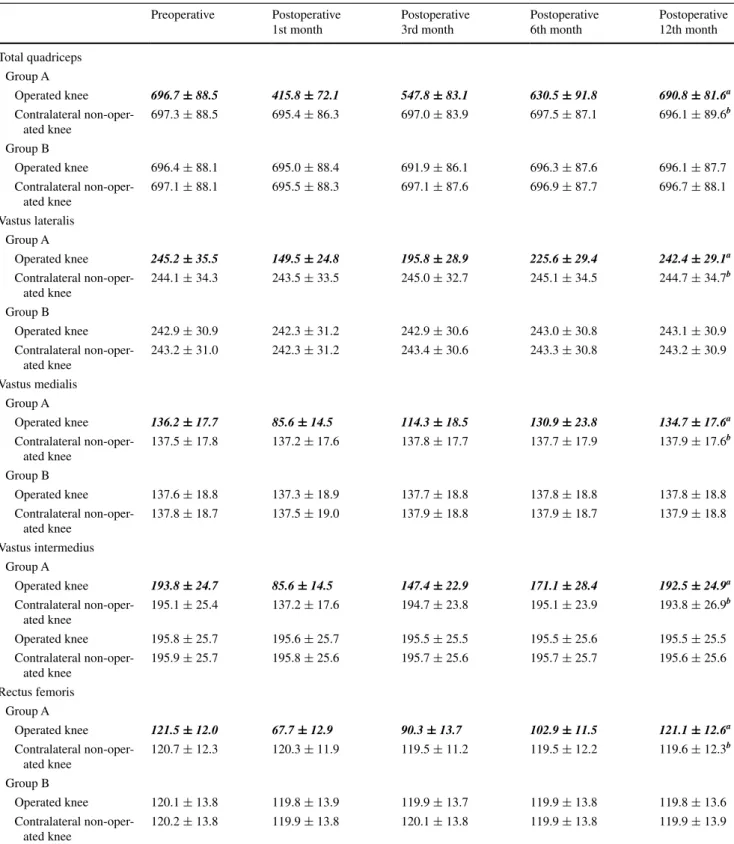
Preoperative Postoperative 1st month Postoperative 3rd month Postoperative 6th month Postoperative 12th month Total quadriceps Group A Operated knee 696.7 ± 88.5 415.8 ± 72.1 547.8 ± 83.1 630.5 ± 91.8 690.8 ± 81.6a Contralateral non-oper-ated knee 697.3 ± 88.5 695.4 ± 86.3 697.0 ± 83.9 697.5 ± 87.1 696.1 ± 89.6 b Group B Operated knee 696.4 ± 88.1 695.0 ± 88.4 691.9 ± 86.1 696.3 ± 87.6 696.1 ± 87.7 Contralateral non-oper-ated knee 697.1 ± 88.1 695.5 ± 88.3 697.1 ± 87.6 696.9 ± 87.7 696.7 ± 88.1 Vastus lateralis Group A Operated knee 245.2 ± 35.5 149.5 ± 24.8 195.8 ± 28.9 225.6 ± 29.4 242.4 ± 29.1a Contralateral non-oper-ated knee 244.1 ± 34.3 243.5 ± 33.5 245.0 ± 32.7 245.1 ± 34.5 244.7 ± 34.7 b Group B Operated knee 242.9 ± 30.9 242.3 ± 31.2 242.9 ± 30.6 243.0 ± 30.8 243.1 ± 30.9 Contralateral non-oper-ated knee 243.2 ± 31.0 242.3 ± 31.2 243.4 ± 30.6 243.3 ± 30.8 243.2 ± 30.9 Vastus medialis Group A Operated knee 136.2 ± 17.7 85.6 ± 14.5 114.3 ± 18.5 130.9 ± 23.8 134.7 ± 17.6a Contralateral non-oper-ated knee 137.5 ± 17.8 137.2 ± 17.6 137.8 ± 17.7 137.7 ± 17.9 137.9 ± 17.6 b Group B Operated knee 137.6 ± 18.8 137.3 ± 18.9 137.7 ± 18.8 137.8 ± 18.8 137.8 ± 18.8 Contralateral non-oper-ated knee 137.8 ± 18.7 137.5 ± 19.0 137.9 ± 18.8 137.9 ± 18.7 137.9 ± 18.8 Vastus intermedius Group A Operated knee 193.8 ± 24.7 85.6 ± 14.5 147.4 ± 22.9 171.1 ± 28.4 192.5 ± 24.9a Contralateral non-oper-ated knee 195.1 ± 25.4 137.2 ± 17.6 194.7 ± 23.8 195.1 ± 23.9 193.8 ± 26.9 b Operated knee 195.8 ± 25.7 195.6 ± 25.7 195.5 ± 25.5 195.5 ± 25.6 195.5 ± 25.5 Contralateral non-oper-ated knee 195.9 ± 25.7 195.8 ± 25.6 195.7 ± 25.6 195.7 ± 25.7 195.6 ± 25.6 Rectus femoris Group A Operated knee 121.5 ± 12.0 67.7 ± 12.9 90.3 ± 13.7 102.9 ± 11.5 121.1 ± 12.6a Contralateral non-oper-ated knee 120.7 ± 12.3 120.3 ± 11.9 119.5 ± 11.2 119.5 ± 12.2 119.6 ± 12.3 b Group B Operated knee 120.1 ± 13.8 119.8 ± 13.9 119.9 ± 13.7 119.9 ± 13.8 119.8 ± 13.6 Contralateral non-oper-ated knee 120.2 ± 13.8 119.9 ± 13.8 120.1 ± 13.8 119.9 ± 13.8 119.9 ± 13.9
WOMAC score and KSS
The total WOMAC score and subscores for pain, stiff-ness, and physical function were significantly lower after the operation in both Group A and Group B (p < 0.001 for all) (Table 4, Fig. 6). The decrease was significant throughout the 12-month follow-up period (Table 4). The total WOMAC score was significantly higher in Group A than in Group B postoperatively at 1–6 months (p < 0.001 for all) but not at 12 months (Table 4). The physical function scores postoperatively at 1–6 months, stiffness scores postoperatively at 3 months, and pain scores postoperatively at 3 to 6 months were also signifi-cantly different between the two groups (p < 0.001 for all) (Table 4).
The KSS knee and functional scores were significantly higher in the operated knees of both groups during the 12-month postoperative period than the preoperative period (p < 0.001 for all) (Table 5, Fig. 7). The KSS for Group A was significantly lower than that for Group B postopera-tively at 1–6 months (Table 5). The 95 % confidence inter-vals for the independent measurements of each parameter were similar among the surgeons, indicating that there was no significant interobserver variability. The measurements derived from the images were also reproducible.
Discussion
The most important finding of the study was that tourniquet use during TKA had a negative impact on the thigh muscles and could postoperatively delay the functional recovery of patients.
Because of the low quality of evidence among the large number of clinical studies on the perioperative tourniquet use in TKA, many meta-analysis studies have recently been published. All these meta-analyses showed that the poten-tial complications of tourniquet use in TKA outweigh its benefits and that the long-term outcome of tourniquet use is uncertain [1, 9, 23, 31, 33, 34]. These studies suggested that although tourniquet use saves time, it increases throm-boembolic complications and does not significantly reduce blood loss [1, 9, 23, 31, 33, 34]. Li et al. [13] indicated that tourniquet use may promote postoperative hidden blood loss and prevent patients’ participation in early reha-bilitation exercises. Ejaz et al. [4] showed that TKA with-out tourniquet use results in better functional and clinical outcomes, i.e. faster recovery and wider range of motion. Recent studies have also suggested tourniquet use at a lower pressure [19, 27] or for a limited time during the operation, for example, during cementation [5, 25] or wound closure [32, 34], to eliminate the risks associated with prolonged
Fig. 5 Preoperative and postoperative tissue measurements in operated and contralateral non-operated knees of patients who underwent TKA with a tourniquet (Group A). On the operated side, p < 0.001 for change with time for all tissues
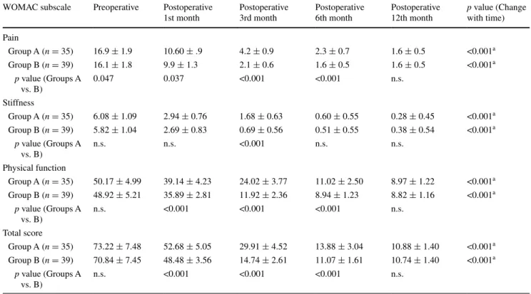
Table 4 Preoperative and postoperative Western Ontario and McMaster Universities Osteoarthritis Index (WOMAC) scores of operated knee of study groups
Data are given as mean ± standard deviation
n.s. not significant
a p < 0.001 for preop versus postop 1st, 3rd, 6th, and 12th months
WOMAC subscale Preoperative Postoperative 1st month Postoperative 3rd month Postoperative 6th month Postoperative 12th month p value (Change with time) Pain Group A (n = 35) 16.9 ± 1.9 10.60 ± .9 4.2 ± 0.9 2.3 ± 0.7 1.6 ± 0.5 <0.001a Group B (n = 39) 16.1 ± 1.8 9.9 ± 1.3 2.1 ± 0.6 1.6 ± 0.5 1.6 ± 0.5 <0.001a p value (Groups A vs. B) 0.047 0.037 <0.001 <0.001 n.s. Stiffness Group A (n = 35) 6.08 ± 1.09 2.94 ± 0.76 1.68 ± 0.63 0.60 ± 0.55 0.28 ± 0.45 <0.001a Group B (n = 39) 5.82 ± 1.04 2.69 ± 0.83 0.69 ± 0.56 0.51 ± 0.55 0.38 ± 0.54 <0.001a p value (Groups A vs. B) n.s. n.s. <0.001 n.s. n.s. Physical function Group A (n = 35) 50.17 ± 4.99 39.14 ± 4.23 24.02 ± 3.77 11.02 ± 2.50 8.97 ± 1.22 <0.001a Group B (n = 39) 48.92 ± 5.21 35.89 ± 2.81 11.92 ± 2.36 8.94 ± 1.23 8.82 ± 1.16 <0.001a p value (Groups A vs. B) n.s. <0.001 <0.001 <0.001 n.s. Total score Group A (n = 35) 73.22 ± 7.48 52.68 ± 5.05 29.91 ± 4.52 13.88 ± 3.04 10.88 ± 1.40 <0.001a Group B (n = 39) 70.84 ± 7.45 48.48 ± 3.56 14.74 ± 2.61 11.07 ± 1.61 10.74 ± 1.40 <0.001a p value (Groups A vs. B) n.s. <0.001 <0.001 <0.001 n.s.
Fig. 6 Preoperative and postoperative Western Ontario and McMaster Universities Osteoarthritis Index (WOMAC) scores for the operated knees of patients who underwent TKA with (Group A) and without (Group B) a tourniquet
tourniquet use. In addition, instead of tourniquet use, other measures that have been shown to be effective and safe in reducing blood loss have been proposed, including the use of tranexamic acid [3, 11]. However, all aspects of tourni-quet use should be extensively evaluated to reach a final conclusion on the benefits and risks of perioperative tour-niquet use in TKA.
Tourniquets can cause tissue ischaemia and reperfusion, but the outcome of ischaemia/reperfusion on the muscles
and soft tissues of the thigh remains incompletely under-stood. Therefore, in this study, the effect of tourniquet use on the volume of the entire thigh, the quadriceps femoris, and other thigh muscles was investigated.
Hocker et al. [8] obtained biopsies from the vastus lateralis during TKA and found increased endoplasmic reticulum stress in muscle tissue with tourniquet use. The biopsies from the quadriceps femoris muscle in TKA with tourniquet use indicated that tourniquet-induced ischaemia/
Table 5 Preoperative and postoperative Knee Society Score (KSS) of operated knee of study groups
Data are given as mean ± standard deviation
n.s. not significant
a p < 0.001 for preop versus postop 1st, 3rd, 6th, and 12th months
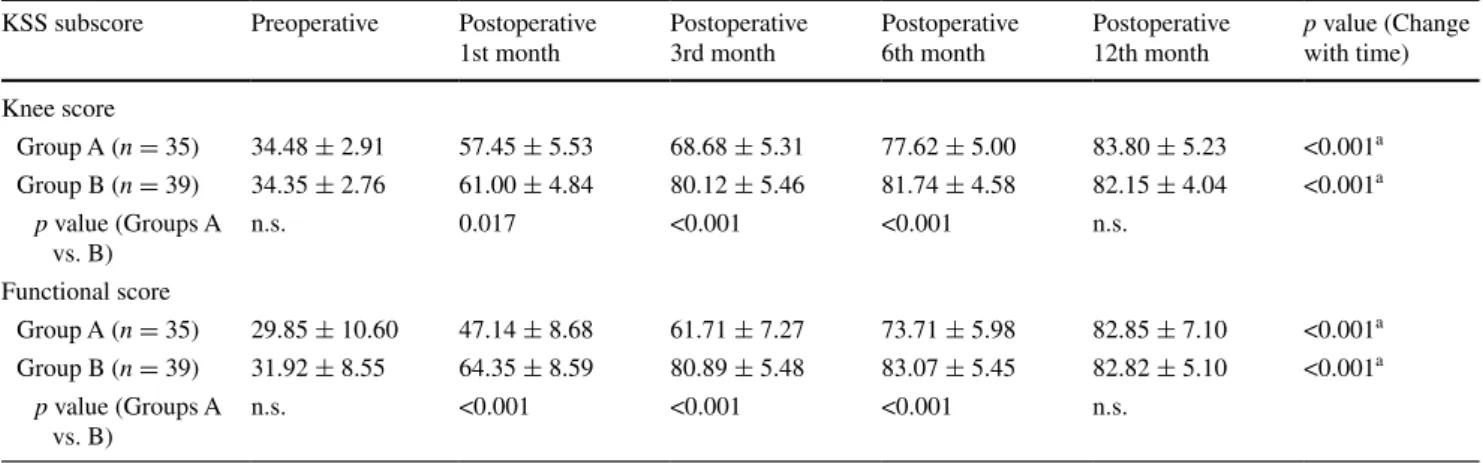
KSS subscore Preoperative Postoperative 1st month Postoperative 3rd month Postoperative 6th month Postoperative 12th month p value (Change with time) Knee score Group A (n = 35) 34.48 ± 2.91 57.45 ± 5.53 68.68 ± 5.31 77.62 ± 5.00 83.80 ± 5.23 <0.001a Group B (n = 39) 34.35 ± 2.76 61.00 ± 4.84 80.12 ± 5.46 81.74 ± 4.58 82.15 ± 4.04 <0.001a p value (Groups A vs. B) n.s. 0.017 <0.001 <0.001 n.s. Functional score Group A (n = 35) 29.85 ± 10.60 47.14 ± 8.68 61.71 ± 7.27 73.71 ± 5.98 82.85 ± 7.10 <0.001a Group B (n = 39) 31.92 ± 8.55 64.35 ± 8.59 80.89 ± 5.48 83.07 ± 5.45 82.82 ± 5.10 <0.001a p value (Groups A vs. B) n.s. <0.001 <0.001 <0.001 n.s.
Fig. 7 Preoperative and postoperative Knee Society Score (KSS) of the operated knee in patients who underwent TKA with (Group A) and without (Group B) a tourniquet
reperfusion induces changes in glutathione metabolism, signifying oxidative stress [30]. Liu et al. [14] compared quadriceps function using electromyography following TKA with and without tourniquet use in 20 patients with osteoarthritis. They found that the tourniquet group had significantly more pain and limited quadriceps function than the non-tourniquet group postoperatively at 6 months. Consistent with the previous studies, it was found that the quadriceps and other thigh muscles were negatively affected by the perioperative tourniquet use. In the group with tourniquet use, in comparison with the non-operated knees, the operated knees exhibited significantly lower thigh and all-muscle volumes for the total quadriceps, vas-tus lateralis, vasvas-tus medialis, vasvas-tus intermedius, and recvas-tus femoris. However, in patients who underwent TKA without tourniquet use, no significant difference was observed in tissue measurements between the operated and non-oper-ated knees. It was considered that the negative effect of a tourniquet on thigh muscle volume is caused by tourniquet-induced tissue ischaemia and reperfusion, which initiates a cascade of tissue reactions leading to oxidative stress, as previously shown [8, 14, 21].
A tourniquet-induced decrease in thigh muscle volume is not without clinical effects. The patients’ postoperative WOMAC score and KSS were evaluated because these are common indicators of pain and physical functional-ity in operated knees. Our results indicated that regard-less of tourniquet use, the functionality of operated knees increased significantly with TKA from the first month after the operation. However, the improvement in functional-ity, which was evaluated by both the WOMAC score and KSS, was greater in patients who underwent TKA with-out tourniquet use than in those who underwent TKA with tourniquet postoperatively up to 12 months. However, both groups were found to have a similar level of functional-ity at the 12-month follow-up. Our findings regarding late functional recovery following TKA with tourniquet use are in line with those of previous studies, which reported that functional recovery following TKA is closely associ-ated with the strength of the thigh muscles, particularly the quadriceps femoris muscle [21]. In a meta-analysis of 13 randomized clinical trials involving 689 patients, the post-operative knee range of motion in the tourniquet group was less than that in the non-tourniquet group during the early stages after surgery; therefore, tourniquet use may hinder patients’ early postoperative rehabilitation exercises [33].
The present study primarily showed that tourniquet use during TKA had negative effects on the knee functions and patients’ quality of life, as shown by WOMAC score and KSS; therefore, caution should be exercised for tourniquet use during TKA in daily clinical practice.
The retrospective design and small sample size were the main limitations of this study. Furthermore, the decision of
tourniquet use was solely based on each surgeon’s clinical and personal experiences, which represented a selection bias. Furthermore, all surgeries were performed by two sur-geons, which was another source of bias. Because the deci-sion of tourniquet use was based on each surgeon’s prefer-ence, not defined by any randomization, and sample size would be small if only one surgeon’s patients were included, two surgeons performed all surgeries. Another limitation was that intraobserver reliability could not be assessed. Despite these limitations, which prevented us from reach-ing a definitive conclusion, this was the first study to show an association between a decrease in thigh muscle volume and tourniquets use in TKA. Our results were significantly important because it is well known that the surgical factors such as approach and tourniquet use affecting the function and strength of the quadriceps femoris muscle should be understood and eliminated by surgeons to obtain the best TKA outcome [7, 26]. Further prospective studies with a larger sample size are needed to reach a final conclusion on the effect of tourniquet use in TKA on patients’ thigh muscles and functionality and to suggest alternative clinical measures to prevent intraoperative blood loss.
Conclusion
In conclusion, the thigh and quadriceps muscle volumes are decreased, and recovery of knee functions is delayed post-operatively by tourniquet use in TKA. Therefore, the use of alternative methods for tourniquet use to prevent intraop-erative blood loss is recommended.
Acknowledgments The authors would like to thank Enago (www. enago.com) for the English language review.
Compliance with ethical standard
Conflict of interest The authors declare no conflicts of interest.
References
1. Alcelik I, Pollock RD, Sukeik M, Bettany-Saltikov J, Armstrong PM, Fismer P (2012) A comparison of outcomes with and with-out a tourniquet in total knee arthroplasty: a systematic review and meta-analysis of randomized controlled trials. J Arthroplasty 27:331–340
2. Bellamy N (1989) Pain assessment in osteoarthritis: experience with the WOMAC osteoarthritis index. Semin Arthritis Rheum 18:14–17
3. Bidolegui F, Arce G, Lugones A, Pereira S, Vindver G (2014) Tranexamic acid reduces blood loss and transfusion in patients undergoing total knee arthroplasty without tourniquet: a prospec-tive randomized controlled trial. Open Orthop J 8:250–254 4. Ejaz A, Laursen AC, Kappel A, Laursen MB, Jakobsen T,
Ras-mussen S, Nielsen PT (2014) Faster recovery without the use of a tourniquet in total knee arthroplasty. Acta Orthop 85:422–426
5. Fan Y, Jin J, Sun Z, Li W, Lin J, Weng X, Qiu G (2014) The limited use of a tourniquet during total knee arthroplasty: a rand-omized controlled trial. Knee 21:1263–1268
6. Gielen M (1991) Cardiac arrest after tourniquet release. Can J Anaesth 38:541
7. Greene KA, Schurman JR 2nd (2008) Quadriceps muscle func-tion in primary total knee arthroplasty. J Arthroplasty 23(7 Suppl):15–19
8. Hocker AD, Boileau RM, Lantz BA, Jewett BA, Gilbert JS, Dreyer HC (2013) Endoplasmic reticulum stress activation dur-ing total knee arthroplasty. Physiol Rep 1:e00052
9. Jiang FZ, Zhong HM, Hong YC, Zhao GF (2015) Use of a tourniquet in total knee arthroplasty: a systematic review and meta-analysis of randomized controlled trials. J Orthop Sci 20:110–123
10. Jorgensen HR (1987) Myoglobin release after tourniquet ischemia. Acta Orthop Scand 58:554–556
11. Kelley TC, Tucker KK, Adams MJ, Dalury DF (2014) Use of tranexamic acid results in decreased blood loss and decreased transfusions in patients undergoing staged bilateral total knee arthroplasty. Transfusion 54:26–30
12. Kellgren JH, Lawrence JS (1957) Radiological assessment of osteo-arthrosis. Ann Rheum Dis 16:494–502
13. Li B, Wen Y, Wu H, Qian Q, Lin X, Zhao H (2009) The effect of tourniquet use on hidden blood loss in total knee arthroplasty. Int Orthop 33:1263–1268
14. Liu D, Graham D, Gillies K, Gillies RM (2014) Effects of tour-niquet use on quadriceps function and pain in total knee arthro-plasty. Knee Surg Relat Res 26:207–213
15. Lohmann-Jensen R, Holsgaard-Larsen A, Emmeluth C, Over-gaard S, Jensen C (2014) The efficacy of tourniquet assisted total knee arthroplasty on patient-reported and performance-based physical function: a randomized controlled trial protocol. BMC Musculoskelet Disord 15:110
16. Maden-Wilkinson TM, Degens H, Jones DA, McPhee JS (2013) Comparison of MRI and DXA to measure muscle size and age-related atrophy in thigh muscles. J Musculoskelet Neuronal Interact 13:320–328
17. McGrath BJ, Hsia J, Epstein B (1991) Massive pulmonary embo-lism following tourniquet deflation. Anesthesiology 74:618–620 18. Olivecrona C, Blomfeldt R, Ponzer S, Stanford BR, Nilsson BY
(2013) Tourniquet cuff pressure and nerve injury in knee arthro-plasty in a bloodless field: a neurophysiological study. Acta Orthop 84:159–164
19. Olivecrona C, Ponzer S, Hamberg P, Blomfeldt R (2012) Lower tourniquet cuff pressure reduces postoperative wound complica-tions after total knee arthroplasty: a randomized controlled study of 164 patients. J Bone Joint Surg Am 94:2216–2221
20. Pfitzner T, von Roth P, Voerkelius N, Mayr H, Perka C, Hube R (2014) Influence of the tourniquet on tibial cement mantle
thickness in primary total knee arthroplasty. Knee Surg Sports Traumatol Arthrosc. doi:10.1007/s00167-014-3341-6
21. Saleh KJ, Lee LW, Gandhi R, Ingersoll CD, Mahomed NN, Sheibani-Rad S, Novicoff WM, Mihalko WM (2010) Quadriceps strength in relation to total knee arthroplasty outcomes. Instr Course Lect 59:119–130
22. Saunders KC, Louis DL, Weingarden SI, Waylonis GW (1979) Effect of tourniquet time on postoperative quadriceps function. Clin Orthop Relat Res 143:194–199
23. Tai TW, Lin CJ, Jou IM, Chang CW, Lai KA, Yang CY (2011) Tourniquet use in total knee arthroplasty: a meta-analysis. Knee Surg Sports Traumatol Arthrosc 19:1121–1130
24. Tamvakopoulos GS, Toms AP, Glasgow M (2005) Subcutaneous thigh fat necrosis as a result of tourniquet control during total knee arthroplasty. Ann R Coll Surg Engl 87:W11–W13
25. Tarwala R, Dorr LD, Gilbert PK, Wan Z, Long WT (2014) Tour-niquet use during cementation only during total knee arthro-plasty: a randomized trial. Clin Orthop Relat Res 472:169–174 26. Thienpont E (2013) Faster quadriceps recovery with the
far medial subvastus approach in minimally invasive total knee arthroplasty. Knee Surg Sports Traumatol Arthrosc 21:2370–2374
27. Unver B, Karatosun V, Tuncali B (2013) Effects of tourniquet pressure on rehabilitation outcomes in patients undergoing total knee arthroplasty. Orthop Nurs 32:217–222
28. Wakankar HM, Nicholl JE, Koka R, D’Arcy JC (1999) The tourniquet in total knee arthroplasty. A prospective, randomised study. J Bone Joint Surg (Br) 81:30–33
29. Wauke K, Nagashima M, Kato N, Ogawa R, Yoshino S (2002) Comparative study between thromboembolism and total knee arthroplasty with or without tourniquet in rheumatoid arthritis patients. Arch Orthop Trauma Surg 122:442–446
30. Westman B, Weidenhielm L, Rooyackers O, Fredriksson K, Wer-nerman J, Hammarqvist F (2007) Knee replacement surgery as a human clinical model of the effects of ischaemia/reperfusion upon skeletal muscle. Clin Sci (Lond) 113:313–318
31. Yi S, Tan J, Chen C, Chen H, Huang W (2014) The use of pneu-matic tourniquet in total knee arthroplasty: a meta-analysis. Arch Orthop Trauma Surg 134:1469–1476
32. Zan PF, Yang Y, Fu D, Yu X, Li GD (2015) Releasing of tour-niquet before wound closure or not in total knee arthroplasty: a meta-analysis of randomized controlled trials. J Arthroplasty 30:31–37
33. Zhang W, Li N, Chen S, Tan Y, Al-Aidaros M, Chen L (2014) The effects of a tourniquet used in total knee arthroplasty: a meta-analysis. J Orthop Surg Res 9:13
34. Zhang W, Liu A, Hu D, Tan Y, Al-Aidaros M, Pan Z (2014) Effects of the timing of tourniquet release in cemented total knee arthroplasty: a systematic review and meta-analysis of rand-omized controlled trials. J Orthop Surg Res 9:125