Journal of Minimal Access Surgery | October-December 2015 | Volume 11 | Issue 4 267
A novel reconstruction method for giant incisional
hernia: Hybrid laparoscopic technique
Ozturk G1, Malya FU2, Ersavas C1, Ozdenkaya Y1, Bektasoglu H2, Cipe G2, Citgez B2, Karatepe O2
1Department of Surgery, Medipol University, 2Department of Hepatobiliary Surgery, Bezmialem Vakif University, Istanbul, Turkey Address for Correspondence: Asc. Prof. Oguzhan Karatepe, Bezmialem Vakif University, Department of Surgery, Adnan Menderes Bulvarı,
Vatan Cad., Istanbul-34715, Turkey. E-mail: [email protected]
Abstract
BACKGROUND AND OBJECTIVES: Laparoscopic reconstruction of ventral hernia is a popular technique today. Patients with large defects have various difficulties of laparoscopic approach. In this study, we aimed to present a new reconstruction technique that combines laparoscopic and open approach in giant incisional hernias. MATERIALS AND METHODS: Between January 2006 and August 2012, 28 patients who were operated consequently for incisional hernia with defect size over 10 cm included in this study and separated into two groups. Group 1 (n = 12) identifies patients operated with standard laparoscopic approach, whereas group 2 (n = 16) labels laparoscopic technique combined with open approach. Patients were evaluated in terms of age, gender, body mass index (BMI), mean operation time, length of hospital stay, surgical site infection (SSI) and recurrence rate. RESULTS: There are 12 patients in group 1 and 16 patients in group 2. Mean length of hospital stay and SSI rates are similar in both groups. Postoperative seroma formation was observed in six patients for group 1 and in only 1 patient for group 2. Group 1 had 1 patient who suffered from recurrence where group 2 had no recurrence. DISCUSSION: Laparoscopic technique combined with open approach may safely be used as an alternative method for reconstruction of giant incisional hernias.
Key words: Combine approach, giant hernia, laparoscopic
ventral hernia repair
INTRODUCTION
Laparoscopic approach has been performed since 1990s in reconstruction of incisional hernias.[1,2] Then its use was
increased due to advances in laparoscopy and mesh materials. Today, laparoscopy almost became the first-step approach in reconstruction of abdominal wall defects.[2] Laparoscopic
technique has advantages of reduced postoperative pain, surgical site infection and length of hospital stay; therefore, it is commonly preferred to open approach.[3,4] However,
laparoscopy usually carries some difficulties especially in a subdivision of patients who had a previous abdominal surgery wherein postoperative adhesions exists.[4,5] In this
study, we aimed to present a novel technique that abdominal wall is prepared via open approach then surgery goes on with laparoscopic technique in such patients who had large abdominal wall defects and in case of a total laparoscopic approach could be difficult as mentioned above.
MATERIALS AND METHODS Patients
Between January 2006 and August 2012, 28 patients who were operated consequently for giant ventral hernia included in this study. The data of those patients is retrospectively analyzed from prospectively collected database.
Patients were divided into two groups. Group 1 (n = 12) identifies patients operated with standard laparoscopic approach, whereas group 2 (n = 16) labels laparoscopic technique combined with open approach. Patients who had defects smaller than 10 cm, recurrences of previous laparoscopic ventral hernia repairs, patients with ASA 4 and over were excluded from this study. All patients had the same follow-up protocol that was on 1st, 6th and 12th months.
How I do it differently?
Access this article online Quick Response Code: Website:
www.journalofmas.com
DOI:
10.4103/0972-9941.142403
Journal of Minimal Access Surgery | October-December 2015 | Volume 11 | Issue 4 268
Ozturk, et al.: Laparoscopic giant hernia repair
Both groups were compared in terms of age, gender, BMI, mean operation time, length of hospital stay, SSI and recurrence rate.
Surgical Technique
Conventional laparoscopic technique
Two of 5-mm and one 10-mm laparoscopic trocars were inserted from left mid or lower quadrant according to facial defect to keep 7-10 cm away from and first trocar was inserted via versaport. Thirty degree rigid laparoscope was used in all cases. Omental and intestinal adhesions were dissected to isolate the hernia sac. Then, EPTF mesh was inserted into abdomen and laid to cover and exceed fascial defect at least 5 cm all around without incision to peritoneum. Six prolane fixation sutures used at each side and additionally mesh was fixed to the inner wall via tacker. A drain was inserted into abdomen at the end of the operation.
Combined laparoscopic technique
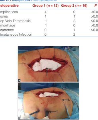
Fascial defect was exposed via incision over previous surgical scar and hernia sac incised to access into the abdomen. Adhesions were dissected and three laparoscopic trocars were inserted into the abdomen as mentioned above under direct vision without using versaport. EPTF mesh was laid under the fascial defect in abdomen and six prolane fixation sutures were used at each side. Then, mesh material left in abdomen and fascial defect was primarily closed. Afterwards, laparoscope inserted and mesh material was fixed to inner abdominal wall via tacker. Preoperative and intraoperative view of coombine technique [Figures 1 and 2].
Statistical method
The means and standard deviations of data were calculated for statistical analysis via SPSS Windows 16.0. The
P-value “< 0.05” was accepted as statistically significant.
RESULTS
Age, gender, length of hospital stay, mean operation time, mean size of fascial defect, ASA score and BMI values of both groups were shown in Table 1.
There is no significant difference in age, gender, BMI, length of hospital stay between two groups.
Postoperative complications were compared and shown in Table 2. In group 1, 4 patients suffered from persisting seroma formation. Three of them recovered after a long follow-up period. The difference is not found as statistically significant. The remaining one needed multiple drainages and then got recovered. In group 2, no seroma formation was identified. Three patients suffered from SSI in group 2, while no SSI
Table 1: Patients demographic data
Group 1 (n = 12) Group 2 (n = 16) P
Age (years) 56.58 (43-69) 59 (39-74) >0.05
Gender (M/F) 3/9 1/15 >0.05
BMI (kg/m2) 30 (26-33) 29.9 (25-38) >0.05
Operation time (min) 76.4 (45-110) 77.31 (50-120) >0.05 Length of hospital
stay (days)
2.4 (2-6) 2.8 (2-8) >0.05 Size of fascia defect (cm) 12.5 (10-18) 12.8 (10-19,5) >0.05
Table 2: Postoperative complications
Postoperative Group 1 (n = 12) Group 2 (n = 16) P
Complications 4 0 <0.05
Seroma 1 1 >0.05
Deep Vein Thrombosis 1 2 >0.05
Hemorrhage 1 0 >0.05
Recurrence 0 1 >0.05
Subcutaneous Infection 0 2
Figure 1: Preoperative view of giant incisional hernia
Figure 2: (a) The abdomen was opened by laparotomy and hernia sac dissected. Dual mesh was implanted (b) The trocars were inserted in an open techniques (T) and transfixation sutures were placed (S)
a
b
Journal of Minimal Access Surgery | October-December 2015 | Volume 11 | Issue 4 269 Ozturk, et al.: Laparoscopic giant hernia repair
was seen in group 1. One patient got myocardial infarct in group 2 at early postoperative period and managed with coronary stenting. Three patients, 1 in group 1 and remaining 2 in group 2, had hemorrhage with short-term decrease in hemoglobin values without need of blood transfusion. When compared of the late complication there was one evident deep vein thrombosis in group 1 and managed with short-term use of oral anticoagulants. In group 2, we did not see any thrombosis cases. No postoperative mortality seen in both groups.
Mean operation time was 76.4 (45-110) in group 1 and 77.31 (50-120) in group 2 that the difference is not found as statistically significant.
DISCUSSION
Incisional hernia is the term defines protrusion of peritoneum trough out a fascia defect emerges following an abdominal operation including a fascia repair. Incisional hernia develop in 2-23% of patients who had an abdominal surgery[1-7] and
classified as small (<5 cm), medium sized (5-10 cm) and large (>10 cm) incisional hernias according to defect size. Urgent surgical intervention may become mandatory for a significant rate of patients and some patients need to modify their life styles or even encounter to change their jobs. This is much likely to appear in patients who have large incisional hernias as those patients are mostly obese or have a history of many previous operations and additional medical challenges may usually accompany them. Numerous methods are used for reconstruction of incisional hernia varying from simple fascial closure to mesh hernioplasty or autodermal plasty.[8-11] As yet, no technique or approach has become the
gold standard for repair of an incisional hernia.[8] In recent
years, laparoscopic repair, even of large incisional hernias, has been performed at many centers, with similar morbidity to open repair.[11] However, long-term follow-up is needed to
elucidate whether laparoscopic repair of incisional hernias is efficacious.[11] Ultimately, the choice of technique is generally
determined by the surgeon’s preference, surgical tradition or even by the hospital’s economic situation.[8] Onlay mesh
hernioplasty is the commonly preferred method among surgical residents due to its technical convenience and short learning period. However, this method carries an increased risk of postoperative complications such as seroma formation, SSI, skin necrosis when compared to other methods. It is reported that Kingsnorth relaxation incisions may reduce recurrence rates and fibrin sealants may reduce seroma formation.[12,13]
Recurrence rate of Onlay technique is reported as 7-15% and postoperative complication rate is 10-58% without classification according to size of defect.[8,9,14] In large incisional
hernia, Baradan et al.[11] reported 7% as recurrence and 12% as
total complication rate in which they prefer Onlay mesh with relaxation incisions. Poelman et al. reported 16% as recurrence, 28% as seroma formation and 21% as SSI rate. Additionally, Gleysteen[14] reported 20% as recurrence rate and 22.4% as
surgical site complication rate. Finally, Trivelliet al.[15] reported
16.7% as mortality rate, 8.3% as SSI rate and 8.3% as recurrence rate in their study including patients who had large incisional hernias where abdominal curve was disappeared. In hybrid technique here we report, there is quite lower recurrence rate in our short-term follow-up period.
Laparoscopic technique for hernia repair became popular in recent years and carries many advantages such as reduced postoperative pain, length of hospital stay and recurrence rates in comparison with conventional open approach.[16] Although various modifications exist for
laparoscopic technique, postoperative morbidity has still been reported as 3-18% of cases.[17,18] Seroma formation is the
leading complication seen at postoperative period and usually arises in the cavity that is formed among dual mesh material, abdominal wall and skin due to intra-abdominal preparation of hernia sac. Seroma could sometimes be confused with recurrence early period. Besides, resolution of seroma may take a long time and affects the patient’s quality of life. Another challenging issue about a giant incisional hernia is high risk of recurrence. The success of an incisional hernia repair primarily depends on recurrence rates. Sixty-six to 90% of recurrences arise in first 2 years.[18] However, they may
be seen up to 10 years following the operation. Therefore, at least 3-5 years follow-up is necessary to make a precise analysis of recurrence rates. SSI is the dominant factor for recurrence.[9] Obesity, chronic pulmonary obstructive disease
(CPOD), large hernia, recurrent hernia, weight lifting activities during a day as a job, chronic constipation, inefficient fixation or inappropriate placement of mesh material are other risk factors affecting the recurrence rates. It is advised to lay mesh material as exceeds the defect at least 3 cm all around. Another advantage of laparoscopic technique is its anatomical concordance due to intra-abdominal placement of mesh material.[19] A total laparoscopic approach may not
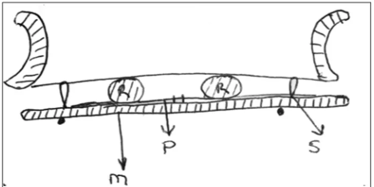
be feasible in large incisional hernia repair where intra-abdominal adhesions frequently seen and have difficulties on insertion of trocars and laying the mesh appropriately. Due to difficulties mentioned above, laparoscopy is not preferred in such cases. The hybrid technique here we report is primarily deals with giant hernias or those possess multiple sacs. The fundamental advantages of this technique are reduced risk of recurrence, overcome the difficulty of dissection and insertion of trocars in laparoscopic approach alone. Mesh position in the abdominal cavity [Figure 3].
Journal of Minimal Access Surgery | October-December 2015 | Volume 11 | Issue 4 270
Ozturk, et al.: Laparoscopic giant hernia repair
A few disadvantages of laparoscopic intraperitoneal placement of mesh material were reported in literature such as hernia sac left inside, the risk of visceral injuries while dissection of adhesions and no reconstruction of abdominal wall. An additional advantage of our technique is reduced risk of visceral injuries while dissection of adhesions.
SSI rate is higher in our technique when compared to conventional laparoscopic approach. However, higher SSI rates do not yield mesh removal due to intra-abdominal placement of mesh material similar to laparoscopic approach.
In addition, SSI rates usually increase following abdominal wall reconstruction. Thus, it is higher in our technique as a disadvantage of open approach combined with laparoscopy. The modified method reported here provides opportunity for abdominal wall reconstruction and has an advantage on facilitated insertion of thick dual-mesh materials into abdomen via open incision.
In conclusion, laparoscopic technique combined with open approach (hybrid approach) can safely be preferred in giant incisional hernias where laparoscopic approach would not be convenient. New studies are mandatory on this title.
REFERENCES
1. Flum DR, Horvath K, Koepsell T. Have outcomes of incisional hernia repair improved with time? A population–based analysis. Ann Surg 2003;237:129-35.
2. LeBlanc KA. Incisional hernia repair: Laparascopic techniques. World J Surg 2005;29:1073-9.
3. Olmi S, Scaini GC, Erba L, Croce E. Laparascopic versus open insicional hernia repair. Surg Endosc 2007;21:555-9.
4. Agarwal BB, Agarwal S, Mahajan KC. Laparascopic ventral hernia repair: Innovative anatomical closure mesh insertion without 10 mm transmyofascial port, and atraumatic mesh fixaation: A preliminary experience of a new technique. Surg Endosc 2009;23:900-5.
5. Palanivelu C, Rangarajan M, Parthasathi R, Madankumar MV, Senthilkumar K. Laparascopic repair of suprapubic insicional hernias: Suturing and intraperitoneal composite mesh onlay. A retrospective study. Hernia 2008;12:251-6.
6. Schoenmaeckers E, Wassenaar EB, Raymakers JT, Rakic S. Bulging of the mesh after laparascopic repair of ventral and insicional hernias. JSLS 2010;14:541-6.
7. Sharma D, Jindal V, Pathania OP, Thomas S. Novel technique for closure of defect in laparascopic ventral hernia repair. J Minim Access Surg 2010;6:86-8.
8. Shrivastava A, Gupta A, Gupta A, Shrivastava J. Erosion of small intestine with necrotising fasciitis of over lying abdominal wall after expanded poly-tetrafluoroethylene mesh implantation: A rare complication after laparoscopic incisional hernia repair. J Minim Access Surg. 2013;9:138-40. doi: 10.4103/0972-9941.115381. PubMed PMID: 24019695.
9. Sajit MS, Parampalli U, McFall MR. A meta-analysis comparing tacker mesh fixation with suture mesh fixation in laparascopic insicional and ventral hernia repair. Hernia 2013;17:159-66.
10. Clapp ML, Hicks SC, Awad SS, Liang MK. Trans-cutaneus Closure of Central Defect (TCCD) in laparascopic ventral hernia repair(LVHR). World J Surg 2012;37:42-51.
11. Baradaran H, Peyvandi H, Hallaj-Mofrad HR, Ahmadi-Amoli H, Yazdankhah-Konari A. Combined fascia and mesh closure of large incisional hernias. Arch Iran Med 2008;11:282-5.
12. Kingsnorth A. The management of incisional hernia. Ann R Coll Surg Engl 2006;88:252-60.
13. Poelman MM, Langenhorst BL, Schellekens JF, Schreurs WH. Modified onlay technique for the repair of the more complicated incisional hernias: Single-centre evaluation of a large cohort. Hernia 2010;14:369-74.
14. Gleysteen JJ. Mesh-reinforced ventral hernia repair: Preference for 2 techniques. Arch Surg 2009;144:740-5.
15. Trivellini G, Bagni CM, Sollini A, Senni M, Leone S, Contessini Avesani E. Repair of giant hernias using more prosthesis. Hernia 2001;5:124-8. 16. Hamy A, Pessaux P, Mucci-Hennekinne S, Radriamananjo S, Regenet
N, Arnaud JP. Surgical treatment of large ıncisional hernias by an intraperitoneal Dacron mesh and an aponeurotic graft. J Am Coll Surg 2003;196:531-4.
17. Chowbey PK, Sharma A, Khullar R, Mann V, Baijal M, Vashistha A. Laparoscopic ventral hernia repair. J Laparoendosc Adv Surg Tech A 2000;10:79-84.
18. Heniford BT, Park A, Ramshaw BJ, Voeller G. Laparoscopic ventral and incisional hernia repair in 407 patients. J Am Coll Surg 2000;190:645-50. 19. Cassar K, Munro A. Surgical treatment of incisional hernia. Brit J Surg
2002;89:534-45.
Cite this article as: Ozturk G, Malya FU, Ersavas C, Ozdenkaya Y, Bektasoglu H, Cipe G, et al. A novel reconstruction method for giant incisional hernia: Hybrid laparoscopic technique. J Min Access Surg 2015;11:267-70.
Date of submission: 20/11/2013, Date of acceptance: 18/12/2013 Source of Support: Nil, Conflict of Interest: None declared. Figure 3: Schematic drawing of the repair. Dual mesh (M) was placed below
the rectus(R) muscles and peritoneum, and hernia defect, which is covered by peritoneum and hernia sac(P), transfixation sutures were placed (S)