IDENTIFICATION OF
VERY LOW DENSITY LIPOPROTEIN RECEPTOR (VLDLR) MUTATIONS IN CEREBELLAR HYPOPLASIA AND QUADRUPEDAL LOCOMOTION
(UNERTAN SYNDROME) IN HUMANS
A THESIS SUBMITTED TO
THE DEPARTMENT OF MOLECULAR BIOLOGY AND GENETICS AND THE INSTITUTE OF ENGINEERING AND SCIENCE OF
BILKENT UNIVERSITY
IN PARTIAL FULFILLMENT OF THE REQUIREMENTS FOR THE DEGREE OF MASTER OF SCIENCE
BY ŞAFAK ÇAĞLAYAN JULY 2008
I certify that I have read this thesis and that in my opinion it is fully adequate, in scope and in quality, as a thesis for the degree of Master of Science.
Prof. Tayfun Özçelik
I certify that I have read this thesis and that in my opinion it is fully adequate, in scope and in quality, as a thesis for the degree of Master of Science.
Prof. Nurten Akarsu
I certify that I have read this thesis and that in my opinion it is fully adequate, in scope and in quality, as a thesis for the degree of Master of Science.
Assoc. Prof. Işık Yuluğ
Approved for the Institute of Engineering and Science
Director of Institute of Engineering and Science Prof. Mehmet Baray
i
ABSTRACT
IDENTIFICATION OF
VERY LOW DENSITY LIPOPROTEIN RECEPTOR (VLDLR) MUTATIONS IN CEREBELLAR HYPOPLASIA AND QUADRUPEDAL LOCOMOTION
(UNERTAN SYNDROME) IN HUMANS
ġafak Çağlayan
M.S in Molecular Biology and Genetics Supervisor: Prof. Tayfun Özçelik
July 2008, 134 Pages
Cerebellar hypoplasia and quadrupedal locomotion in humans, also known as Unertan syndrome, is a severe neurodevelopmental condition accompanied by dysarthria and impaired cognitive skills. The molecular underpinnings of development of the brain structures required for bipedal gait in humans can be established through identification of the gene(s) associated with this disorder. Four consanguineous families from Turkey exhibiting this autosomal recessive trait were studied. In two families (A and D), affected individuals shared homozygosity in a critical 1.032-Mb region in chromosome 9p24. Sequence analysis linked the disease to two distinct mutations in the very low density lipoprotein receptor (VLDLR) gene; the nonsense change R257X in family A and the single nucleotide deletion leading to frameshift I780TfsX3 in family D. VLDL receptor is a co-receptor of reelin molecule. Reelin signaling pathway is involved in neuronal migration and lamination to form brain cortices during embryonic development. Mutant VLDL receptors are truncated proteins that cannot be inserted into the membrane. Homozygosity mapping linked the disease locus in family B to chromosome 17p13. Family C does not share homozygosity in neither of the loci.
ii
ÖZET
ĠNSANDA SEREBELLAR HĠPOPLAZĠ VE QUADRUPEDAL YÜRÜMEDE (ÜNERTAN SENDROMU) DÜġÜK YOĞUNLUKLU LĠPOPROTEĠN
RESEPTÖRÜ (VLDLR) MUTASYONLARININ TESPĠT EDĠLMESĠ
ġafak Çağlayan
Moleküler Biyoloji ve Genetik Yüksek Lisans DanıĢman: Prof. Tayfun Özçelik
Temmuz 2008, 134 Sayfa
Serebellar hipoplazi ve quadrupedal yürüme şiddetli bir nörogelişimsel hastalıktır. Unertan sendromu olarak da bilinen bu duruma disartirik konuşma ve zeka geriliği eşlik etmektedir. İnsanda bipedal yürüme için gerekli beyin yapılarının gelişmesinin moleküler temelleri, bu bozukluğa yol açan gen(ler)in bulunması ile kurulabilir. Çalışmada, otozomal resesif hastalığı taşıyan Türkiye’deki dört akraba aile incelenmiştir. Ailelerin ikisinde (A ve D), hasta bireyler 9p24 kromozomunda 1.032-Mb büyüklüğündeki bir kritik bölgede homozigosite göstermektedirler. Dizi analizi, hastalığı düşük yoğunluklu lipoprotein reseptörü (VLDLR) genindeki iki farklı mutasyona bağlamıştır; A ailesindeki R257X anlamsız değişim ve D ailesindeki I780TfsX3 çerçeve kaymasına yol açan tek nükleotidlik delesyon. VLDL reseptörü reelin molekülünün reseptörlerinden birisidir. Reelin sinyal yolağı embriyonik gelişim sürecinde beyin korteksinin oluşması için nöronların taşınmasında ve tabakalanmasında rol oynamaktadır. Mutant VLDL reseptörleri hücre zarına eklenemeyecek kesik proteinler olarak oluşmaktadırlar. Homozigosite haritalaması B ailesindeki hastalık bölgesini 17p13 kromozomuna bağlamıştır. C ailesi iki bölgenin hiçbirisi ile homozigosite paylaşmamaktadır.
iii
TO MY PARENTS, ZEHRA AND AZĠZ ÇAĞLAYAN FOR THEIR LOVE AND SUPPORT
iv
ACKNOWLEDGEMENTS
It would not be easy to thank enough all people with whom I did this study. Yet, I will try…
Foremost, I would like to thank and express my sincerest gratitude to my supervisor Prof. Tayfun Özçelik for his guidance, encouragement, and support throughout my thesis work. I have learned a lot from his scientific and personal
advices. Without his vision and commitment, the project would have never reached the great result it did.
I am indebted to Prof. Nurten Akarsu since she did a great part of this study with performing genomic mapping studies. More than that, her cheerful ambiance always motivated me during our studies.
I am really very lucky for meeting Elif Uz who was my second advisor. I learnt a lot about life from her and I wish she would continue to be one of my best fellows with her immense compassion.
I would like to thank Chigdem Mustafa Aydın, Melda Kantar, Emre Onat and Süleyman Gülsüner. We shared several things that I cannot write down all along with benches and desks.
I have been very happy to be a member of Bilkent MBG family. It would be very difficult to find such a lovely people in such a peaceful environment.
I am very grateful to all my friends for whom I need a chapter to mention all. Nevertheless, my special “big thanks” go to Hande Koçak, Ceren Sucularlı, Tolga Acun, Bala-Gur Dedeoglu and Zeynep Keskin-Tokcaer for their sincere friendship.
It is impossible to express my endless love and thanks to my family. I will forever grateful to them…
v
TABLE OF CONTENTS
ABSTRACT I
ÖZET II
DEDICATION PAGE III
ACKNOWLEDGEMENTS IV
TABLE OF CONTENTS V
LIST OF TABLES XI
LIST OF FIGURES XII
ABBREVIATIONS XIV
PART I: INTRODUCTION 1
1.1 Quadrupedal Locomotion in Humans: Unertan Syndrome 1
1.1.1 Families described in Turkey 1
1.2 Ataxia 5
1.2.1 Autosomal dominant ataxias 9
1.2.2 Autosomal recessive ataxias 12
vi
1.3 Nervous System Development 16
1.3.1 Neuronal migration and brain development 16 1.3.1.1 Formation of the cerebral cortices 16
1.3.1.2 Laminating the cerebellum 18
1.3.2 Mouse models with neurodevelopmental defects 20
1.3.2.1 Reeler mouse 20
1.3.2.2 Scrambler and yotari mice 25
1.3.2.3 Identification of reelin and Dab1 genes 26 1.3.2.4 VLDLR and ApoER2 knock-out mice 26
1.3.3 Reelin signaling pathway 28
1.4 Aim and Strategy 29
PART II: MATERIALS and METHODS 30
2.1 Recruitment of Families 30
2.2 DNA and RNA Samples 30
2.2.1 Sample collection 30
2.2.2 DNA isolation from blood samples 31
2.2.3 Amplification of purified genomic DNA 31
2.2.4 RNA isolation from blood samples 31
2.2.5 cDNA synthesis 31
2.3 Genetic Mapping 32
2.3.1 Genome-wide linkage analysis 32
vii
2.4 Candidate Gene Analysis 39
2.4.1 Polymerase Chain Reaction (PCR) 39
2.4.1.1 Primers 39
2.4.1.2 PCR conditions 39
2.4.1.3 Agarose gel electrophoresis 41
2.4.2 Mutation search 42
2.4.2.1 Sequencing reactions 42
2.4.2.2 Data analysis 42
2.4.3 Restriction digestion assay 42
2.4.4 Quantitative real-time reverse transcriptase PCR (Q-PCR) 43
2.5 Solutions and Buffers 44
PART III: RESULTS 45
3.1 Clinical Assessment 45
3.2 Chromosomal Loci Associated With Quadrupedal Locomotion 52 3.2.1 Genetic heterogeneity; Unertan syndrome type I 57
3.3 Candidate Gene Analysis in 9p24 57
3.3.1 VLDLR as a candidate gene 57
3.3.2 VLDLR sequencing in families A and D 58 3.3.3 Restriction endonuclease based mutation screening in 60 families A and D
3.3.4 VLDLR sequencing in families B and C 62 3.3.5 Restriction enzyme based mutation screening in 62 control individuals
3.4 Quantitative Analysis of VLDLR Expression in Affected and 63 Unaffected Individuals
viii
PART IV: DISCUSSION and CONCLUSION 66
4.1 Unertan Syndrome vs. Disequilibrium Syndrome 69 4.2 Is Quadrupedal Locomotion an Environmental Adaptation 72
4.3 Future Prospects 73
PART V: REFERENCES 74
PART VI: APPENDICES 86
xi
LIST OF TABLES
Table 1.1 Sporadic spinocerebellar syndromes 7
Table 1.2 Summary of the molecular features of spinocerebellar ataxias 11 Table 1.3 Summary of the molecular features of autosomal recessive ataxias 14
Table 1.4 Summary of the molecular features of X-linked ataxias 15
Table 2.1 Part of pedigree file for family A 35
Table 2.2 Part of data file for family A 36
Table 2.3 Part of map file for family A 37
Table 2.4 Part of model file for family A 38
Table 2.5 Primers that were used for sequencing of VLDLR gene 40 Table 3.1 Physical and neurological findings of the patients in families A and D 48 Table 4.1 Physical, radiological, and genetic characteristics of the 70
xii
LIST OF FIGURES
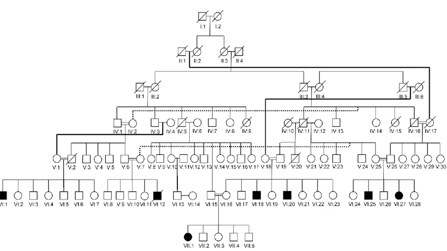
Figure 1.1 Pedigree of family A residing in southeastern Turkey. 2
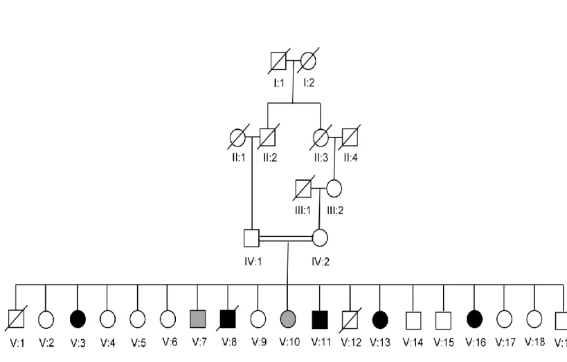
Figure 1.2 Pedigree of family B. 3
Figure 1.3 Pedigree of family C. 4
Figure 1.4 Pedigree of family D. 5
Figure 1.5 Development of the cerebral cortex. 17
Figure 1.6 Neuronal migrations during development of the cerebellum. 19 Figure 1.7 Malpositioning of neurons in the reeler neocortex. 22 Figure 1.8 Histological appearance of the normal and reeler telencephalon. 23 Figure 1.9 Schematic and histological appearance of the normal, reeler 24
and scrambler cerebellum
Figure 2.1 Workflow of GeneChip® mapping 10K and 500K assays 33 Figure 2.2 Sizes of the fragments of pUC Mix Marker 8 and appearance 41
on agarose gel electrophoresis.
Figure 3.1 Quadrupedal palmigrate walking of Unertan syndrome patients 46 Figure 3.2 Neuroradiological images of affected individuals. 51 Figure 3.3 Homozygosity mapping of cerebellar hypoplasia and 53
quadrupedal locomotion to chromosome 9p24.
Figure 3.4 Haplotype analysis of family A using SNP markers from 15q 54 residing between the 55- and 65-Mb region.
xiii
Figure 3.5 Exclusion of chromosome 9p24 in family C with haplotype 55 Figure 3.6 Haplotype and exclusion analyses of family C using polymorphic 56
markers from 17p13 critical region.
Figure 3.7 Chromatograms presenting VLDLR c769C->T mutation in 59 family A and VLDLR c2339delT mutation in family D.
Figure 3.8 Restriction enzyme based mutation screening in family A 61 and family D.
Figure 3.9 Screening of c769C->T and c2339delT mutations in control 63 individuals.
Figure 3.10 PCR amp/cycle graph for VLDLR. 64
Figure 3.11 Melt curve graph for VLDLR. 64
Figure 3.12 Quantitative analysis of VLDLR expression in patients and controls. 65 Figure 4.1 Schematic representation of the mutations in exons and 68
xiv
ABBREVIATIONS
ApoER2 apolipoprotein E receptor 2
bp base pair
ddH2O deionized water
del deletion
DNA deoxyribonucleic acid
dNTP deoxynucleotide triphosphates
E embryonic-day
MgCl2 magnesium chloride
mM milimolar
μl microliter
mRNA messenger RNA
PCR polymerase chain reaction Q-PCR Quantitative real-time PCR
RNA ribonucleic acid
RT-PCR reverse transcriptase PCR
SCA spinocerebellar ataxia
SNP single nucleotide polymorphism
TAE tric-acetic acid-EDTA
1
PART I: INTRODUCTION
1.1 Quadrupedal Locomotion in Humans: Unertan Syndrome
This unique phenotype was first described by Prof. Uner Tan in a consanguineous family living in southern Turkey. Non-progressive cerebellar hypoplasia, quadrupedal locomotion, dysarthric speech and mental retardation are the hallmarks of this condition, which is named as Unertan syndrome based on the name of discoverer (Tan, 2005).
1.1.1 Families described in Turkey
Initially we studied family A, a consanguineous kindred living in southeast region of Turkey (Figure 1.1). There are seven affected individuals in this family. All affected individuals walk quadrupedally. There is one girl who can walk bipedally, but she prefers quadrupedal locomotion. Other main features of the affected individuals are cerebellar hypoplasia, impaired cognitive skills, and primitive language (Tan et al., 2008a; Ozcelik et al., 2008a).
Family B is the first identified family (Figure 1.2). Five of 19 children walk in a quadrupedal fashion. Two children using bipedal locomotion have a neurological phenotype similar to that of their quadruped siblings (Tan, 2005; Turkmen et al., 2006).
Another family exhibiting Unertan syndrome is family C residing in southern Turkey (Figure 1.3). There are four affected individuals and two of them ambulate quadrupedally. One of the remaining two is a biped ataxic man who was a quadruped during childhood, and the other patient, who was also a quadruped, is no longer able to ambulate now (Tan, 2006b).
2
Figure 1.1 Pedigree of family A residing in southeastern Turkey. Filled symbols represent the affected individuals. Squares indicate males, and circles indicate females. All affected individuals are resulting from consanguineous marriages.
3
Figure 1.2 Pedigree of family B. Parents of the affected children are second cousins. Two children, who use bipedal locomotion, but have a neurological phenotype similar to that of their quadruped siblings are shown with gray symbols.
4
5
Family D was encountered during the course of our studies (Figure 1.4). This consanguineous family is from western Turkey and all of the three affected individuals are quadrupeds (Tan, 2008b; Ozcelik et al., 2008a).
Figure 1.4 Pedigree of family D.
1.2 Ataxia
The word ataxia means “absence or loss of order” and the term ataxia is generally used to describe uncoordinated walking. Actually, ataxia is neither a specific disease nor a disorder. It is a set of symptoms affecting coordination of gait, speech (dysarthria), hands, arms, and eye movements (nystagmus).
Atrophy of cells in the cerebellum or its connections, diseases causing cerebellar or spinocerebellar degeneration, or damages to cerebellar/spinal structures are the most common causes of ataxia.
6
There are several medical and neurological conditions that can lead to ataxia. According to the National Ataxia Foundation of the USA, ataxia can appear in people suddenly as a result of one or more of these conditions: head trauma, stroke, brain hemorrhage, brain tumor, congenital abnormality, severe viral infection, exposure to certain drugs or toxins (i.e. alcohol, anti-convulsive medicines), cardiac or respiratory arrest. Additionally there are several conditions that may result in ataxia over time. These are hypothyroidism, deficiencies of certain vitamins (E, B12), exposure to certain drugs or toxins (heavy metals, anti-convulsive medicines, chronic alcohol exposure, and certain cancer drugs), congenital abnormality, multiple sclerosis, syphilis (locomotor ataxia), hereditary disorders, idiopathic (unknown cause) cerebellar degeneration disorders (Official website of the National Ataxia Foundation of the USA; http://www.ataxia.org/).
Cerebellar/spinocerebellar syndromes are a heterogenous group of neurological disorders causing ataxia. Patients with spinocerebellar syndromes exhibit classical ataxia features like disequilibrium, progressive incoordination of gait and limbs, and speech and eye movement disturbances. Other neurological symptoms including pyramidal or extrapyramidal signs, spasticity,
ophthalmoplegia, and dementia can be also found in patients (Mariotti & Di Donato, 2001).
Ataxic individuals can display different symptoms and severity of the disease can be variable among patients. Because of this variability in phenotypes and neuropathological findings, making a proper clinical classification and differential diagnosis is very difficult. Nevertheless, certain types of ataxias can be differentiated with the help of additional specific symptoms. Also other affected family members can be useful in diagnosis in the case of hereditary ataxias. Hence, spinocerebellar syndromes can be distinguished into two forms: 1) sporadic ataxias of unknown cause or that are acquired due to associated illness and 2) inherited spinocerebellar ataxias (Mariotti & Di Donato, 2001).
7
It is a challenging task for clinicians to diagnose sporadic ataxia since they should rule out every possibility of hereditary ataxia before making a final
diagnosis. Phenotypes with multiple system atrophy are the major group in the set of sporadic spinocerebellar syndromes of unknown cause. Acquired or non-hereditary spinocerebellar syndromes can be due to certain environmental factors including alcoholism or vitamin deficiencies. Multiple sclerosis, vascular
diseases, and tumors or paraneoplastic syndromes can also cause cerebellar
ataxias (Table 1.1) (Mariotti & Di Donato, 2001). Conditions resulting in sporadic ataxia can affect only the cerebellum (“pure cerebellar” form) or they can cause additional symptoms such as neuropathy (dysfunction of the peripheral nerves), dementia (impaired intellectual function), weakness, rigidity, or spasticity of the muscles (“cerebellar plus” form) (Official website of the National Ataxia
Foundation of the USA; http://www.ataxia.org/).
Table 1.1 Sporadic spinocerebellar syndromes. (Mariotti & Di Donato, 2001) Unknown causes
Multiple system atrophy
Olivopontocerebellar atrophy Striatonigral degeneration Shy-Drager syndrome Immune
Multiple sclerosis
Glutamic acid decarboxylase antibody Paraneoplastic syndromes
Systemic diseases Celiac diseases
8
Hereditary ataxias are neurological disorders caused by a defect in a certain gene that is inherited through families. This group of diseases is
characterized by progressive degeneration of the cerebellum and spinocerebellar tracts of the spinal cord accompanied by various pathologies in central and peripheral nervous systems (Di Donato et al., 2001).
Since clinical phenotypes of different genotypes of inherited ataxias show significant overlap, it is difficult to make a classification based on clinical or histopathological findings (Banfi & Zoghbi, 1994). So, it is more useful to divide hereditary cerebellar/spinocerebellar syndromes according to the inheritance mode as autosomal dominant, autosomal recessive, X-linked and mitochondrial
disorders (Mariotti & Di Donato, 2001).
There is no specific treatment for directly curing diseases that cause ataxia. Aims of therapies are generally to diminish the severity of symptoms. For
example if ataxia is due to a stroke, vitamin deficiency, or exposure to a toxic drugs or chemicals, then treatment would target specific ailment. Physical therapy is a widely used option for people with impaired gait. Furthermore, ataxic
individuals can use adaptive devices (i.e. a cane, crutches, walker, or wheelchair to help for walking; devices to assist feeding, and self cares for patients having difficulties in hand and arm coordination; and communication devices if speech is impaired) to be able to live independently.
9 1.2.1 Autosomal dominant ataxias
Autosomal dominant cerebellar/spinocerebellar ataxias are a complex group of neurodegenerative diseases. These disorders are clinically heterogeneous since affected individuals exhibit several overlapping neurological symptoms. These symptoms include loss of balance and motor coordination, dysarthric speech, ophthalmoplegia, pyramidal and extrapyramidal signs, dementia, pigmentary neuropathy, peripheral neuropathy, oculomotor disturbances of cerebellar and supranuclear genesis, retinopathy, optic atrophy, spasticity, sphincter disturbances, cognitive impairment, and epilepsy (Duenas et al., 2006; Zoghbi, 2000; Schols et al., 2004).
The prevalence of the autosomal dominant ataxias is assumed as three cases per 100,000 individuals, although real number can be higher. Different ethnic and continental populations show diverse prevalence rates for specific types because of heterogeneity of the disorders (Schols et al., 2004). Onset of ataxia symptoms is generally between 30 and 50 years of age. However, there are individuals with disease onset in childhood or at later decades after 60 years (Duenas et al., 2006).
10
Disease onset in adulthood, occurrence of anticipation (disease onset becomes at earlier ages and disease become more severe in successive
generations) and atrophy of cerebellum are common clinical features of autosomal dominant ataxias. Nonetheless, variable and unpredictable association of signs of central nervous system and peripheral nervous system can accompany common symptoms. Because of this, clinical classification is complicated. Yet, autosomal dominant ataxias have been divided into three groups based on clinical findings: ADCA (Autosomal dominant cerebellar ataxia) I, ADCA II and ADCA III (Harding, 1993; Duenas et al., 2006). This type of classification and diagnosis is especially useful for patient management.
On the other hand identification of genetic basis of most of these disorders have had a significant impact on nosology, diagnostic procedures and the
management of patients. It led to the development of a new classification system based on molecular genetic data, and improvements in diagnosis and patient management (Di Donato, 1998). In this systematic, autosomal dominant ataxias are distinguished into 29 defined subtypes, of which the causative genes and pathogenic mutations are known in 15 (Table 1.2) (Duenas et al., 2006, Schols et
al., 2004). Molecular genetics approaches have revealed that the main pathogenic
mechanisms of these disorders include abnormal proteins or RNAs, which “gain a toxic function”, and lead to neuronal and systemic dysfunction (Gatchel &
11
Table 1.2 Summary of the molecular features of spinocerebellar ataxias. (Duenas et al., 2006)
SCA subtype Genomic location Gene/locus Protein Mutation
SCA1 6p23 ATXN1 Ataxin 1 CAG repeat
SCA2 12q24 ATXN2 Ataxin 2 CAG repeat
SCA3 14q24.3-q31 ATXN3 Ataxin 3 CAG repeat
SCA4 16q24-qter SCA4 U U
SCA5 11q13 SPTBN2 Beta-III spectrin D, MM
SCA6 19p13 CACNAIA CACNAIA CAG repeat
SCA7 3p21.1-p12 ATXN7 Ataxin 7 CAG repeat
SCA8 13q21 KLHLIAS Kelch-like I CTG repeat
SCA9 Reserved U U U
SCA10 22q13 ATXN10 Ataxin 10 ATTCT repeat
SCA11 15q15.2 TTBK2 TTBK2 D, I
SCA12 5q31-q33 PPP2R2B PPP2R2B CAG repeat
SCA13 19q13.3-q13.4 KCNC3 KCNC3 MM
SCA14 19q13.4 PRKCG PRKCG MM
SCA15 3p24.2-pter U U U
SCA16 8q23-q24.1 U U U
SCA17 6q27 TBP TBP CAG repeat
SCA18 7q22-q32 U U U SCA19* 1p21-q21 U U U SCA20 11p13-q11 U U U SCA21 7p21.3-p15.1 U U U SCA22* 1p21-q23 U U U SCA23 20p13-p12.3 U U U SCA24 1p36 U U U SCA25 2p21-p13 U U U SCA26 19p13.3 U U U SCA27 13q34 FGF14 FGF14 MM SCA28 18p11.22-q11.2 U U U SCA29 3p26 U U U
DRPLA 12p13.31 ATN1 Atrophin 1 CAG repeat
Undefined** 16q22.1 PLEKHG4 Puratrophin 1 5’SNS
* SCAs 19 and 21 are likely allelic forms of the same gene.
** The gene encoding Puratrophin 1 lies on the same chromosomal region where the SCA4 gene localizes.
Genes in genomic location are noted according to Ensembl. D, deletions; I, insertions; MM, missense mutations; SCA, spinocerebellar ataxia; SNS, single-nucleotide substitutions; U, unknown.
12 1.2.2 Autosomal recessive ataxias
Autosomal recessive ataxias are a group of rare and clinically heterogeneous neurological diseases. The most important symptom of these disorders is spinocerebellar ataxia, involving the cerebellum, brainstem, or spinocerebellar long tracts. Patients exhibit poor balance with falls, imprecise hand coordination, postural or kinetic tremor of the extremities or trunk,
dysarthria, dysphagia, vertigo, and diplopia (Perlman, 2004). Various neurologic, ophthalmologic and systemic pathologies affecting both central and peripheral nervous system, and in some cases other systems and organs can accompany ataxia (Palau & Espinos, 2006).
These phenotypic features can be so rare. Yet, they are important to make a differential diagnosis and to decide on further therapies. Moreover, since these disorders are clinically heterogeneous, detailed assessments and additional diagnostic studies (i.e. neuroimaging or electrophysiological examinations) are required to make a proper differentiation. For example atrophy of cerebellum is a useful distinguishing feature and neuroimaging can be particularly useful to confirm clinical findings. Performing genetic tests/mutation analysis in diagnosis is also possible for the diseases for which causative genes were identified (Di Donato et al., 2001; Fogel & Perlman, 2007). Ataxia associated with deficiency of coenzyme Q10 and abetalipoproteinemia are the only types of recessive ataxias that have specific treatments (Espinos-Armero et al., 2005).
13
Until recent years characterization of autosomal recessive ataxias
remained poor when compared to dominant ataxias. Several conditions have been genetically described in the last few years. It has been found that most of the recessive ataxias are associated with a “loss of function” of specific cellular proteins involved in metabolic homeostasis, cell cycle, and DNA repair/protection processing (Di Donato et al., 2001). Generally each disease of this group is
caused by defect(s) in a specific gene. Nonetheless, some recessive ataxias are genetically heterogeneous and can be caused by mutation(s) in more than one gene (Palau & Espinos, 2006).
The prevalence of autosomal recessive cerebellar ataxias has been
estimated to be 7 in 100,000 individuals (Espinos-Armero et al., 2005). Among a large number of rare recessive disorders, Freidriech ataxia is the most common type in Caucasian populations (estimated prevalence is 2-4 cases in 100,000). Other frequent types include ataxia telangiectasia (1-2.5/100,000) and early onset cerebellar ataxia with retained tendon reflexes (1/100,000) (Palau & Espinos, 2006). Remaining types are much rare and prevalence of some can be variable among different regions (Di Donato et al., 2001). For most of the autosomal recessive ataxias disease onset is before age 20 years (Fogel & Perlman 2007).
Based on pathogenic mechanisms five main types of autosomal recessive ataxias can be distinguished: 1) congenital (developmental disorder); 2) ataxias associated with metabolic disorders including ataxias caused by enzymatic defects; 3) ataxias due to DNA repair defects; 4) degenerative and progressive ataxias that include ataxias with known cause and pathogenesis (such as Friedrich ataxia), and ataxias of unknown etiology; 5) ataxias associated with
other/additional features (Table 1.3) (Espinos-Armero et al., 2005; Palau & Espinos, 2006).
14
Table1.3 Summary of the molecular features of autosomal recessive ataxias (Palau & Espinos, 2006)
Protein (Gene or Locus) Location Congenital ataxias
Joubert syndrome
JBTS 1 (cerebelloparenchymal disorder IV, CPD IV) U 9q34.3
JBTS 2 (CORS2) U 11p12-q13.3 JBTS 3 AHI1 6q23.3 JBTS 4 (nephronophthisis 1) NPHP1 2q13 JBTS 5 CEP290 or NPHP6 12q21.3 JBTS 6 TMEM67 8q21.13-q22.1 JBTS 7 RPGRIP1L 16q12.2
Cayman ataxia Cayataxin (ATCAY) 19p13.3
Metabolic ataxias
Ataxia with isolated vitamin E deficiency (AVED) α-TTP (TTPA) 8q13.1-q13.3
Abetalipoproteinemia MTP 4q22-q24
Cerebrotendinous xanthomatosis CYP27A1 2q33-qter
Refsum disease PAHX or PHYH 10pter-p11.2
PEX7 6q22-q24
DNA repair defects
Ataxia telangiectasia ATM 11q22.3
Ataxia with oculomotor apraxia 1 (AOA 1) Aprataxin (APTX) 9p13.3
Ataxia with oculomotor apraxia 2 (AOA 2) or SCAR1 Senataxin (SETX) 9q34
Ataxia telangiectasia-like disorder (ATLD) MRE11A 11q21
Spinocerebellar ataxia with axonal neuropathy (SCAN1) TDP1 14q31-q32
Xeroderma pigmentosum (XP) NPHP6 12q21.32
XP of complementation group A XPA 9q22.3
XP of complementation group B XPB/ERCC3 2q21
XP of complementation group C XPC 3p25
XP of complementation group D XPD/ERCC2 19q13.2-q13.3
XP of complementation group E XPE (DDB2) 11p12-p11
XP of complementation group F XPF/ERCC4 16p13.3-p13.13
XP of complementation group G XPG/ERCC5 13q33
XP variant (XPV) or XP with normal DNA repair rates POLH 6p21.1-p12
Degenerative ataxias
Friedrich ataxia Frataxin (FRDA) 9q13
Mitochondrial recessive ataxic syndrome (MIRAS) Polymerase γ (POLG) 15q25
Charlevoix-Saguenay spastic ataxia Sacsin (SACS) 13q12
Early onset cerebellar ataxia with retained tendon reflexes
(EOCARR) 13q11-q12
Infantile onset spinocerebellar ataxia (IOSCA) Twinkle (C10ORF2) 10q24
Marinesco-Sjögren syndrome(MSS)
Classical MSS SIL1 5q31
MSS with myoglobinuria CTDP1 18qter
Coenzyme Q10 deficiency with cerebellar ataxia ?
15 1.2.3 X-linked ataxias
X-linked ataxias are a group of rare and poorly characterized disorders. Relatively well described diseases in this group include fragile X tremor/ataxia syndrome, which is caused by an RNA gain-of-function-based mechanism (Orr & Zoghbi, 2007), and sideroblastic anemia with spinocerebellar ataxia (Table 1.4).
Table 1.4 Summary of the molecular features of X-linked ataxias
Disease Genomic location Gene/locus Protein Mutation
SCAX1 Xp11.21-q21.3 U U U SCAX2 U U U U SCAX3 U U U U SCAX4 U U U U SCAX5 Xq25-q27.1 U U U FXTAS Xq27.3 FMR1 FMRP CGG repeat
XLSA/A Xq13.1-q13.3 ABC7 ABC7 MM
FXTAS, Fragile X tremor/ataxia syndrome; MM, missense mutations; SCAX, X-linked spinocerebellar ataxia; U, unknown; XLSA/A, X-X-linked sideroblastic anemia and ataxia
16 1.3 Nervous System Development
1.3.1 Neuronal migration and brain development
There are several distinct structures, like cerebellum, neocortex and
thalamus, in the brain, which constitutes the central nervous system along with the spinal cord. These specific brain regions are assembled through connections between morphologically and functionally different neurons. Migration of similar neurons into discrete layers and their positioning in these layers during embryonic development is a key step in construction of unique neuronal structures.
1.3.1.1 Formation of the cerebral cortices
During embryogenesis, six-layered structure of the mammalian cortical plate is established through the serial migration of neurons from their proliferative regions to a final position within the cortex. This corticogenesis process is
comprised of three stages (Figure 1.5; Gleeson & Walsh, 2000). The first step of the neocortical development is formation of the preplate or primordial plexiform layer. Preplate is established above the ventricular zone and below the pial surface around embryonic-day (E) 10-12 in mice and weeks 8-9 in humans. Ventricular zone contains proliferative cells which give rise to cortical neurons. Second step is construction of the cortical layers. This step begins with migration of first group of cortical neurons to divide the preplate into a deeper sub-plate and a superficial marginal zone, forming cortical plate between these two layers (Marin-Padilla, 1998). During cortical plate stage which takes place in E 10-17 in mice and weeks 10-18 in humans, cortical neurons exit ventricular zone, penetrate through the sub-plate and form sequential cortical layers. This lamination step occurs in an inside-out fashion. Newly-generated neurons pass the sub-plate and older cortical plate neurons, and settle below Cajal-Retzius cells of the marginal zone. So, early-born cells of cortical plate remain in deeper layers and younger cells populate
17
more superficial layers (Angevine & Sidman, 1961). Degeneration of the sub-plate and lamination of the cortical sub-plate into six-layered neocortex is the last step of corticogenesis (Gleeson & Walsh, 2000). Cells migrate radially and
tangentially during development of these neuronal layers (O’Rourke et al., 1992). Cortical neurons most often migrate radially along glial cells spanning cerebral wall to reach their final layers (Hatten, 1999). Meanwhile, a fraction of cells turn within the intermediate zone and migrate orthogonal to the radial fibers
(O’Rourke et al., 1992).
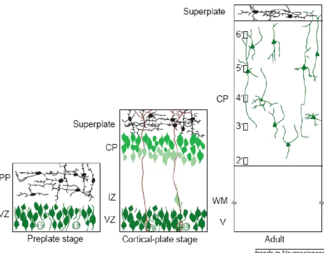
Figure 1.5 Development of the cerebral cortex. Neurons are deposited into cortical plate in an “inside-out” fashion (indicated by the progressive lighter shades of green in more superficial cortical plate neurons). Note radial alignment of the cortical neurons and the relatively cell-free marginal zone. CP, cortical plate; IZ, intermediate zone; MZ, marginal zone; PP, preplate; SP, subplate; VZ, ventricular zone; WM, white matter (Gleeson & Walsh, 2000)
18 1.3.1.2 Laminating the cerebellum
Cerebellum development is also carried through distinct steps. There are two different germinative zones where the cerebellar neurons are generated. Ventricular zone (also known as the ventricular germinative matrix) gives rise to precursor cells of the deep cerebellar nuclei and the Purkinje cells of the cerebellar cortex. Neurons of deep nuclei are the first generated cells at about E 10-12 in mice (Goldowitz & Hamre, 1998) and week 8 in human embryogenesis (Wang & Zoghbi, 2001). Afterwards, precursors of the Purkinje cells are produced between E 11-13 in mice (Goldowitz & Hamre, 1998) and in week 9 in humans (Hatten, 1999; Wang & Zoghbi, 2001). These precursor cells evolve to mature Purkinje cells. Maturation process continues until postnatal period. Projection of the Purkinje cells to the deep nuclei cells and refinement of input they receive from the inferior olivary complex also take place during this maturation. Purkinje cell precursors start to migrate towards the surface of cerebellum through the wall of cerebellar anlage. In the meantime precursors of the cerebellar granule neurons arise from the rhombic lip. Starting from week 11 in humans, the rhombic cells sweep across the cerebellar anlage, migrate over surface of the cerebellum and establish the external granular layer on the periphery of roof (Wang & Zoghbi, 2001). Beneath the external granular layer, the Purkinje cells establish the Purkinje cell plate and wait for inward migration of the granule cells (Miyata et
al., 1996, Hatten, 1999). External granular layer above the Purkinje cell plate is
the second germinative region of the cerebellum and gives rise to the granule cells. These cells migrate deeper into the cortex along the radially oriented Bergmann glial cells through the field of differentiating Purkinje cells (Wang & Zoghbi, 2001). Granule cells are arrested at their final destination deeper to the Purkinje cells. Three layers are formed at the end of these orchestrated migrations; an outer molecular layer of the granule cell axons and Purkinje cell dendrites, a layer of the Purkinje cells and an inner layer of the granule cells (Figure 1.6). During the course, Purkinje cell plate overlying the germinative zone functions as
19
a scaffold for proper lamination of the cerebellar cortex (Hatten, 1999). Inward migration of the granule cells continues after birth and the external granular layer disappears within the first year of life in humans (Wang & Zoghbi, 2001). The cerebellar nuclei, which are likely comprised of cells moved through nuclear transitory zone, take their position beneath the Purkinje cell plate. Remarkably, the Purkinje cells are also important in regulating cell proliferation in the external granular layer. Sonic hedhehog, a mitogenic factor, is produced and released by the Purkinje cells and act on precursors of the granule neurons to control
proliferation (Wechsler-Reya & Scott, 1999).
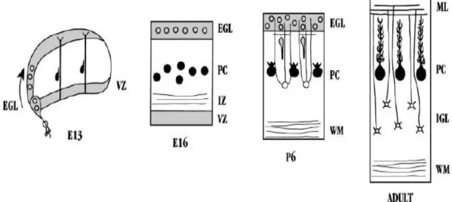
Figure 1.6 Neuronal migrations during development of the cerebellum. At embryonic period, while Purkinje cell precursors (filled circles) are migrating through the wall of the cerebellar anlage, precursors of the granule cells (unfilled circles) sweep across the roof. Afterwards, in the perinatal period, inward
migration of the granule cells takes place. Connections between laminated neurons are established during adulthood. EGL, external granular layer; IGL, internal granular layer; PC, Purkinje cells (Hatten, 1999)
20
1.3.2 Mouse models with neurodevelopmental defects
Reeler and trembler mice are the classic examples of mouse models with
congenital defects in the nervous system. These mice arose spontaneously, and were described more than 50 years ago (Falconer, 1951). They have been widely used to investigate role of neuronal migration in brain development, and to dissect molecular pathways regulating the choreographed migrations of neurons.
1.3.2.1 Reeler mouse
Reeler mouse was recognized with its “reeling” gait (Falconer, 1951).
Incoordination of motor skills and ataxic condition of reeler mouse was due to its disrupted cortical assembly. The principal defect is neuronal malpositioning resulting from abnormal migration (Rakic & Caviness, 1995; D’Arcangelo et al., 1995). Inside-out cell positioning during neurogenesis fails, and layers in the cerebral cortex, cerebellum, hippocampus and other laminated brain regions develop abnormally (Rakic & Caviness 1995; Rice & Curran, 1999).
Neurons of reeler mice arise from their birthplace (ventricular zone, subventricular zone and rhombic lip) without any problem. Also cells start their migration on their normal program. However, they cannot establish normal
architectural structures because they cannot find their proper location at the end of migration and fail to align into appropriate cell layers (Caviness, 1982; Rice & Curran, 1999). Yet, the rest of their differentiation program is unaffected. Neurons can differentiate to their destined classes, normal dendritic and axonal branching can be achieved, cells can establish successful connections with their targets, and gliogenesis and myelination are not directly affected. On the other hand since neurons do not stay at their normal location, the dendritic trees and axonal pathways are often formed abnormally. The cerebral cortex, the cerebellum, and the hippocampus are the most affected organs of reeler mice. Additionally,
21
abnormal development is observed in several other structures involving the spinal cord, brain stem, thalamus, midbrain, olivary complex, olfactory bulb, cochlear nuclei, facial nerve nucleus, retina, and tectum (Goffinet et al., 1984; Goffinet, 1984; Frost et al., 1986; Rakic & Caviness, 1995; Yip et al., 1998; Rice & Curran, 1999; Rice et al., 2001; Tissir & Goffinet, 2003).
Abnormal development of the reeler cerebrum can be obviously seen from beginning of cortical plate formation at E 16. In the reeler cortex, all major
neuron classes are generated on time in the ventricular zone and neuronal cohorts follow their normal migration schedule (Rice & Curran, 2001). Preplate of reeler mouse develops as in wild type mouse. Afterwards, however, the inside to outside neurogenesis in the cortical plate stage does not take place (Figure 1.7). Preplate cannot be divided into the marginal zone and the subplate due to failure in
invasion of the preplate by the first cohort of migrating cortical neurons (Hoffarth
et al., 1995; Ogawa et al., 1995; Sheppard & Pearlman, 1997; Rice & Curran,
1999). Subsequent waves of cortical neurons can start to migrate along radial glial cells in the intermediate zone. But, they cannot bypass their predecessors,
possibly because they do not penetrate the subplate or because they do not dissociate from radial cells properly (Pinto-Lord et al., 1982; Gleeson & Walsh, 2000). Instead, they populate the region beneath the subplate leading to an
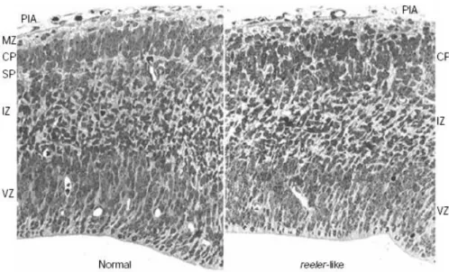
inversely laminated cortex (Ogawa et al., 1995). At the end of serial migrations in a wild type mouse cortex, the marginal zone remains relatively cell-free except for Cajal-Retzius and several other neurons. The six-layered cortical plate contains tightly packed cells which are aligned radially and the subplate assumes the position beneath the cortical plate (Rice & Curran 1999; 2001). On the other hand, in the reeler mouse the cortical plate is inverted and its radial alignment is lost (Hunter-Schaedle, 1997). Although neurons seem morphologically normal, their axons run obliquely in the cerebral wall and their dendrites follow tortuous paths (Goffinet, 1979). The Cajal-Retzius cells and subplate cells remain superficial to this disorganized cortical plate in a cell-dense region known as the superplate (Figure 1.8) (Derer, 1985; Rice & Curran 1999; 2001).
22
Figure 1.7 Malpositioning of neurons in the reeler neocortex. Development of the
reeler cerebral cortex is characterized with inability of neurons in penetrating
through the subplate. Instead, cortical cells remain underneath the preplate and late generated cells do not migrate past their predecessors (indicated by
progressive lighter shades of green in deeper cortical plate neurons). Superficial layer of the cortical plate is invaded by several neurons forming superplate structure. 6’-2’ numbers represent inverted layers of the cortical plate. Note
23
Figure 1.8 Histological appearance of the normal and reeler telencephalon. Disorganized cortical plate at embryonic day 14.5 is populated with cells in an oblique direction. (Tissir & Goffinet, 2003)
Most obvious phenotypic feature of reeler mouse is impaired gait coordination. This is probably due to severe developmental defect in its cerebellum. Number of the Purkinje cells in the reeler cerebellum is decreased when compared to the wild type. Radially migrating Purkinje cells stop
prematurely on their migration path. They cannot establish the Purkinje cell plate and instead, reside in ectopic clusters deep in the cerebellum (Figure 1.9)
(Goldowitz et al., 1997; Gallagher et al., 1998, Rice & Curran, 2001). Since Purkinje cells cannot migrate close enough to the granule cells, granule cells do not receive proliferation signals from the Purkinje cells and suffer a secondary degeneration. Moreover, the Purkinje cells cannot function as a structural scaffold for construction of other regions. Disruption of foliation of the cerebellar
structures leads to functional deficits in the cerebellum (Rice & Curran 1999; Tissir & Goffinet, 2003).
24
Figure 1.9 Schematic and histological appearance of the normal, reeler and
scrambler cerebellum. A) Cells in the external granular layer (egl) and in the
nuclear transitory zone (ntz) are reelin secreting cells (black circles) at early phases of development. Purkinje cell precursors (pp) express reelin receptors and Dab1, and migrate towards reelin secreting cells. B-C) Several days later,
Purkinje cell plate (pcp) is established underneath the egl as can be seen in the micrographs (arrowheads show pcp and arrows indicate egl). In the reeler and
scrambler cerebellum, Purkinje cells are unable to form the Purkinje cell plate.
25
Development of the reeler hippocampus is also characterized by abnormal migration and malpositioning of neurons. Hippocampus remains poorly
laminated; pyramidal cell layer cannot be established and granule cells reside in a disorganized dentate gyrus (Sweet et al., 1996; Tissir & Goffinet, 2003).
1.3.2.2 Scrambler and yotari mice
More than 10 years ago two new mutant mice with neurodevelopmental defects were discovered. The one arose spontaneously was described at the Jackson Laboratory (Ben Harbor, ME) and named as scrambler (Sweet et al., 1996). Yotari that appeared during generation of knock-out mice is the second mutant mouse exhibiting a reeler-like phenotype (Sheldon et al., 1997;
Yoneshima et al., 1997).
Scrambler and yotari mice were phenotypically indistinguishable from reeler mice. They exhibit ataxia at 2 weeks after birth: trembling, walking with a
wide gait, dragging hind limbs and flipping onto backs are certain signs (Howell
et al., 1997). Impaired gait of these mice is because of defects in their cerebellum
which develop smaller than normal and without foliation. Purkinje cells aggregate into a disorganized group at ectopic positions and their dendrites run at random directions. They fail to form a distinct layer. Granule cell population is smaller than normal and inward migration can be achieved by only few of them. Most of the granule cells reside in a superficial position to the Purkinje cells (Figure 1.9) (Howell et al., 1997; Goldowitz et al., 1997).
Like in reeler mice, neurons arise and start their migration on time in
scrambler. However, they fail to form the inside-out pattern during neurogenesis
of laminated structures (Gonzalez et al., 1997). Abnormal migration and
malpositioning of neurons affect a wide range region involving the cerebral cortex and the hippocampus. The cortical plate is inversely laminated and the marginal zone is invaded by neurons. Vestiges of the normal structures are present in the
scrambler hippocampus. Nevertheless, the large pyramidal neurons are dispersed
26
1.3.2.3 Identification of reelin and Dab1 Genes
The reeler phenotype was found to be caused by deletion in reelin gene. Identification of reelin gene was achieved through a transgene insertion into the
reeler locus (D’Arcangelo et al., 1995). Shortly after this discovery, it was found
that disruption of the mouse disabled1 (Dab1) gene causes the scrambler and
yotari phenotypes (Howell et al., 1997; Sheldon et al., 1997; Ware et al., 1997).
In the meantime, mutations in Cdk5 (cyclin dependent kinase 5) gene or its neuronal specific activator p35 were linked to similar neurodevelopmental disorders in mice (Ohshima et al., 1996; Chae et al., 1997).
1.3.2.4 VLDLR and ApoER2 knockout mice
Disrupted VLDLR and ApoER2 genes, two members of the LDL receptor family, cause a phenotype characterized by similar behavioral and
neuroanatomical defects observed in the reeler mouse (Trommsdorff et al., 1999).
ApoER2 and VLDLR knock-out mice are smaller than normal and are
ataxic at 2 weeks age. Cerebellum is smaller than normal and lacks foliation. Purkinje cells remain in ectopic clusters deep in the cerebellum. Pyramidal cells of the hippocampus are dispersed into multiple layers. Granule cells do not establish histotypical layers of dentate gyrus. Although corticogenesis begins normally, neurons migrate abnormally during construction of the cerebral wall. Observation of similar defects in reelin or Dab1 knock-out mice led to the conclusion that these proteins function on the same signaling pathway (Trommsdorff et al., 1999).
27
On the other hand if only VLDLR or only ApoER2 gene is targeted, mouse has less severe defects than double knock-out mouse. For instance, the marginal zone can be observed as a distinct layer in the cerebral cortex of ApoER2-/- or
VLDLR-/- mice although the inner layer of the cortex is still disorganized. In contrast, similar to reeler and scrambler mice, in ApoER2-/-;VLDLR-/- mice there is no visible marginal zone since it is invaded by ectopically positioned neurons (Sweet et al., 1996; Goldowitz et al., 1997; Gonzalez et al., 1997; Howell et al., 1997; Sheldon et al., 1997; Ware et al., 1997; Trommsdorff et al., 1999).
When neuroanatomical deficits of ApoER2-/-, VLDLR-/- and
ApoER2-/-;VLDLR-/- mice are compared, it would be observed that the cerebellum is the most affected organ in VLDLR-/- mice and the cerebral cortex is the most affected structure in ApoER2-/- mice. Only mice lacking both receptors exhibit an identical phenotype of reeler or scrambler mice which have extensive disruptions in both the cerebellum and cerebral cortex (Trommsdorff et al., 1999).
In the cerebellum of VLDLR-/- mouse, the Purkinje cells do not appropriately receive Reelin signal sent from the granule cells in the external granular layer. So the Purkinje cells do not complete their migration and fail to form the Purkinje cell plate. Nevertheless, Purkinje cells of ApoER2-/- mouse can establish a distinct layer in the cerebellum although they are smaller than normal cells and their projections are randomly organized (Trommsdorff et al., 1999).
During development of the cerebral cortex of ApoER2 deficient mouse, cortical layers are formed inversely since most of the neurons fail to reach their normal positions and are horizontally aligned. Yet, in VLDLR-/- mouse, cortical neurons can migrate in a radial alignment and find their normal layer. But VLDLR deficiency prevents them to distribute properly in their determined layer
28
Moreover, in a recent study it was shown that ApoER2 and VLDLR are involved in different steps of neuronal migration during cortical development; whereas proper migration of late generated neurons depends on functional
ApoER2 protein, VLDLR is required to prevent migrating neurons from invading
the marginal zone (Hack et al., 2007).
All these findings demonstrate that ApoER2 and VLDLR have similar functions and one receptor can partially compensate for the loss of other receptor. Yet, since they can have divergent functions at some points, both of them are important and required for proper development of the laminated brain regions (Rice & Curran, 1999; 2001).
1.3.3 Reelin signaling pathway
After identification and characterization of reelin, several studies were performed to constitute a linear signaling pathway guiding neurons on their way during development of nervous system.
These studies revealed that Reelin is a large glycoprotein (D’Arcangelo et
al., 1997) secreted by Cajal-Retzius cells in the marginal zone of the cerebral
cortex (D’Arcangelo et al., 1995; Hirotsune et al., 1995; Ogawa et al., 1995), and by the external granular layer and deep cerebellar nuclei neurons in the
cerebellum (D’Arcangelo et al., 1995, Miyata et al., 1996). Reelin binds to
VLDLR and ApoER2 and is internalized subsequently (D’Arcangelo et al., 1999,
Hiesberger et al., 1999). Binding of Reelin to these lipoprotein receptors induces a tyrosine signaling cascade that phosphorylates Dab1 (Howell et al., 1999). Dab1 is an adaptor protein and interacts with NPxY motifs in the cytoplasmic tails of
VLDLR and ApoER2 (Howell et al., 1999; Rice & Curran, 2001). Hence, VLDL
receptor and ApoER2 are necessary to transmit Reelin signal to intracellular molecules for proper positioning of cells in brain development.
29
Cadherin-related neuronal receptors (Senzaki et al., 1999) and α3β1 integrin (Dulabon et al., 2000) are other transmembrane proteins that Reelin can bind.
Dab1 is phosphorylated by Src family tyrosine kinases (Arnaud et al.,
2003; Bock & Herz, 2003). Activation of Dab1 promotes two different signaling pathways. One of them is a kinase cascade modulating activity of tau protein, a microtubule stabilizing protein (Hiesberger et al., 1999). The other pathway regulates actin cytoskeleton through Crkl/C3G/Rap1 cascade (Ballif et al., 2004). Lis1, which is an important regulator of cell positioning during cortical lamination is another intracellular partner of Reelin pathway and binds to phosphorylated
Dab1 upon Reelin stimulation (Assadi et al., 2003). Pafah1b complex formed by
Lis1 and two other subunits was also found to be interacting with VLDL receptor and was recently integrated into Reelin pathway (Zhang et al., 2007).
Reelin may dictate different and comparable signals on migrating neurons. It can be a stop signal arresting migration of neurons (Dulabon et al., 2000), a chemoattractant for migrating neurons (Gilmore & Herrup, 2000), or it can be necessary for the timely detachment of migrating neurons (Hack et al., 2002; Sanada et al., 2004).
1.4. Aim and Strategy
The aim of this study is to identify the gene(s) that are associated with quadrupedal locomotion (Unertan syndrome) in humans. Linkage genome scan using high-density microarray platform, homozygosity mapping and candidate gene analysis strategies were employed to achieve this goal.
30
PART II: MATERIALS AND METHODS
2.1 Recruitment of Families
Four consanguineous families, family A, family B, family C and family D, all from Turkey and exhibiting Unertan syndrome, were studied. Healthy and affected members of these families were referred to Bilkent University, Faculty of Science, Molecular Biology and Genetics Department (Ankara, Turkey) by collaborating physicians at Cukurova University, Faculty of Science (Adana, Turkey). Patients and other unaffected individuals were enrolled after obtaining informed consent according to the protocols approved by the Ethics Committees of Baskent and Cukurova Universities with the decision numbers KA07/47, 02.04.2007 and 21/3, 08.11.2005, respectively.
2.2 DNA and RNA Samples 2.2.1 Sample collection
Peripheral blood was obtained from available patient and healthy members of the families. Samples were collected in tubes containing EDTA. They were divided into 1 ml aliquots in 1.5 ml eppendorf tubes. The samples have been stored at -80°C.
31 2.2.2 DNA isolation from blood samples
200 µl blood was used for each DNA isolation reaction. To prepare DNA from blood, Nucleospin™ Blood kit (Macherey-Nagel Inc., PA, USA) (#740 951.250) was used according to the manufacturer’s instructions. Concentration of the DNA was determined by using NanoDrop™ ND-1000 UV-Vis
Spectrophotometer.
2.2.3 Amplification of purified genomic DNA
2.5 µl purified genomic DNA was amplified with REPLI-g™ Midi kit (Qiagen Inc.) (#150043) according to the manufacturer’s guidelines.
Concentration of amplified DNA was checked by using NanoDrop™ ND-1000 UV-Vis Spectrophotometer.
2.2.4 RNA isolation from blood samples
For extraction of RNA from blood, QlAamp™ RNA Blood Mini kit (Qiagen Inc.) (#52304) was used with 1.5 ml blood. After performing isolations according to the manufacturer’s instructions, NanoDrop™ ND-1000 UV-Vis Spectrophotometer was used to quantify concentration of RNA.
2.2.5 cDNA synthesis
cDNA was prepared from 11 µl RNA with RevertAid™ First Strand cDNA Synthesis kit (MBI Fermentas, Amh, NY, USA) (#K1622) according to the guidelines of the manufacturer.
32 2.3 Genetic Mapping
Determining mode of inheritance is the first and one of the critical steps in genetic modeling of human disorders. Hereditary human disorders have
chromosomal, Mendelian (single gene disorders) or complex inheritance patterns. Conditions of the complex inheritance include reduced penetrance, imprinting effect, mitochondrial inheritance, dynamic mutations, epistasis, and
gene-environment interaction. On the other hand, diseases obeying Mendelian rules can be inherited over autosomes or sex chromosomes. Autosomal characters can be dominant or recessive, and sex linked characters are basically divided into X-linked dominant, X-linked recessive and Y-linked (Strachan & Read, 2004).
Pedigree analysis of the families diagnosed with Unertan syndrome
revealed that the disorder has not been observed over generations. Instead, disease arises in one generation implying vertical transmission, and affected children are resulting from consanguineous marriages. Because of these reasons the mode of inheritance was decided as autosomal recessive.
Second step of genetic modeling studies is to determine how to approach mapping the disease. One approach is candidate gene analysis which was not used initially in our studies since there was no obvious candidate. So, the other method, whole genome genotyping, was performed.
2.3.1 Genome-wide linkage analysis
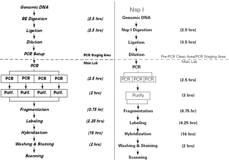
Whole-genome SNP genotyping was performed with the commercial release of the GeneChip® 250K (NspI digest) or GeneChip® 10K Affymetrix arrays as described (Matsuzaki et al., 2004). Guidelines in GeneChip® mapping assay manuals provided by Affymetrix were followed to carry out experiments (Figure 2.1).
33
34
The third step of genetic mapping is to decide statistical analysis strategy. According to presence or absence of an inheritance mode, parametric or non-parametric statistics method is used to analyze genome wide data obtained from the SNP chips. Parametric analysis is used in linkage studies, and need a pre-determined inheritance mode and large families exhibiting the phenotype. There is no need to know inheritance mode while using non-parametric statistic method and because of this it is used in association studies in disorders demonstrating complex inheritance. Since our pedigrees perfectly fit autosomal recessive
inheritance with full penetrance, we used parametric, linkage, approach to analyze the data generated from SNP chips.
To analyze SNP chip data computer based statistical analysis programs are used. These programs include LINKAGE, GENE HUNTER, ALLEGRO and MERLIN programs. MERLIN program was used in our analysis since it allows the user to handle very large numbers of markers rapidly and efficiently (Abecasis et al., 2002). It also facilitates fast multipoint analysis which contains number of markers in an interval, i.e relevant markers in 1 cM interval. Hence, multipoint linkage analysis was done with the parametric component of the MERLIN Package v1.01 (Abecasis et al., 2002; Abecasis & Wigginton, 2005).
Next step is preparing the input files which describe the pedigree, analytic parameters, map information and marker list included in the analysis. There are four files used by the MERLIN program. These are pedigree, data, map, and
model files. Pedigree file, i.e UTS.ped, describes relationships between
individuals and their disease status (Table 2.1). Data file, i.e UTS.dat, contains the list of markers and describe the affection status, i.e rare disorders with a model estimation (Table 2.2). Map file, i.e UTS.map, contains the order and genetic positions of the SNP markers (Table 2.3). Disease model is described in the model file, i.e UTS.model, in which we specified the disorder as very rare disease (Table 2.4).
35
Table 2.1 Part of pedigree file for family A. Family A was divided into two parts for convenience as shown with numbers 1 and 2 in the family column. In sex column, numbers 1 and 2 indicates male and female respectively. Data for SNP markers of each individual are given in genotypes column. In status column, numbers 1 and 2 indicates unaffected and affected individuals respectively. F, family; FID, father identification number; MID, mother identification number; PID, personal identification number; S, sex
F PID FID MID S Genotypes Status
1 1 0 0 2 1/1 2/2 1/1 1/1 0/0 1/1 1 1 2 0 0 1 1/1 0/0 1/1 1/1 1/1 1/1 1 1 3 2 1 2 1/1 1/2 1/1 1/1 0/0 1/1 1 1 4 2 1 1 1/1 2/2 1/1 1/1 1/1 1/1 2 1 5 2 1 1 1/1 0/0 1/1 1/1 0/0 1/1 1 1 6 2 1 2 1/1 2/2 1/1 1/1 1/2 1/1 2 1 7 2 1 2 1/1 2/2 1/1 1/1 1/1 1/1 1 2 1 0 0 2 1/1 2/2 1/1 1/1 1/1 1/1 1 2 2 0 0 1 1/1 2/2 2/2 1/1 1/2 1/2 1 2 3 2 1 1 1/1 2/2 1/1 1/1 1/2 1/2 2 2 4 2 1 2 1/1 2/2 0/0 1/1 1/2 1/2 1 2 5 2 1 1 1/1 2/2 1/2 1/1 1/1 1/2 1 2 6 2 1 2 1/1 2/2 1/2 1/1 1/1 1/2 1 2 7 2 1 1 1/1 2/2 0/0 1/1 1/2 1/1 2 2 8 2 1 2 1/1 2/2 1/1 1/1 1/1 1/2 1 2 9 0 0 1 1/1 2/2 2/2 1/1 1/1 1/1 1 2 10 2 1 2 1/1 2/2 2/2 1/1 1/2 1/2 1 2 11 9 10 2 1/1 1/2 2/2 1/1 1/2 1/1 2 2 13 9 10 2 1/1 2/2 2/2 1/1 1/1 0/0 1 2 14 9 10 1 1/1 2/2 2/2 1/1 1/1 1/2 1 2 15 9 10 1 1/1 2/2 2/2 1/1 1/2 0/0 1
36
Table 2.2 Part of data file for family A. Second column are the list of the
markers. Very rare disease describes the disease and refers to the model file where penetrance values of the disease are given. A, affection status; M, marker
M SNP_A-1513154 M SNP_A-1511366 M SNP_A-1509154 M SNP_A-1514257 M SNP_A-1518033 M SNP_A-1516287 M SNP_A-1516246 M SNP_A-1514307 M SNP_A-1509247 M SNP_A-1515558 M SNP_A-1511470 M SNP_A-1507812 M SNP_A-1509562 M SNP_A-1518487 M SNP_A-1509919 M SNP_A-1512266 M SNP_A-1510062 M SNP_A-1512471 M SNP_A-1511505 M SNP_A-1510562 M SNP_A-1519343 M SNP_A-1515796 M SNP_A-1516100 M SNP_A-1515465 M SNP_A-1515289 M SNP_A-1519085 M SNP_A-1509293 M SNP_A-1518948 M SNP_A-1508116 . . . . . . . . A VERY_RARE_DISEASE E END-OF-DATA
37
Table 2.3 Part of map file showing a sample of SNP markers from
chromosome 1, for family A. SNP names and their genetic positions are given in the second and third columns respectively. CHR, chromosome number
CHR MARKER LOCATION 1 SNP_A-1509443 3.859.407 1 SNP_A-1518557 5.848.183 1 SNP_A-1517286 8.655.963 1 SNP_A-1516024 8.853.158 1 SNP_A-1514538 9.387.403 1 SNP_A-1516403 9.510.903 1 SNP_A-1518687 12.351.566 1 SNP_A-1509959 12.353.228 1 SNP_A-1515791 13.890.000 1 SNP_A-1513560 13.891.000 1 SNP_A-1512212 13.892.000 1 SNP_A-1519671 14.865.946 1 SNP_A-1515942 15.275.197 1 SNP_A-1512107 20.442.669 1 SNP_A-1514390 21.496.830 1 SNP_A-1518041 24.577.258 1 SNP_A-1508673 28.395.543 1 SNP_A-1519660 28.663.799 1 SNP_A-1511922 28.677.756 1 SNP_A-1510413 28.678.445 1 SNP_A-1509189 29.025.883 1 SNP_A-1518353 29.446.071 1 SNP_A-1509509 29.753.222 1 SNP_A-1516958 30.998.694 1 SNP_A-1515835 31.231.667 1 SNP_A-1516239 31.907.546 . . . . . . . . . . . .
38
Table 2.4 Part of model file for family A. We specified the disorder as very rare disease, and determined penetrance as 0.0001, 0.0001 and 0.9999 for individuals carrying, respectively, 0, 1 and 2 copies of the disease allele
VERY_RARE_DISEASE 0.0001 0.0001, 0.0001, 0.9999 Rare_recessive
After description of the input files, the analysis was carried out along a grid of locations equally spaced at 1 cM, which allows multipoint analysis in 1 cM interval. And the graphic was plotted as a pdf output which is given at results part. The syntax used in the analysis is:
Prompt> merlin –d UTS.dat –p UTS.ped –m UTS.map -- model UTS.model -- grid1 -- markerNames -- pdf
2.3.2 Homozygosity mapping and haplotype analysis
Haplotypes were constructed and homozygosity regions were determined through visual inspection of the genotype data. Haplotype analysis was performed on chromosomal regions with positive lod scores. Lod score is logarithm of the odds ratio which is ratio of probability of linkage of a trait and a marker over probability of no-linkage between the disease and the marker. Lod scores are calculated for several estimates of recombination frequency values and a lod score greater than 3.0 is considered as evidence for linkage. In addition to MERLIN analysis, MLINK component of the LINKAGE program (FASTLINK, version 3) was used for pairwise linkage analysis (Lathrop & Lalouel, 1984;
Cottingham et al., 1993; Schaffer et al., 1994).
Polymorphic markers from the critical intervals of chromosomes 9p24 (D9S1779 [0.4 Mb] and D9S1871 [3.7 Mb]) and 17p13 (D17S1298 [3.51 Mb]) were used to test for homozygosity to the respective chromosomal loci.
39 2.4 Candidate Gene Analysis
2.4.1 Polymerase Chain Reaction (PCR) 2.4.1.1 Primers
Primers covering exons of VLDLR gene in NC_000009 sequence were designed by using Primer3 (http://frodo.wi.mit.edu/) and BLAST
(http://www.ncbi.nlm.nih.gov/blast/Blast.cgi) websites (Table 2.5). Primers were purchased from Iontek Inc. (Istanbul, Turkey).
2.4.1.2 PCR conditions
4 µl DNA (100-150 ng) was used as a template with 2.5 µl PCR buffer (10X), 1.5 µL MgCl2 (25 mM), 0.3 µl dNTPs (10mM), 1 µl (10pmol/µl) from
each primer and 0.2 µl Taq DNA Polymerase (5u/µl) (MBI Fermentas, Amh, NY, USA) (#EP0402). Volume of the sample was adjusted to 25 µl with ddH2O.
Reactions were performed in the Techne™ TC-512 and Techgene thermal cyclers. Reaction conditions were 5 min initial denaturation at 94°C, followed by 35 cycles of 94°C for 30 sec, 58°C – 62°C (A.T.) for 30 sec and 72°C for 30 sec or 40 sec and 5 min final extension at 72°C. A.T. is the annealing temperature at which the primer pair binds to its specific target sequence.
40
Table 2.5 Primers that were used for sequencing of VLDLR gene.
Primer
Name Forward Primer (5’ to 3’) Reverse Primer (5’ to 3’)
Amplified Region
Product Size (bp)
Primer Pair 1 ACTCACGCACGCTCACACT TCCGAAAGGAGGAAGAAGGT Exon 1 495 Primer Pair 2 GGAGGAGACTGTGCAAGTTGT GTGGGCAAACGGAGACCTAC Exon 1 366 Primer Pair 3 TCCCCATCCATGGGTATTAG TGATAACCCCACGTCAAACA Exon 2 365 Primer Pair 4 TGAGCCCTCATGTGAAGCTA TGTTGGACCAGGGAGAACAT Exon 3 388 Primer Pair 5 GCAGCAGCTTTGCATTGAT AGAAACACCAAGCGATGGTC Exon 4 328 Primer Pair 6 TGAACGGACCAATCTTGATG GTCGCATACCCAGCTGATG Exon 5 243 Primer Pair 7 CCCATGAGTTCCAGTGCAG CTTTCAGGGGCTCATCACTC Exons 5-6-7 659 Primer Pair 8 CCGATGAAGTCAACTGCAAA CCTACCTGGGCTTTTAAGTCA Exon 7 396 Primer Pair 9 GGTAACTTGCCGAGGAGTTAGA CAGAATTAGTCTTGCTGCTCCT Exon 8 398 Primer Pair 10 GCAGGTGATGGGAAAGGATA CAACAGCCATACCAGTCCAA Exon 9 231 Primer Pair 11 TGGGAGGAGGTGGTTTAGAA CGATGCTAGATGGGGCTAGA Exon 10 355 Primer Pair 12 TGAGCTGTGGTGTTAACTGGA TCCTGACCTACACAGATACCATTC Exon 11 400 Primer Pair 13 TGCCTTGAGTTTTCTGCTCA AGTTGAGTGGGTGGTCGAGA Exon 12 258 Primer Pair 14 GCTTCGCAAGGTTTATGGTG AAGCCATGTTCAGCTGCTCT Exon 13 389 Primer Pair 15 TGTCCCAGTTCAGCATTCAG GGGTACAGGAGGGCAAAAGT Exon 14 389 Primer Pair 16 GGCAAGGACTCAGGTCTTCA CCCGGCATACAATAGCAGAT Exon 15 378 Primer Pair 17 ACAGCTAGCCATGCTGGAAC CCAGGAACAACTCTGGCTTA Exon 16 391 Primer Pair 18 GCCAGAGTTGTTCCTGGTGT ACAGCATAAAGGCCCATGAA Exon 17 325 Primer Pair 19 GGCCCATGTGTATTCCAACT CACCCAGGTCTCCTTTCTGA Exon 18 392 Primer Pair 20 CAACTCAAAAGCAAGGTCCA GGTAACCACATCCAAAGCTGA Exon 19 338 Primer Pair 21 CTCTCGGCTGGAAGAACATC CCTATTGCCATTGTCCCAAC Exon 19 360 Primer Pair 22 CTTCAGCTTTGGATGTGGTT CCAGCCCAATTACAGGCTTA Exon 19 540 Primer Pair 23 AGGACTGGTAACTTGTCGTGCGGAG GCAGCCAGAGCGCCCAGAGCG Repeats in 5’ UTR 106
41 2.4.1.3 Agarose gel electrophoresis
1X TAE electrophoresis buffer was used to dissolve agarose (Basica LE, EU) with final percentage of 1.5 % and ethidium bromide with final concentration of 30 ng/µl.
PCR product was mixed with 1/5 volume of 6X loading dye solution before loading onto agarose gel. 1X TAE was used as running buffer for gel with different voltage and time parameters depending on size of the fragment. pUC Mix Marker 8 (0.1 µg/µl) (MBI Fermentas, Amh, NY, USA) (#SM0303) was used as DNA marker. GelDoc imaging system (Bio-Rad, CA, USA) with
MultiAnalyst software version 1.1 (Bio-Rad, CA, USA) was used to visualize gel and take photographs.
Figure 2.2 Sizes of the fragments of pUC Mix Marker 8 and appearance on agarose gel electrophoresis.