Images in Clinical Neurology / Klinik Görünüm
DO I:10.4274/tnd.10693 Turk J Neurol 2018;24:171-174
Central Nervous System Angiosarcoma: A Case Report
Santral Sinir Sistemi Anjiyosarkomu: Bir Olgu Sunumu
Ümmü Serpil Sarı1, Ayşin Kısabay Ak2, Gülgün Yılmaz Ovalı3, Hatice Mavioğlu2, Sinem Zeybek2, Erkin Özgiray4, Muhammed Fatih Sarı5, Yeşim Ertan6
1Balikesir University Faculty of Medicine, Department of Neurology, Balikesir, Turkey 2Celal Bayar University Faculty of Medicine, Department of Neurology, Manisa, Turkey 3Celal Bayar University Faculty of Medicine, Department of Radiology, Manisa, Turkey 4Ege University Faculty of Medicine, Department of Neurosurgery, Izmir, Turkey 5Balikesir State Hospital, Clinic of Neurosurgery, Balikesir, Turkey 6Ege University Faculty of Medicine, Department of Pathology, Izmir, Turkey
171
Dear Editor,
Angiosarcomas are rare malignant tumors originating from endothelial cells in the walls of blood or lymph vessels. The local recurrence of primary central nervous system (CNS) sarcomas is common, whereas the metastatic CNS angiosarcomas are more likely to originate from the heart. Clinically, CNS angiosarcomas are characterized by a rapid onset of neurological symptoms, aggressive progression and a short patient’s life span (1,2).
A 42-year-old male patient presented with a sudden onset of slurred speech, numbness in the left side, and truncal ataxia. The patient had two primary generalized tonic-clonic seizures. He was admitted to the department of neurology of our university hospital.
The neurologic examination revealed lethargy, limited cooperation, dysarthria, ptosis, peripheral facial paralysis, and hyperactive deep tendon reflexes and ataxia on the right side, as well as mild paresis of the left upper limb. Antiedema therapy with dexamethasone and antiepileptic therapy (phenytoin and levetiracetam) were started immediately. Magnetic resonance imaging (MRI), diffusion, perfusion, and MRI spectroscopy revealed multiple lesions of various sizes in the multiple areas and multiple scattered hyperintensity areas containing
hemosiderin on the T2-weighted images (Figures 1A, 1B, 1C, 1D, 1E, 1F).
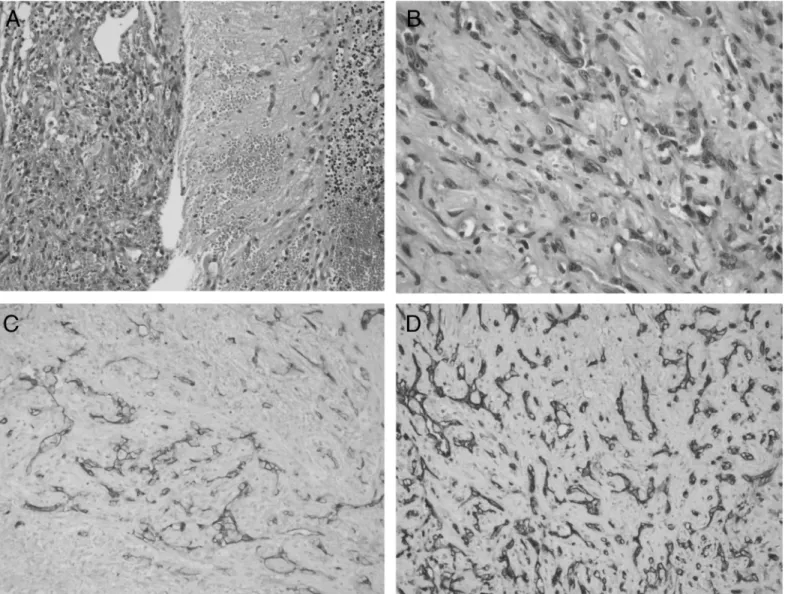
Cerebral metastases, lymphoma and parasitic infections were considered in the differential diagnosis of the patient. Hepatic, renal, and thyroid function tests, tests for vasculitis, scrotal and abdominal ultrasound, thoracic and abdominal computed tomography, and echocardiography were normal. The serum toxocara, toxoplasma, brucella, syphilis, hepatitis markers, and human immunodeficiency virus serology were all negative. The patient was referred to a specialized stereotaxic neurosurgery center for a stereotaxic biopsy (Figure 2A, 2B, 2C, 2D). On the fourth postoperative day, the patient died of respiratory insufficiency due to elevated intracranial pressure.
We did not consider angiosarcoma in our differential diagnosis when we first saw the MRI scan from the patient. The fact that the tumors contained high amounts of hemosiderin drew our attention but instead we considered more common pathologies. The definitive diagnosis could only be made pathologically.
Primary and secondary angiosarcomas are usually seen as well-defined, highly vascular, highly enhanced, and digitally edematous lesions on neuroimaging (3,4).
Ad dress for Cor res pon den ce/Ya z›fl ma Ad re si: Ayşin Kısabay Ak MD, Celal Bayar University Faculty of Medicine, Department of Neurology, Manisa, Turkey
Phone: +90 536 256 68 09 E-mail: [email protected] ORCID ID: orcid.org/0000-0002-5728-9824
Re cei ved/Ge lifl Ta ri hi: 13.11.2016 Ac cep ted/Ka bul Ta ri hi: 06.12.2017 ©Copyright 2018 by Turkish Neurological Society
Turkish Journal of Neurology published by Galenos Publishing House.
Keywords: Angiosarcoma, primary, metastatic, central nervous system, pathology, neuroimaging Anahtar Kelimeler: Anjiyosarkom, primer, metastaz, santral sinir sistemi, patoloji, nörogörüntüleme
Turk J Neurol 2018;24:171-174 Sarı et al.; Central Nervous System Angiosarcoma: Case Report
Histologically, the tumor contained malignant cells with variable levels of differentiation and vascular structures covered with clustered or blister-like malignant endothelial cells, and highly differentiated solid regions consisting of malignant fusiform cells in collagenous and myxoid stroma. The vascular channels were covered with tufted or blister-like papillary malignant endothelial cells. This feature can be helpful for differentiation from other malignancies. The cells are immunohistochemically positive for CD31, which is a sensitive and specific endothelial marker (5).
In our case, the origin of the metastasis could not be found, and the lesions were multiple and different from each other; this gave the impression that they were metastatic lesions.
We reported this case because of the extreme rarity of angiosarcomas as a malignant tumor in the CNS and in order to remind physicians to keep the diagnosis of angiosarcoma in mind in patients with a similar clinical presentation.
172
Figure 1. A, B, C) Neuroimaging showing multiple lesions of various sizes in the left basal ganglia as well as in the bilateral occipital, frontal, parietal and temporal lobes and right cerebellum. They were centrally and peripherally enhanced and digitally edematous. Hypointense signal changes appear in the T1-T2 sequences.
Turk J Neurol 2018;24:171-174 Sarı et al.; Central Nervous System Angiosarcoma: Case Report
173
Figure 1. D) Diffusion magnetic resonance imaging showing restricted peripheral diffusion of the lesions along with artefacts due to hemorrhage, E) In perfusion magnetic resonance imaging, hyperperfusion was seen in all lesions, F) Magnetic resonance spectroscopy did not show a choline peak or decrease in NAA, suggesting a glial tumor
Turk J Neurol 2018;24:171-174 Sarı et al.; Central Nervous System Angiosarcoma: Case Report
Ethics
Informed Consent: A consent form was completed by the
patient.
Peer-review: Internally peer-reviewed. Authorship Contributions
Surgical and Medical Practices: Ü.S.S., E.Ö., S.Z., M.F.S., Y.E., G.Y.O., Concept: Ü.S.S., A.K.A., H.M., M.F.S., Design: M.F.S., Ü.S.S., G.Y.O., Data Collection or Processing: Ü.S.S., H.M., A.K.A., E.Ö., S.Z., Y.E., G.Y.O., Analysis or Interpretation: H.M., Ü.S.S., S.Z., Literature Search: H.M., A.K.A., Ü.S.S., M.F.S., Y.E., Writing: H.M., A.K.A., Ü.S.S., E.Ö.
Conflict of Interest: No conflict of interest was declared by
the authors.
Financial Disclosure: Preparation for publication of this
article is partly supported by Turkish Neurosurgical Society.
References
1. Galanis E. Chapter 18: Malignant Central Nervous System Neoplasms of Mesenchymal Origin. Pg: 325-340. Brain Tumors Practical Guide to Diagnosis and Treatment. Informa. 2007.
2. Kuratsu J, Seto H, Kochi M, Itoyama Y, Uemura S, Ushio Y. Metastatic angiosarcoma of the brain. Surg Neurol 1991;35:305-309.
3. Araoz PA, Eklund HE, Welch TJ, Breen JF. CT and MR imaging of primary cardiac malignancies. Radiographics 1999;19:1421-1434.
4. Gallo P, Dini LI, Saraiva GA, Sonda I, Isolan G. Hemorrhage in cerebral metastasis from angiosarcoma of the heart: case report. Arq Neuropsiquiatr 2001;59:793-796. 5. Mena H, Ribas JL, Enzinger FM, Parisi JE. Primary angiosarcoma of the
central nervous system. J Neurosurg 1991;75:73-76.
174
Figure 2. A) Tumor showing angiosarcoma morphology; it is uniformly separated from the cerebellum, B) Pleomorphic tumor cells having distinctive giant nucleoli, C) Immunohistochemistry positive for CD34 in the tumor cells, D) Immunohistochemistry positive for CD31