Malignant and Borderline Ovarian Neoplasms with Evaluation by Ultrasonography, Ca125 Level and Age
Burak Karadag
1*, M Kocak
2, F Kayıkcioglu
3, F Ercan
4, B Dilbaz
3, MF Kose
5, A
Haberal
6Asian Pac J Cancer Prev, 15 (19), 8489-8493
Introduction
Adnexal masses are common in gynecology practice and the decision to treat the patient by observation, laparoscopy, or laparotomy will often depend upon basic evaluation in various age groups of women. In the United States, a woman has a 5-10% lifetime risk of undergoing surgery for an adnexal mass and, of these, benign neoplasms constitute a significant number, but 4% of new ovarian cancers expected to be diagnosed at local or regional states in 2007 (Jemal et al., 2007). Although most adnexal masses are benign, the goal of the diagnostic evaluation is to exclude ovarian cancer for which 5 year
1Department of Obstetrics and Gynecology,Ankara Teaching and Research Hospital, Ankara, 2Private Edremit Gulf Hospital,
Edremit, Balıkesir, 4Department of Obstetrics and Gynecology, Meram Faculty of Medicine, Necmettin Erbakan University, Konya, 5Department of Obstetrics and Gynecology, Faculty of Medicine, Bahçeşehir University, İstanbul, 6Department of Obstetrics and
Gynecology, Faculty of Medicine, Başkent University Ankara, 3Etlik Zübeyde Hanım Women’s Health Teaching and Research
Hospital, Ankara Turkey *For correspondence: [email protected]
Abstract
Objective: To verify the basic preoperative evaluation in the discrimination between benign and malignant adnexal masses in our clinical practice. Materials and Methods: Data were collected on the records of 636 women with adnexal masses who had undergone surgery either by open or endoscopic approaches. Those with obvious signs of malignancy, any history of cancer, emergency surgeries without basic evaluation were excluded. The preoperative features by age, ultrasound and serum Ca125 level were compared with final histopathological diagnosis at the four departments of the institution. These are the general gynecology (Group 1: exploratory laparotomy), the gynecologic endoscopy (Group 2: laparoscopy and adnexectomy), the gynecological oncology (Group 3: staging laparotomy) and the gynecologic endocrinology and infertility (Group 4: laparoscopy and cystectomy). Results: There were simple and complex cyst rates of 22.3% and 77.2%, respectively. There were 86.3% benign, 4.1% (n:20) borderline ovarian tumor (BOT) and 6.4% (n:48) malignant lesions. There were 3 BOT and 9 ovarian cancers in Group 1 and one BOT and two ovarian cancer in the Group 2. During the surgery, 15 BOT (75%) and 37 ovarian cancer (77%) were detected in the Group 3, only one BOT was encountered in the Group 4. The risk of rate of unsuspected borderline or focally invasive ovarian cancer significantly increased by age, size, complex morphology and Ca125 (95% CI, OR=2.72, OR=6.60, OR=6.66 and OR=4.69, respectively). Conclusions: Basic preoperative evaluation by comprehensive ultrasound imaging combined with age and Ca125 level has proved highly accurate for prediction of unexpected malignancies. Neither novel markers nor new imaging techniques provide better information that allow clinicians to assess the feasibility of the planned surgery; consequently, the risk of inadvertent cyst rupture during laparoscopy may be significantly decreased in selected cases.
Keywords: Adnexal masses - ultrasonography - risk of ovarian cancer - laparoscopic surgery
RESEARCH ARTICLE
Risk for Malignant and Borderline Ovarian Neoplasms
Following Basic Preoperative Evaluation by Ultrasonography,
Ca125 Level and Age
relative mortality rate approaches 50%. Management decisions often are influenced by imaging characteristic of the mass, serum Ca125 assay, the age and family history of the patient (Mancuso et al., 2004; Heilbrun et al., 2009; Shaaban et al., 2009).
The differential diagnosis of an adnexal mass includes both gynecologic and nongynecologic sources and, when arising from the ovary, may be a benign or malignant and or borderline tumor. Numerous studies have been looking at the risk of malignancy in adnexal masses, comparing imaging morphology and serology with either histopathology or mode of surgical management (Ghezzi et al., 2008; Yen et al., 2009; Arab et al., 2012;
Simple cysts, less than 10cm in diameter in the presence of a normal Ca125 levels are have a low risk of malignancy and the laparoscopic management of diameter with a low risk of malignancy index often can be managed conservatively in reproductive-age and even in postmenopausal women (RCOG 2003). Conversely, those that are more likely to be malignant are best managed with prompt surgery by a physician with advanced training and expertise in the management of ovarian cancer, such as a gynecologic oncologist.
The differentiation between benign and malignant masses is, a clinical challenge in women with complex cysts who require surgical management; however, many can be managed laparoscopically. Conservative surgical procedures such as ovarian cystectomy is a method of choice in reproductive age women who desire to preserve their future fertility. Furthermore, laparoscopic diagnosis of suspicious cysts on clinical presentation prevents many laparotomies for the treatment of benign conditions in premenopausal women. Patient advantages as compared with laparotomy include superior cosmetic results, shorter recovery and at least equvalent results. However, one concern associated with laparoscopic cystectomy is inadvertent cyst rupture in an occult, early stage ovarian cancer with the presence of normal Ca125 level in women who desire future fertility as well as a safe approach to their lesions. Intraoperative rupture of malignant cysts may cause dissemination of its content and may have some impact on survival. When malignancy is detected immediate surgical staging by laparoscopy or by converting laparotomy is indicated to prevent adverse outcomes in such cases (Gocmen et al., 2009).
The most appropriate surgical management of complex adnexal masses still controversial, depends on the clinical circumstances for women with varying risk of malignancy in the literature (Rangel et al., 2005).
Our aim was to investigate the relationship between preoperative features and modes of management in our series of adnexal mass and to review our institutional evaluation and management efforts on these lesions by patient’s age, ultrasonographic imaging and serologic means.
Materials and Methods
Six hundred and thirty six women who underwent surgical exploration for an adnexal mass at four referral departments of our institution were included for two years, from January 2008 to December 2009. Individual history of breast or ovarian cancer, cases with malignant ascites and with evidence of abdominal or distant metastasis or any emergency adnexal surgery without basic evaluation were excluded from the study.
The basic preoperative assessment to permit the distinction of benign from malignant were the patient history, pelvic examination, serum Ca125 levels and Transvaginal/Transabdominal ultrasound (TV-USG/TA-USG) either with or without Doppler imaging studies was performed. Morphology of the masses was defined either as simple or as complex according to presence of papillary
Modes of surgical management were determined regarding to the review of the preoperative imaging findings, Ca125 levels and age of patients and then they were admitted to the departments for the adjusted adnexal surgeries. For the initial clinical presentation, adnexal masses were categorized into one of four departments: General Gynecology (Group 1), Gynecological Endoscopy (Group 2), Gynecologic Oncology (Group 3), or Gynecologic Endocrinology and Infertility ( Group 4). Patients with large masses without high risk of malignancy and large masses or with high index of malignancy suspicion were operated in the General Gynecology (Group 1) and Gynecologic Oncology department (Group 3) s via laparotomies, respectively. These categories, along with notation regarding the patient’s age and ultrasonographic features and serum Ca125 levels were helpful criterias in order to indicate whether the mass was likely to be benign or not. Typically benign, simple and small masses, were managed in the Gynecological Endoscopy (Group 2) and Gynecologic Endocrinology and Infertility department (Group 4) s via laparoscopic surgery.
During the laparoscopic surgeries, peritoneal washings were obtained and set aside before proceeding with insicion or resection of any suspicious adnexal mass in the each groups. The decision to perform cystectomy or oopherectomy dependent on the clinical circumstances such as future fertility, apperance of the mass during surgery, and the age of the women. The entire adnexa had removed once a malignant mass was diagnosed and the laparoscopic surgeons had requested intraoperative assistance with staging from a gynecologic oncologist immediately after frozen-section evaluation in all cases.
Preoperative findings and modes of management were compared with intraoperative findings, frozen section and final histopathological diagnosis in the four groups.
Statistical analysis
Data were collected and transferred into SPSS statistical database [SPSS Inc. Chicago, IL (Statistical Package for Social Science), Version 17 for Windows] on computer enviorenment. The Shapiro-Wilk test was used to assess the conformity of the continuous variables in the study to normal distribution. Continuous variables were expressed as mean, median, mini¬mum and maximum, whereas percentages and frequencies were used for categorical variables. Student’s t-test was used if it complied with the normal distribution and the Mann-Whitney U-test was used if it did not comply. Intergroup differences for categorical values were assessed with chi square test. Logistic Regression test was performed to assess the risk of malignancy by the basic preoperative features of the study population. A P value lower than 0.05 was accepted to be statistically significant.
Results
Mean age, gravida, maximum diameter of mass and Ca 125 levels of the study population were 39.1± 13.3 , 2.4± 2.4, 80.6± 44.3mm, 67.4± 230.4 U/ml, respectively. Number of patients operated in the General Gynecology,
Malignant and Borderline Ovarian Neoplasms with Evaluation by Ultrasonography, Ca125 Level and Age
Gynecological Endoscopy, Gynecologic Oncology and Gynecologic Endocrinology and Infertility departments were 139(22%), 225(35%), 114(18%) and 158(25%), respectively. Total number of patients underwent laparoscopic surgery was 383 and 253 patients were operated by laparotomy.
Preoperative patient characteristics and final histopathologic results are summarized in Table 1. There were simple and complex cyst rates of 22.3 % and 77.2 %, respectively. Final histopathologic results revealed that the majority (89,3%) were benign, 3.1% of (n:20) were borderline ovarian tumors (BOT) and 7.6% of (n:48) were malignant lesions.
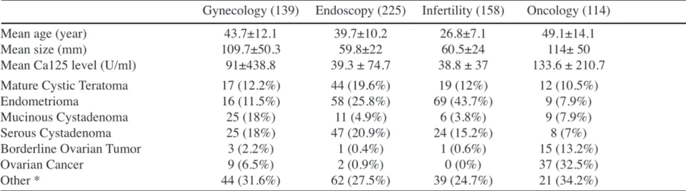
The mean age, diameter of the mass, Ca 125 levels and the morphology of the mass were compared in the groups. The mean age was 43.7±12.1, 39.7±10.2, 49.1±14,1 and 26.8±7.1 years in the groups, respectively. The mean diameter of the mass was 109.7 ± 50.3, 59.3±22, 114±50 and 60.5±24 mm, respectively The mean Ca125 level was 91±438.8, 39.3±74.7, 133.6±210,7 and 38.8±37 U/ ml, respectively. The variance of means of age, maximum diameter of mass and CA125 levels were found to be statistically significant (p=0.0003, p=0.0001, p=0.0007) that all values were higher in patients selected on the Gynecologic Oncology department. Comparisons of the morphology revealed a statistical difference between the women operated on the Gynecologic Endocrinology and Infertility and the Gynecological Endoscopy departments. Other group comparisons didn’t reach statistical significance (p>0.05).
There were 3 BOTs and 9 ovarian cancers in the General Gynecology and one BOT and two ovarian cancers in the Gynecological Endoscopy. During the surgery, 15 BOTs (75 %) and 37 ovarian cancers ( 77%) were detected in the Gynecologic Oncology, only one BOT was encountered in the Gynecologic Endocrinology and Infertility, above mentioned rates of malignancy in the groups was statistically significant (P<0,05). Seventy-five (75%) of the malignant masses and ninety (90%) of the borderline masses operated via laparotomy, respectively. The rate of conversion to laparotomy for endoscopic suspicion of cancer was actually ~1% for the whole endoscopic surgeries. Without any other diagnostic tool, only by basic evaluation, unexpected borderline or focally invasive ovarian cancer rate was 2.5 % in our study. Based on the basic preoperative evaluation in 636
patients showed a sensitivity of 76.4%, and specificity of 89% in differentiating benign and malign adnexal masses. Basic features of the study population; age (>40), tumor size (>10cm), morphology and Ca125 level (>35) were analysed by Logistic Regression and The Odd’s Ratios (OR) for malignancy risk were calculated as OR=2.72, OR=6,60, OR=6,66 and OR=4.69, 95% CI, respectively. The complex morphology gave the highest malignancy risk.
Although lower Ca125, younger age and smaller cyst size, it was noticed that each mass with BOT had a complex morphology wheras two masses with mucinous cystadenocarcinoma had simple cyst morphology on the basic ultrasound imaging.
Discussion
Adnexal masses affects as many as 10% of female population, one to 0.1% of them were found to be with malignant neoplasms. Most clinicians have followed a policy of surgical treatment based on benign or malignant features of adnexal masses to recognize the potentials of laparoscopic approach for optimal management. The possibility of tumor spillage on invasive lesion is a major concern, especially with the laparoscopic surgery. Current basic evaluation for a pelvic mass palpaple on pelvic examination include a pelvic sonogram by either the abdominal or transvaginal approach and serum Ca125 determination. Optional preoperative studies include other imaging techniques such as MRI, CT, PET studies and novel tumor markers. Whatever the preoperative work-up, many unexpected, early stage ovarian cancers or borderline tumors have been reported in clinical practice.
Ovarian neoplasms are common in women of all ages and large screening series demonstrate that the majority of adnexal masses are benign. As many as %10 women in the United States will undergo surgery in their lifetime (Jemal et al., 2007). The laparoscopic management of adnexal masses appears a gold standard in most countries, but is still controversial in suspicious masses for in stage 1 ovarian malignancy in which iatrogenic cyst rupture may be a significant prognostic factor. Accurate preoperative and intraoperative selection of the cases is mandatory, the diagnosis is usually based on histologic findings in surgical specimens of unexpected borderline or focally invasive ovarian cancers after tumor spill and malignant
Table 1. Preoperative Characteristics and Final Pathological Results of Patients
Gynecology (139) Endoscopy (225) Infertility (158) Oncology (114) Mean age (year) 43.7±12.1 39.7±10.2 26.8±7.1 49.1±14.1 Mean size (mm) 109.7±50.3 59.8±22 60.5±24 114± 50 Mean Ca125 level (U/ml) 91±438.8 39.3 ± 74.7 38.8 ± 37 133.6 ± 210.7 Mature Cystic Teratoma 17 (12.2%) 44 (19.6%) 19 (12%) 12 (10.5%) Endometrioma 16 (11.5%) 58 (25.8%) 69 (43.7%) 9 (7.9%) Mucinous Cystadenoma 25 (18%) 11 (4.9%) 6 (3.8%) 9 (7.9%) Serous Cystadenoma 25 (18%) 47 (20.9%) 24 (15.2%) 8 (7%) Borderline Ovarian Tumor 3 (2.2%) 1 (0.4%) 1 (0.6%) 15 (13.2%) Ovarian Cancer 9 (6.5%) 2 (0.9%) 0 (0%) 37 (32.5%) Other * 44 (31.6%) 62 (27.5%) 39 (24.7%) 21 (34.2%)
*Includes benign paraovarian masses, retroperitoneal masses, intraligamenter myoma, tuboovarian absess, hydrosalpinx, functional ovarian cysts, nongynecologic structures, etc.
endoscopic surgery even in many benign cases.
Today, new technologies such as ultrasonography and tumor marker assay have increased the diagnosis rate for adnexal masses. These combined diagnostic tools , however, frequently do not distinguish benign masses from early stages of an ovarian malignancy, which may result in suboptimal surgery (Mancuso et al, 2004). Murta and Nomelini reviewed that benign and early stage malignant lesions can present similar clinical characteristics and the clinicians can only rely on combination with color Doppler ultrasonography and /or tumor markers in relate to improve accuracy of these techniques to decide optimal management (Murta et al., 2006). The vast majority of adnexal masses can be safely managed laparoscopically provided that there are basic preoperative evaluation, expertise in gynecologic oncology and immediate access to frozen section diagnosis. On the other hand, accumulating scientific data support the laparoscopic approach to the large adnexal mass without any other signs of malignancy, as safe and effective treatment (Panici et al., 2007).
Our data showed that the use of basic preoperative evaluation by age, Ca125 level and the size of the mass resulted in a prediction on 90% of borderline tumors and 75% of ovarian cancers. The rate of unexpected borderline or focally invasive ovarian cancer which were evaluated by only the age, ultrasound imaging and serology was 5.9% in our study.
The study of Riccardo et al. (2004) indicates that the rate of unexpected borderline or early stage ovarian cancer was 1.2% of 683 women under 40 years of age with adnexal masses. By contrast to Riccardo’s series, there is a higher incidence of unexpected borderline or focally invasive ovarian cancer, the rate was 5.9% in the present series in which both premenopausal and postmenopausal women either with simple or complex mass were included in our study.
Ultrasound evaluation of adnexal masses is the most widely used technique and sonographic findings of simple or complex mass, combined with serum Ca125 level and menopausal status provide an important information to determine the optimal surgery. It has also been suggested that colour-flow Doppler studies may be of benefit in assessing adnexal masses. However, several studies have reported that the addition of Doppler sonography resulted in a small improvement in positive predictive values at the cost of a significant decrease in sensitivity (Brown et al., 2007; Jokubkiene et al., 2007). Although, Doppler analysis in small percent of women were not included into the present study , it was noticed that malignant Doppler features were observed in both women with BOT and women with benign conditions. Exacoustos et al. concluded that neither complex cyst with the presence of papillae nor Doppler studies constituted highly sensitive preoperative markers for 33 women with BOT (Exacustos et al., 2005). The incidence of unexpected malignancies at laparoscopic surgery in the present study has been shown to be only ~1% ( 4 of 383 laparoscopies) that is lower than in the Marana et al series, in which frozen section revealed that the rate of BOT was 14.3% in 35 of 667 women with
Accumulating evidence supporting to the specialty care of a gynecologic oncologist on the basis of pelvic examination, imaging studies, family history as well as serum Ca125 determination (Gostout et al., 2006). As a single marker for prediction of ovarian cancer is Ca125 level, in combination with ultrasonographic features, it is used to determine guidelines for the safe use of laparoscopy for adnexal masses. A survey by Robin et al for British Gynaecological Cancer Society (BGCS) reported that the incidence of malignant cases was 4.1% of women with adnexal mass on whom laparoscopic surgery had been performed (Robin et al., 2005). The serum glycoprotein Ca125 concentration is elevated in 80% of women with ovarian cancer; but reported sensitivities for early stage disease were found to decrease to 50% of women with Stage 1 ovarian cancer or with BOT’s. HE4 (human epididymis protein 4) had the highest sensitivity at 72.9% in 259 women with adnexal masses, it seemed to be best single marker for Stage 1 disease, their results is promising for more accurate prediction of occult ovarian cancers (Moore et al., 2008). In addition to unpredictable levels in early stage ovarian carcinoma, it is well known that serum Ca125 values fluctuate during the menstrual cycle, but are rarely greater than 100 to 200U/mL in patients with benign conditions. Risk of malignancy in young females with adnexal masses was found as 14.6% of cases, slightly drops to 6-11% of premenopausal and up to 29-35% of postmenopausal women in the presence of high levels of Ca125 (Bristow et al., 2009).
Benign adnexal lesions affect young women, whereas malignant ovarian neoplasms occurs at more advanced ages. In the present study, laparoscopic management of complex adnexal mass appears to be safe in young women with moderately elevated Ca125 levels which were often associated with endometriosis and inflammatory processes. The incidence of managing an unexpected ovarian cancer by laparoscopy is 6.5 in 1000 women diagnosed with an adnexal mass, in which early stage ovarian carcinomas and tumors of malignant potential are neither often associated with high levels of Ca125 nor older age. After the diagnosis of early stage ovarian cancer or BOT’s, to avoid tumor spillage and incomplete surgery, a converting laparotomy had to be performed in each case of the present study, to ensure an optimal approach. However, several studies have shown that these patients could benefit laparoscopic staging procedures by gynecologic oncologist experienced with advanced laparoscopy and is equivalent to traditional approach (Sagiv et al., 2005; Nezhat et al., 2009 ).
Several techniques have been proposed to differentiate benign and malign cases, especially using ultrasonographic morphological scoring (Arab et al., 2012; Yavuzcan et al., 2013; Tantipalakorn et al., 2014). As a result of these studies; the sensitivity was reported from 68.5% to 82.9% and the specificity 75.6% to 95.3% for prediction of malignancy. Similarly to the literature; in our study, the sensitivity and the specificity values were 76.4% and 89% respectively.
Combined with basic parameters (age, size of tumor, morphology and Ca125 level), current specific markers
Malignant and Borderline Ovarian Neoplasms with Evaluation by Ultrasonography, Ca125 Level and Age
and tools, such as, HE4 (22), proteomics (Vladimir et al., 2008), FDG PET (Fenchei et al., 2002 ) and the ovarian crescent sign (Hillaby et al., 2004) in combination with clinical parameters for the detection of early stage ovarian cancer could provide improvements in mortality rates from late stage ovarian cancers.
Basic preoperative evaluation for the risk of malignancy is simple and fast and does not involve the use of any other complicated diagnostic tool, a comprehensive ultrasound imaging combined with age and Ca125 level has proved highly accurate for prediction of unexpected malignancies in our data.
Our study suggests that age (OR:2.72), size of the mass (OR:6.60) and serum Ca125 levels (OR:4.69), as well as complex morphology (OR:6.66) play essential roles in preoperative evaluation are enough to determine modes of surgery for adnexal masses.
The most appropriate surgical management would vary depending on preoperative evaluation in women with varying risk of malignancy and clinicians should decide the most appropriate place for them to be managed. The results of our study strongly suggest that the basic evaluation offers a valid, safe and discriminative tool for the most cases with adnexal mass.
References
Arab M, Yaseri M, Ashrafganjoi T, et al (2012). Comparison of two ovarian malignancy prediction models based on age sonographic findings and serum Ca125 measurement. Asian
Pacific J Cancer Prev, 13, 4199-202.
Bristow R, Nugent A, Zahurak M, Khouzhami V, Fox H (2009). Impact of surgeon specialty on ovarian-conserving surgery in young females with an adnexal mass. J Adolesc Health,
39, 411-6.
Brown DL, Douglas L (2007). A practical approach to the ultrasound characterization of adnexal masses. Ultrasound
Quarterly, 23, 87-105.
Exacustos C, Romanini ME, Rinaldo D, et al (2005). Preoperative sonographic features of borderline ovarian tumors. Ultrasound Obstet Gynecol, 25, 50-9.
Fenchei S, Grab D, Nuessie K, et al (2002). Asymptomatic adnexal masses:correlation of FDG PET and histopathologic findings. Radiology, 223, 780-9.
Ghezzi F, Cromi A, Bewrgamini V, et al (2008). Should adnexal mass size influence surgical approach? a series of 186 laparoscopically managed large adnexal masses. Intl J Obstet
Gynecol, 115, 1020-7.
Gocmen A, Atak T, Ucar M, Sanlıkal F (2009). Laparoscopy-assisted cystectomy for large adnexal cysts. Arch Gynecol
Obstet, 279, 17-22.
Gostout BS, Brewer Molly AD (2006). Guidelines for referral of the patient with an adnexal mass. Clin Obstet Gynecol,
49, 448-58.
Heilbrun ME, Olpin J, Shaaban A (2009). Imaging of benign adnexal masses: characteristic presentations on ultrasound, computed tomography, and magnetic resonance imaging.
Clin Obstet Gynecol, 52, 21-39.
Hillaby K, Alsam N, Salim R, et al (2004). The value of detection of normal ovarian tissue (the ‘ovarian crescent sign’) in the differential diagnosis of adnexal masses. Ultrasound Obstet
Gynecol, 23, 63-7.
Jemal A, Siegel R, Ward E (2007). Cancer Statistics. Cancer J
Clin, 57, 43-66.
Jokubkiene L, Sladkevicius P, Valentin L (2007). Does three-dimensional power doppler ultrasound help in discrimination between benign and malignant ovarian masses? Obster
Gyenol Survey, 62, 308-9.
Mancuso A, De Vivo A, Triolo O, Irato S (2004). The role of transvaginal ultrasonography and serum Ca125 assay combined with age and hormonal state in the differential diagnosis of pelvic masses. Eur J Gynaecol Oncol, 25, 207-10.
Marana R, Muzii L, Ferrari S, et al (2009). Management of adnexal cystic masses with unexpected intracystic vegetations detected during laparoscopy. J Minim Invasive
Gynecol, 12, 502-8.
Moore RG, Brown AK, Miller C, et al (2008). The use of multiple novel tumor biomarkers fort he detection of ovarian carcinoma in patients with pelvic mass. Gynecol Oncol,
108, 402-8.
Murta EFC, Nomelini RS (2006). Early diagnosis and predictors of malignancy of adnexal masses. Curr Opin Obstet Gynecol,
18, 14-9.
Nezhat FR, Ezzati M, Chuang L, et al (2009). Laparoscopic management of early ovarian and fallopian tube cancers:surgical and survival outcome. Am J Obstet Gynecol,
200, 1-6.
Panici PB, Palaia I, Bellati F, et al (2007). Laparoscopy compared with laparoscopically guided minilaparotomy for large adnexal masses: a randomized controlled trial. Obstet
Gynecol, 110, 241-8.
Riccardo M, Ludovico M, Giovan FC, et al (2004). Laparoscopic excision of adnexal masses. J Am Assoc Gynecol Laparosc,
11, 162-6.
Robin AF, Martin EG, John HS (2005). Ovarian cancers related to minimal access surgery. Int J Obstet Gynaecol, 102, 726-30.
Royal College of Obstetricians and Gynaecologists. Ovarian cysts in postmenopausal women.Guideline No.34. Oct and urokinase plasminogen activator receptor in the sera of lung cancer patients. Oncol Lett, 2, 693-9.
Sagiv R, Golan A, Glezerman M (2005). Laparoscopic management of extremely large ovarian cysts. Obstet
Gynecol, 105, 1319-122.
Shaaban A and Maryam R (2009). Ovarian cancer: detection and radiologic staging. Clin Obstets Gynecol, 52, 73-93 Vladimir N, Malaika A, Feng S et al (2008). The early detection
of ovarian cancer:from traditional methods to proteomics. Can we really do beter than serum Ca125? Am J Obstet
Gynecol, 199, 215-23
Tantipalakorn C, Wanapirak C, Khunamornpong S, et al (2014). IOTA simple rules in differentiating between benign and malignant ovarian tumors. Asian Pac J Cancer Prev, 15, 5123- 6.
Yavuzcan A, Caglar M, Ozgu E, et al (2013). Should cut-off values of the risk of malignancy index be changed for evaluation of adnexal masses in Asian and Pacific populations? Asian Pac J Cancer Prev, 14, 5455-9.