Yazışma Adresi/Address for Correspondence: Dr. Eylül Hatice Bozkurt Yılmaz, Baskent University, Department of Pulmonary Medicine, Adana, Turkey E-mail: [email protected]
Geliş tarihi/Received: 18.06.2017 Kabul tarihi/Accepted: 17.10.2017
ARAŞTIRMA / RESEARCH
Role of plasma asymmetric dimethylarginine levels in diagnosis and
follow-up of acute pulmonary thromboembolism
Akut pulmoner tromboembolizmin tanı ve takibinde plazma asimetrik dimetilarjin
düzeylerinin rolü
Eylül Hatice Bozkurt Yılmaz
1, Mustafa Yılmaz
2, Füsun Öner Eyüboğlu
11Baskent University, Department of Pulmonary Medicine, 2Department of Cardiology, Adana, Turkey
Cukurova Medical Journal 2018;43(2):444-449
Abstract Öz
Purpose: Nitric oxide is an important mediator that plays
a major role in platelet adhesion/aggregation. Asymmetric dimethylarginine (ADMA) is a competitive inhibitor of endothelial nitric oxide synthase. In this study, we aimed to examine the ADMA levels in diagnosis and follow-up of acute pulmonary thromboembolism.
Materials and Methods: The study was designed as a
prospective study. Thirty consecutive patients with acute PTE and 20 healthy volunteers were enrolled into the study. Patients were followed 1 month. Plasma ADMA levels were measured on admission and following the first month.
Results: Ona dmision there was statistically significant
difference between two groups. When we compared serum ADMA levels difference between patients group with resolution and without resolution, there was not any statistically significant difference. There was a significant difference between serum ADMA levels of massive group and the other 2 groups.
Conclusion: Plasma ADMA levels elevated in patients
with pulmonary thromboembolism. This finding supports the idea ADMA levels correlated with thrombus burden.
Amaç: Nitrik oksit trombosit yapışması ve toplanmasında
rol oynayan önemli bir maddedir. Asimetrik dimetilarjinin (ADMA) endotelyal nitrik oksit sentetazın yarışmacı inhibitörüdür ve seviyesi hipokside artar. Bu çalışmada ADMA seviyesin akut pulmoner tromboembolizm tanı ve takibindeki yerini araştırmayı amaçladık.
Gereç ve Yöntem: Çalışma prospektif olarak dizayn
edildi. Akut pulmoner tromboembolizmi olan 30 hasta ve 20 sağlıklı gönüllü çalışmaya dahil edildi. Hastalar 1 ay takip edildi. Plazma ADMA seviyesi başvuruda ve 1. ay takipte ölçüldü.
Bulgular: Hastaneye başvuru sırasındaki hasta ve kontrol
grubunun serum ADMA seviyeleri arasında istatistiksel anlamlı fark vardı. Birinci ay kontrolde rezolüsyonu olan ve olmayan grupların serum ADMA değişimi arasında istatistiksel anlamlı fark bulunmadı. Serum ADMA seviyesi masif olan grupta, submasif ve masif olmayan gruplara göre daha yüksekti.
Sonuç: Plazma ADMA seviyesi akut pulmoner
tromboembolizmi olan hastalarda yükselir ve ADMA seviyesi trombüs yükü ile koreledir.
Key words: Pulmonary embolism, asymmetric
dimethylarginine, nitric oxide Anahtar kelimeler: Pulmoner emboli, asimetrik dimetilarjinin, nitrik oksit
INTRODUCTION
Pulmonary artery embolism is defined as a partial or complete occlusion of a pulmonary arterial branch. It is associated with high morbidity and mortality, which may sometimes be difficult to diagnose. Approximately 70-90% of cases are caused by thrombosis of lower extremity veins1-2. Venous
thromboembolism (VTE) encompasses deep vein thrombosis (DVT) and pulmonary thromboembolism (PTE). It is the third most frequent type of cardiovascular disease3-4. Hypercoagulability, endothelial damage and stasis are known to play role in pathophysiology of PTE 5-6. The main mechanism of nitric oxide (NO) involves that inhibition of proliferation of smooth
445 muscle cells, endothelium derived vasodilatation, cell-cell interaction in blood vessels (platelet adhesion/aggregation and inhibition of monocyte adhesion) so NO plays a key role in PTE pathophysiology7-10. Asymmetric dimethylarginine (ADMA) is competitive inhibitor of endothelial nitric oxide synthase (e-NOS) so plays a regulatory role in the formation of endothelial NO11-14. Plasma ADMA levels increases in hypoxia10. In this study, we aimed to examine the plasma ADMA levels in diagnosis and follow-up of acute PTE.
MATERIALS AND METHODS
Thirty consecutive patients admitted to hospital on an outpatient basis between December 2009 and October 2010 were evaluated for possible enrollment into the study. This prospective study was approved by the local ethics committee (KA09/276). Each patient provided a signed informed consent form.
The following were excluded from the study: patients with known malignancy, liver disease, active infection, diabetes mellitus, congestive heart failure and patients, who did not want to join the study. Twenty healthy volunteers were taken in our study. Patients were followed 1 month. Plasma ADMA levels were measured on admission and first month. Computed tomographic angiography (CTPA) and echocardiography were performed in patients on admission and the end of first month. Status of hypoxemia was evaluated with arterial blood gas on admission. It was evaluated with arterial blood gas or pulse oxymetri at the end of first month. The diagnosis of PTE was confirmed only when CTPA showed a pulmonary vascular filling defect.
Patients received either low- molecular-weight heparin at a fixed dose per kilogram of body weight subcutaneously two times per day or unfractionated heparin at an initial bolus dose of 80IU per kilogram followed by a continuous intravenous infusion at an initial rate of 18 IU per kilogram per hour. The dose was subsequently adjusted so that the activated partial thromboplastin time (aPTT) was two to three times the control value in normal subjects. In each patient, oral anticoagulant therapy (warfarin) was initiated at first day of heparin and continued together for 3 days. Patients were treated for three months with warfarin. The dose was adjusted to
achieve an international normalized ratio (INR) of 2.0 to 3.0.
Biochemical analysis
Plasma ADMA levels were analyzed by using high performance liquid chromatography (HPLC). The method includes reversed phase HPLC analysis by using fluorescence detection (Shimadzu LC 10A fluorescent detector, Japan) in a high-performance liquid chromatography system (Shimadzu RF 10XL, Japan). The plasma concentrations of ADMA were measured by HPLC with precolumn derivatization with ophthaldialdehyde (OPA) and 3-mercaptopropionic acid. Samples and standards were incubated with OPA reagent for exactly 30 s before injection into the HPLC system15.
Statistical analysis
Variables are presented as mean±SD or median (range, interquartile range [IQR]) for continuous data and as proportion for categorical data. The compliance of numerical variables to a normal distribution was assessed using the Shapiro Wilk test. Relationships between categorical variables were assessed by the Chi- Square and Fisher’s exact tests. Non parametric data were compared with the Mann-Whitney U or Kruskall-Walls analyze test as appropriate. Comparisons of pre and post treatment values were performed using the Wilcoxon test. Linear relationships between two variables were assessed using the Pearson (for parametric data) and Spearman (for non parametric data) correlation analyses. Results were evaluated using 95% confidence intervals, and p<0.05 was considered statistically significant. Statistical analysis was performed using commercially available computer program (SPSS version 21.0 for Windows; SPSS, Inc., Chicago, Illinois, US).
RESULTS
A total of 50 participants (30 patients and 20 controls) were enrolled into the study. In the patients group mean age was 64.9±16.5 years and 22 (73.60%) patients were women. In the control group mean age was 64.1±14.3 years and 14 (70%) patients were women. There was no significant difference between baseline demographic characteristics between the two groups (p>0.05). Baseline clinic characterics of study patients on admission are summarized in table 1. There was a statistically
446 significant difference between patient and control group according to serum ADMA levels. In the patients groups serum ADMA levels was 0,35 (0.16-0.62; IQR: 0.46) μmol/L and in the control group serum ADMA levels was 0.18 (0.11-0.29; IQR: 0.18) μmol/L (p=0.022). Comparison of serum ADMA levels on admission are shown in Figure 1. Twenty three patients had thrombus resolution but 7 patients had not thrombus resolution in first month control. When we compared serum ADMA levels difference (ΔADMA) between admission and at the end of firsth month control in patients group with resolution (n:23) and without resolution (n:7) found there was not a different (respectively, ΔADMA was -0.07 0.33-0.09; IQR: 0.42) μmol/L and -0.12 (-0.27- 0.29; IQR: 0.56) μmol/L p=0.666). Comparison of ΔADMA between the groups are shown in Figure 2. When we compared serum ADMA levels in massive, submassive and non massive groups, there was a significant difference
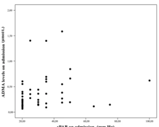
between serum ADMA levels of massive group and the other 2 groups (massive group ADMA levels was 1.4 (1.3-1.59; IQR: 0.29, submassive group ADMA levels was 0.31 (0.17-0.57; IQR:0.4), nonmassive group ADMA levels 0.27 (0.14-0.52; IQR: 0.38) μmol/L (p=0.019). There was no difference between non massive and submassive groups (p=1). Comparison of serum ADMA levels on admission between PTE degree groups are shown in Figure 3. We found positive correlation between serum ADMA levels on admission with systolic pulmonary arterial pressure (sPAP) and right ventricle (RV) systolic function (respectively p=0.002, r=0.53 and p=0.012, r=0.45). Conversely there was a negative correlation between serum ADMA levels on admission and partial oxygen pressure (PaO2) (p=0.021, r= -0.42). We showed that there was a correlation between ADMA levels and sPAP (Figure 4) and ADMA levels and PaO2 (Figure-5).
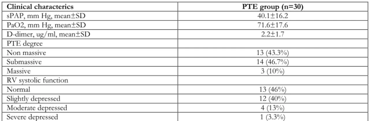
Tablo 1. Baseline clinical characterics of the study patients on admission.
Clinical characterics PTE group (n=30)
sPAP, mm Hg, mean±SD 40.1±16.2
PaO2, mm Hg, mean±SD 71.6±17.6
D-dimer, ug/ml, mean±SD 2.2±1.7
PTE degree Non massive 13 (43.3%) Submassive 14 (46.7%) Massive 3 (10%) RV systolic function Normal 13 (46%) Slightly depressed 12 (40%) Moderate depressed 4 (13%) Severe depressed 1 (3.3%)
PAP: Pulmonary arterial pressure, PaO2: Partial oxygen pressure, PTE: Pulmonary thromboembolism, RV: Right ventricle
Figure-1. Comparison of serum ADMA levels on
admission patient and control groups (p=0.022). Figure 2. Comparison of serum ADMA levels change (ΔADMA) between admission and first month control in patient group with resolution and without resolution (p=0.666).
447
Figure 3. Comparison of serum ADMA levels on
admission between PTE degree groups (p=0.019). Figure 4. Correlation between serum ADMA levels and sPAP (p=0.002, r=0.53).
Figure 5. Correlation between serum ADMA levels and sPaO2 (p=0.021, r = - 0.42).
DISCUSSION
PTE is a disease associated with high morbidity and mortality, which may sometimes be difficult to diagnose. Hypoxia, pulmonary hypertension, and RV systolic dysfunction are well-known risk factors for increased morbidity and mortality16. Ninety percent of PTEs arise from proximal deep veins of lower extremity, and more than 50% of proximal DVTs result in PTE5. Factors responsible for intravascular coagulation were first described by Virchow in 1856. Endothelial dysfunction, hypercoagulopathy, and stasis are the risk factors for intravascular coagulation17. Dysfunction of injured endothelium results in a reduced NO synthesis, decreasing the protective effect of NO8. NO is constantly released under normal conditions; various
substances (acetylcholine, noradrenaline, ATP, bradykinin etc.) and pharmacological agents (L-arginine) increase NO synthesis by stimulating the activity of the e-NOS enzyme8. Dysfunctional endothelial L-arginine/NO pathway constitutes the main mechanism for the detrimental effects of many cardiovascular risk factors (hypercholesterolemia, hypertension, smoking, diabetes mellitus, vascular inflammation etc.) on vessel wall9. In 1992 the discovery of ADMA as the endogenous competitive inhibitor of NOS suggested that this molecule may have a role in the dysfunction of the endothelial L-arginine/NO pathway10. ADMA reduces NO production and its protective bioactivity in a dose-dependent manner. In addition, high ADMA levels leads to production of detrimental superoxide radicals instead of NO by eNOS. As a result, ADMA affects the hemostatic balance between vasodilation and vasoconstriction, thrombocyte activation, cell-to-cell interaction, pro-/antiproliferative balance, pro-/antioxidative stimulation, and pro-inflammatory process11. Lungs are the primary source of NOS and ADMA and they acts as the primary organs that are responsible for the balance between NOS and ADMA18. ADMA is metabolized by dimethyl arginine dimethyl amino hydrolase (DDAH) and its isoform DDAH II19. It has been reported that DDAH II activity is reduced in hypoxia-induced pulmonary hypertension20. Suppressed endothelial DDAH II activity is considered a major cause of pulmonary hypertension21. Many studies to date have demonstrated that lower DDAH I activity is another factor for increased ADMA and decreased NO levels21-22. It is known that ADMA levels elevate in
448 pulmonary hypertension associated with congenital heart defects and in idiopathic pulmonary hypertension13;23. All these information suggest that hypoxia is the main culprit for elevated ADMA levels in PTE.
Dominik G et al. did not find any significant differences between plasma ADMA, L- arginine, and symmetric dimethylarginine levels in patients with DVT and control subjects24. In that study, however, none of the patients had PTE or hypoxemia. In our study ADMA levels were significantly higher in patients with PTE compared to the control subjects, suggesting that ADMA may be an ancillary marker for the diagnosis of PTE. Considering the Virchow triad, it is clear that patients with DVT also has endothelial dysfunction and hypercoagulability. Thrombus in the pulmonary arteries may have led to a higher ADMA levels as a result of hypoxia with hypoxia-induced endothelial injury and increased oxygen radicals. In our study there was a negative correlation between ADMA levels and PaO2.
The lack of any significant difference between patients with and without resolution with respect to the change in ADMA (ΔADMA) levels may be explained by an improvement of hypoxemia also in patients without resolution. These findings may suggest that hypoxemia may be the cause of a higher ADMA levels in PTE. We found a negative correlation between ADMA levels and PaO2 levels on arterial blood gas analysis. These findings may support the hypothesis that hypoxia is the primary mediator of ADMA elevation.
Skoro-Sajer et al. showed that plasma ADMA levels increased in patients with chronic thromboembolic pulmonary hypertension (CTEPH)12. They showed that hemodynamic parameters and the severity of pulmonary vascular disease were significantly correlated to plasma ADMA levels12. The authors also suggested that ADMA was a predictor of mortality and morbidity in patients with CTEPH12. Another study showed elevated ADMA levels in PTE, although it was not a predictor of CTEPH development25. That study, however, also included patients with chronic disease.
There are some limitations of this study. At the control visit first month later hypoxemia was checked with a pulse oximetry, and no arterial blood gas analysis was done when oxygen saturation exceeded 92%. Hence, not all patients underwent
arterial blood gas analysis at the first month visit. In our study, one of the possible mechanisms was hypoxia-induced endothelial injury that is further exacerbated by oxygen radicals, impairing NO metabolism. However, no parameter was evaluated to measure the amount of oxygen radicals. Massive PTE was diagnosed in only 3 of 30 patients in the PTE group. Therefore, one other limitation of this study is a number of patients as low as 3 in this group where ADMA’s effects may have been greatest. This study enrolled a total of 30 patients who are not representative of a general patient population. Therefore, we suggest that more studies with a larger sample size should be conducted in this field. Furthermore, the study is not a long term follow up design, like third or sixth month.
In conclusion, plasma ADMA levels were found elevated in PTE, and ADMA levels were correlated to thrombus burden. ADMA elevation in acute PTE probably occurs through hypoxia-induced mechanisms. Plasma ADMA levels, in addition to biochemical and imaging tests, may be helpful in determination of the severity of PTE. However, plasma ADMA levels may not be a useful parameter in the follow-up of PTE and assessment of thrombus resolution at the first month of therapy.
REFERENCES
1. Agnelli G, Becattini C. Acute pulmonary embolism.
N Engl J Med. 2010;363:266-74.
2. Konstantinides SV, Torbicki A, Agnelli G, Danchin N, Fitzmaurice D, Galiè N et al. Task Force for the Diagnosis and Management of Acute Pulmonary Embolism of the European Society of Cardiology (ESC). 2014 ESC guidelines on the diagnosis and management of acute pulmonary embolism. Eur Heart J. 2014;35:3033-69.
3. Heit JA. The epidemiology of venous thromboembolism in the community. Arterioscler Thromb Vasc Biol. 2008;28:370–2.
4. Cohen AT, Agnelli G, Anderson FA, Arcelus JI, Bergqvist D, Brecht JG et al. Venous thromboembolism (VTE) in Europe. The number of VTE events and associated morbidity and mortality. Thromb Haemost. 2007;98:756-64.
5. Girard P, Sanchez O, Leroyer C, Musset D, Meyer G, Stern JB et al. Deep venous thrombosis in patients with acute pulmonary embolism: prevalence, risk factors, and clinical significance. Chest. 2005;128:1593-600.
6. Riedel M. Acute pulmonary embolism 1: Pathophysiology, clinical presentation, and diagnosis. Heart. 2001;85:229-40.
449 7. Furchgott RF, The Discovery of endothelium
derived relaxing factor and its importance in the identification of nitric oxide. J Am Med Assn. 1996;276:1186-8.
8. Hingorani AD, Cross J, Kharbanda RK, Mullen MJ, Bhagat K, Taylor M et al. Acute systemic inflammation impairs endothelium-dependent dilatation in humans. Circulation. 2000;102:994-9. 9. Sydow K, Munzel T. ADMA and oxidative stress.
Atherosclerosis suppl. 2003;4:41-51.
10. Skoro-Sajer N, Mittermayer F, Panzenboeck A, Bonderman D, Sadushi R, Hitsch R et al. Asymmetric dimethylarginine is increased in chronic thromboembolic pulmonary hypertension. Am J Respir Crit Care Med. 2007;176:1154-60.
11. Böger RH, Bode-Böger SM, Szuba A, Tsao PS, Chan JR, Tangphao O et al. Asymmetric dimethylarginine (ADMA): a novel risk factor for endothelial dysfunction: its role in hypercholesterolemia. Circulation. 1998;98:1842-7. 12. Vallance P, Leone A, Calver A, Collier J, Moncada S.
Accumulation of an endogenous inhibitor of NO synthesis in chronic renal failure. Lancet. 1992;339:572-5.
13. Kielstein JT, Impraim B, Simmel S, Bode-Boger SM, Tsikas D, Frolich JC et al. Cardiovascular effects of systemic nitric oxide synthase inhibition with asymmetrical dimethyl argininein humans. Circulation. 2004;109:172-7.
14. Milat LJ, Whitley J, Leiper JM, Jeimes LD, Siragy MH, Carey RM et al. Evidence for Dysregulation of Dimethyl arginine Dimethyl aminohydrolase I in Chronic Hypoxia induced Pulmonary Hypertension. Circulation. 2003;108:1493-98.
15. Teerlink T. HPLC analysis of ADMA and other methylated L-arginine analogs in biological fluids. J Chromatogr B Analyt Technol Biomed Life Sci. 2007;851:21-9.
16. James E. Dalen. Pulmonary embolism: What have we learned since Virchow? natural history, pathophysiology, and diagnosis. Chest. 2002;122:1440-56.
17. White RH. The epidemiology of venous thromboembolism. Circulation. 2003;107:14-8. 18. Bulau P, Zakrzewicz D, Kitowska K, Leiper J,
Gunther A, Grimminger F et al. Analysis of methylarginine metabolism in the cardiovascular system identifies the lung as a major source of ADMA. Am J Physiol Lung Cell Mol Physiol. 2006;292:18-24.
19. Arrigoni FI, Vallance P, Haworth SG, Leiper JM. Metabolism of asymmetric dimethylarginines is regulated in the lung developmentally and with pulmonary hypertension induced by hypobaric hypoxia. Circulation. 2003;107:1195–201.
20. Pullamsetti S, Kiss L, Ghofrani HA, Voswinckel R, Haredza P, Klepetko W et al. Increased levels and reduced catabolism of asymmetric and symmetric dimethylarginines in pulmonary hypertension. FASEB J. 2005;19:1175-7.
21. Sasaki A, Doi S, Mizutani S, Azuma H. Roles of accumulated endogenous nitric oxide synthase inhibitors, enhanced arginase activity, and attenuated nitric oxide synthase activity in endothelial cells for pulmonary hypertension in rats. Am J Physiol Lung Cell Mol Physiol. 2007;292:1480-7.
22. Leiper J, Nandi M, Torondel B, Murray-Rust J, Malaki M, O’Hara B et al. Disruption of methylarginine metabolism impairs vascular homeostasis. Nat Med. 2007;13:198-203.
23. Gorenflo M, Zheng C, Werle E, Fiehn W, Ulmer HE. Plasma levels of asymmetrical dimethyl-L-arginine in patients with congenital heart disease and pulmonary hypertension. J Cardiovasc Pharmacol. 2001;37:489-92.
24. Haider DG, Bucek RA, Reiter M, Minar E, Hron G, Kyrle PA et al. The cardiovascular risk marker asymmetrical dimethylarginine is not affected by venous thromboembolism. Transl Res. 2006;148:26-9.
25. Altuntaş M, Atalay F, Can M, Altın R, Tor M. Serum asymmetric dimethylarginine, nitrate, vitamin B(12), and homocysteine levels in individuals with pulmonary embolism. Mediators Inflamm.