Gross Pathology of SpliNPVs and Alterations in Spodoptera littoralis
Boisd. (Lepidoptera:Noctuidae) Morphology Due to Baculoviral
Infection
*Umut TOPRAK1 Şerife BAYRAM1 M. Oktay GÜRKAN 1
Geliş Tarihi: 30.09.2004
Abstract: Baculoviruses are invertebrate-specific pathogens and baculoviral infections cause alterations in the physiology, metabolism and morphology of insects. It is important to recognize these physiological and symptomatologic changes to understand baculovirus infection cycle and biology. For this reason, in our study
Spodoptera littoralis Boisd. (Lepidoptera: Noctuidae) larvae were inoculated with the SpliNPV to doses of 3000 and 20,000 OBs for third instars and to concentrations of 106 and 3x 10⁶ OBs/ml for neonates and the alterations due to NPV
infection in larvae were then examined. Bioassays carried out with the third instar S. littoralis larvae revealed that no symptoms were detected during the first three days post-inoculation due to SpliNPV infection. Approximately, in the 4th day, infected larvae began to respond much more slowly than healthy larvae. This symptom was followed by whitening and slimming of cuticula, failure in molting, swelling of the body, diarrhoea, climbing to high places and hanging, liquefaction of the body and death. Infected neonates exhibited limited symptoms of distension and the failure of molting. The larvae died in approximately 8-8.5 days for third instars and in approximately 3-3.5 days for neonates due to baculoviral infection for the both doses and concentrations. On the other hand, different doses or concentrations did not cause an alteration in the occurence time of symptoms.To associate the NPV biology with the genes involved in baculovirus genome and understand their life cycle will improve their efficacy as biopesticides and help to the effective use of baculoviruses.
Key Words: Spodoptera littoralis, NPV, baculovirus, gross pathology, morphology
SpliNPV’lerin Genel Patolojisi ve Bakulovirüs Enfeksiyonundan Dolayı
Spodoptera littoralis
Boisd. (Lepidoptera:Noctuidae) Morfolojisindeki Değişimler
Öz: Bakulovirüsler omurgasızlara spesifik patojenler olup bakuloviral enfeksiyonlar böceklerin fizyolojisi, metabolizması ve morfolojisinde değişimlere neden olmaktadır. Bu fizyolojik ve simptomatolojik değişikliklerin farkına varmak, bakulovirüs enfeksiyon döngüsünü ve biyolojisini anlamak için önemlidir. Bu amaçla, çalışmamızda Spodoptera
littoralis Boisd. (Lepidoptera:Noctuidae) larvaları 3. dönemler için 3000 ve 20,000 OBs dozlarında, birinci dönemler için ise 106 and 3x 10⁶ OBs/ml konsantrasyonlarında SpliNPV ile inokule edilmiş ve daha sonra larvalarda NPV
enfeksiyonundan kaynaklanan değişimler irdelenmiştir. Üçüncü dönem S. littoralis larvalarıyla yapılan denemelerde inokulasyonu takip eden ilk üç gün boyunca SpliNPV enfeksiyonundan kaynaklı simptom görülmediği saptanmıştır. Yaklaşık olarak dördüncü günle birlikte hastalıklı larvalar sağlıklı larvalara göre daha yavaş tepkiler vermiştir. Bu simptomu, kutikulanın beyazlaşması ve incelmesi, deri değiştirememe, vücudun şişmesi, ishal durumu, yüksek yerlere tırmanma ve asma, vücudun sıvılaşması ve ölüm takip etmiştir. Enfekteli birinci dönem larvalar ise şişme ve deri değiştirememeyi içeren kısıtlı simptomlar vermiştir. Bakulovirüs enfeksiyonundan dolayı 3. dönem larvalar 8-8.5 günde, 1. dönem larvalar ise 3-3.5 günde her iki doz ve konsantrasyonda ölmüştür. Diğer taraftan farklı doz ya da konsantrasyonlar, simptomların ortaya çıkış zamanında bir değişikliğe neden olmamıştır. NPV bioyolojisini bakulovirus genomunda yer alan genlerle ilişkilendirmek ve hayat döngülerini anlamak bunların birer biyopestisit olarak etkinliğini artıracak ve daha etkin kullanımlarına yardımcı olacaktır.
Anahtar Kelimeler: Spodoptera littoralis, NPV, bakulovirüs, genel patoloji, morfoloji
* Prepared from Master’s Thesis, This research was funded by Biotechnological Institute of
Ankara University (project number: 25)
1 Univ. of Ankara, Fac. of Agriculture, Department of Plant Protection Ankara
Introduction
The family Baculoviridae consists of enveloped invertebrate pathogenic viruses containing a circular doublestranded DNA genome ranging from 90 to 160 kb (Blissard and Rohrman, 1990). The DNA genomes replicate in the host cell nucleus where they associate with capsid proteins to form the infectious virions (Figueiredo et al. 1999). The family is composed of two genera, Nucleopolyhedrovirus (NPV) and Granulovirus (GV) (Murphy et al. 1995). Baculoviruses occlude their virions in large, proteinaceous occlusion bodies (OBs) which help
the virus to remain viable outside the host for years. The occlusion boddies of NPVs are specifically identified as polyhedra. Baculoviruses generate two different phenotypes in their replicative cycle; the occlusion-derivedvirus (ODV), needed to spread the infection between larvae and the budded virus (BV), needed for the dissemination of the infection within the host. The life cycle of baculoviruses in nature starts with the ingestion of OBs present on contaminated diet by a larva (Kikhno et al. 2002). The OBs are dissolved in the alkaline structure of
the midgut and the virions are released from OBs. The ODVs pass through the peritrophic membrane and infect the larval midgut cells. It has been suggested that there may be some baculoviral proteins incorporated into the ODV which may enhance the ability of the virions to pass through the peritrophic membrane and these proteins include forms of chitinases and metalloproteases (Kalmakoff and Ward 2003). Following the infection of the nucleus of the midgut cell, budded virus is produced and disseminates the virus throughout the host.
The Egyptian cotton leafworm, Spodoptera littoralis Boisd. (Lepidoptera:Noctuidae) is an important pest of a variety of vegetable, fodder and fibre crops all over the world and causes extensive looses in many cultivated plants in greenhouses and fields. The insect is widely distributed throughout Africa, the Middle East and in the Mediterranean region it is regarded as a major pest (Jones et al. 1994).
Several hundred NPV isolates have been described from insects primarily of the order Lepidoptera (Martignoni and Iwai 1986). NPVs cause lethal epizootic diseases in their host-insect populations and because of their widespread occurence among economically important insect pests, they have received considerable attention as microbial pesticides (Payne 1988). Different SpliNPV isolates have also been isolated from egyptian cotton worm, S. littoralis to date and these isolates were designated SpliMNPV-A, SpliMNPV-B (Cherry and Summers 1985) and SpliMNPV-C (Maeda et al. 1990) according to their restriction-fragment profiles. The ability of SpliMNPV-B to successfully infect several Spodoptera species including Spodoptera exigua Hübner (Merdan et al. 1977), Spodoptera exempta Walker, Spodoptera
frugiperda Smith (McKinley et al. 1981) and Spodoptera
litura Fabricius (Okada 1977), make it a suitable candidate
for use as a microbial pest control agent (Faktor et al. 1997). SpliNPV-B variants have been isolated from diseased S. littoralis larvae collected in Israel, Egypt and Morocco and Japan (Cherry and Summers 1985, Kislev and Edelman, 1982, Maeda et al. 1990). The fifth sample of a SpliNPV-B variant was isolated from diseased S.
littoralis larvae in 2002 in Turkey. These samples were
collected from cotton fields in the South region of Turkey, Mersin and were named SpliNPV-TR1 to denote the first Turkish NPV isolate of S. littoralis by Toprak and Gürkan (2004).
Baculoviral infections cause alterations in the physiology, metabolism (Tanada and Kaya, 1993) and morphology of insects. It is important to recognize these physiological and symptomatologic changes to understand baculovirus infection cycles and biology. However, is notable that there are few symptomatologic studies specifically containing SpliNPV infection of S. littoralis. In this study, the symptomatology of the NPV disease of S.
littoralis, the principal stages of host invasion and viral
spread to the major tissues were investigated.
Materials and Methods
S. littoralis larvae are reared at the Department of Plant Protection, Faculty of Agriculture, University of
Ankara as a continuously maintained culture that was first brought to department in 2002. The larvae were reared on the lettuce leaves in plastic cages (24x33x15 cm) and lettuce leaves were sterilized with 1 % NaOCl before being given to larvae. S. littoralis were reared under controlled conditions with 16:8 h (L:D) photoperiod, 27±0.5˚ C temperature and 70 % relative humidity.
The wild type SpliNPV was isolated from field-infected S. littoralis larvae in 2002 and defined as a Turkish isolate (TR1) of SpliNPV-B by Toprak and Gürkan (2004). Production of SpliNPV-TR1 was performed in third instar S. littoralis larvae with virus occlusion bodies to a dose of 20,000 OBs and virus concentrations were quantified with an improved haemocytometer (Hausser Scientific, improved neubauer haemocytometer, 0.100 mm deep) under a light microscope. Six counts per haemocytometer were measured to reduce the counting errors.
For isolation of OBs, cadavers were treated with 0.1 % sodium dodeycl sulphate
(
SDS) (1 ml per cadaver) for one night at 4 C˚ and filtered through five layers of cheesecloth. OBs were pelleted by centrifugation at 3600 g for 10 minutes at room temperature in 50 ml centrifuge tubes. The pellet was resuspended in 0.5 % SDS and centrifugation and resuspension repeated with 0.3 M NaCl before final resuspension of OBs in distilled water (Modified from O’Reilly et al., 1992).Bioassays were carried out with the neonate larvae using a droplet feeding technique (Hughes et al., 1986) and with the third instar larvae using a leaf disk assay. In droplet feeding bioassays the larvae were starved for 3 h to encourage them to take up virus droplets and were then allowed to drink from an aqueous suspension containing 10 % (w/v) sucrose, 0.001 % blue food coloring dye and polyhedra at concentrations 0 (control), 106 and 3x10⁶ polyhedra/ml. After 10 minutes larvae that had ingested from these solutions were transferred to individual wells of insect rearing plates containing fresh lettuce leaves and supplied with additional lettuce leaves daily. In the leaf disk assays, carried out with the third instars, 0(control), 3000, and 20000 polyhedra were given to third instar larvae on individual 5 cm diameter lettuce leaf disks. Larvae that consumed the whole disk contaminated with virus were transferred to new cups and supplied with additional lettuce leaves daily. All bioassays were conducted at a constant temperature of 27.5±0.5 ° C and 32 larvae were used per one dose or per one concentration. All larvae were observed untill death. Infection symptoms were photographed during infection period for the doses and concentrations.
Results and Discussion
Baculovirus diseases are primarily diseases of the larval stages and the progression and signs of disease depend on several factors including the instar in which NPV infection becomes apparent infective dose, nutrition, temperature, degree of compatibility of the virus with its host, and the physical characteristics of the larva (Federici 1997).
To our results, different doses or concentrations did not cause an alteration in the occurence time of symptoms. In addition, no deaths were detected in controls due to NPV infection and the NPV doses or concentrations ingested by larvae produced 100 % mortality. 100 % mortality occured approximately at the end of 8 days post-inoculation for third instar inoculations at 3000 and 20,000 OBs and approximately at the end of 3.5 days post-inoculation for neonate inoculations at 106
and 3x10⁶ OBs/ml. Bioassays carried out with the third instars revealed that the S. littoralis larvae showed no symptoms during the first three days post-inoculaton for both doses. Thus, infected larvae normally do not show obvious signs of disease 1-3 days post-inoculation (OECD, 2002). Not only did Aizawa (1963) report that most lepidopteran larvae infected with NPVs show no external signs or symptoms for 2 to 5 days after viral ingestion but Federici (1997) also reported that in typical NPV infections, such as the diseases caused by the NPVs of Autographa californica Speyer (Lepidoptera:Noctuidae) (AcMNPV), Trichoplusia ni Hübner (Lepidoptera: Noctuidae) (TnSNPV) and Helicoverpa zea Boddie (Lepidoptera: Noctuidae) (HzMNPV) in noctuid larvae, there are few gross signs of disease during the first 3 days of infection. On or near the 4th day, infected larvae begin to respond much more slowly than healthy larvae to tactile stimuli such as touching (Federici 1997). In our study, it was also noted that when healthy larvae were dropped from a low height, they collected themselves on their legs, but the infected larvae gave no such response and instead lay on their dorsal side. Indeed, this situation can be related to the infection of central nervous system and muscle cells. Thus, when a larva started to lose control of the central nurveous system, It was not possible to respond to its surroundings. As a matter of fact, Tanada and Kaya (1993) also reported that the NPVs cause systemic infections, multiplying in major tissues and organs (polyorganotropic), particularly the fat body, hypodermis, trachea, blood cells, malpighian tubules, reproductive organs, salivary glands, midgut, pericardium and also muscle and nervous tissues.
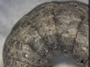
By the 4th days of post-inoculation, infected larvae’s cuticula showed a pale, whitening colour and thinning (fig 1a-b). Following this pale colouring on the cuticula, speckled colour of cuticula became more distinctive in appearence (fig 2). This symptom can be associated with the beginning of infection of hypodermal cells. The most typical symptoms are noted in the larval stages where either whitening or yellowing of the gut and /or the remainder of the body organs is associated with infection and replication by Evans and Shapiro (1997). The initial signs were the gradual changes in colour and luster of the integument with an increase in opaqueness, milkiness and glossiness (Tanada and Kaya, 1993). Tanada and Kaya (1993) also reported that the larva became less active and generally lost its appetite, though some were known to continue to feed up to a few days before death. Thinner appearance of cuticula can be associated with the activities of cathepsin and chitinase genes in the baculovirus genome. First and foremost in the baculovirus biological cycle, release of the polyhedra to environment is the most important step. For this step, the BV phenotype turns into ODV at the end of infection to form polyhedra.
Release of the polyhedra to environment is only possible with the liquefaction of the host. Indeed, the slimming of the cuticula is the beginning of the process that will bring the host to liquefaction. Both slimming of cuticula and liquefaction are associated with the activities of cathepsin and chitinase genes. These genes are classified under the auxiliary genes of baculoviruses and they have the possible role of damaging the peritrophic membrane to aid in the initial infection (Kalmakoff and Ward 2003).
In our study, It was observed that some of the larvae can not molt (figure 3). This could be due to the activity of the ecdysteroidglucosyltransferase (egt) gene. Ecdysone is the best characterized insect hormone responsible for regulating multiple genes that drive the growth, metamorphosis, and sexual maturation of the insect (Karim and Thummel, 1992; Schwartz and Truman, 1983). Many baculoviruses sequenced to date have been shown to contain egt gene that encodes for an egt enzyme (Ahrens et al. 1997, Ijkel et al. 1999, Kuzio et al. 1999) and this enzyme inactivates insect ecdysteroid hormones (Bianchi et al. 2000, O’Reilly and Miller 1989). This inactivation leads to a cessation of metamorphosis, keeping the insect in its larval, voracious state and therefore increasing the production of viral progeny (Pinedo et al. 2003). Briefly, presence of sufficient quantities of egt enzyme delays or prevents molting but in some cases, there can be a balance between the ecdysone concentration excreted by the larvae and egt concentration. In such a case, the larva can molt only to a certain degree. Federici (1997) also associated the failure of the larva to molt after infection with the production by the NPV of an ecdysteroid UDP-glucosyl transferase that glucosylates the molting hormone, ecdysone and commented that this situation is rarely observed.
a
b
Figure 1. (a,b) Whitening of the cuticula due to NPV infectionFollowing the fourth day, infected larvae showed swollen boddies (figure 2a). This could be due to the infection of nuclei and the hypertrophy that had occured in the cell. Thus, Tanada and Kaya (1993) reported that the nucleus increased in size due to the baculovirus infection. In NPV infections, at day 4 or 5, the larva will begin to appear swollen and the cuticle can appear glossy (Federici 1997). In the final stage of infection, occlusion boddies are formed and the nuclei are packed with occlusion bodies which causes the cellular hypertrophy and swollen appearence of the infected larvae (OECD 2002). Thus, the hypertropy in the nucleus is an important sign of NPV infection and causes insect swollen.
Another important symptom, diarrhoea was detected in the larvae. These larvae secrete a dark-brown fluid from their anus (data not shown). A similar symptom was reported for sawflies by Aizawa (1963). This situation can be due to the infection of proctodaeum or malpighian tubules.
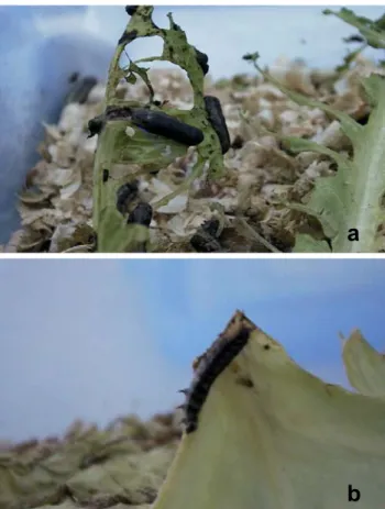
By the sixth day, some of the diseased larvae crawl to the top of the twigs on which they were feeding and hung from the bottom of the twigs by their abdominal legs (figure 4a,b). It is reported that shortly before dying, the larvae may move away from the food, disperse or climb an elevated location to hang from a branch or tree top by their abdominal and procaudal legs (Tanada and Kaya, 1993, Federici 1997). This stuation was noted as a negative geotropism in OECD reports (2002), but the reason for this behaviour can not be explained sufficiently to date. One possible explanation could be the result of an evolution which provides for virus to be spread by winds, rains etc. more effectively.
In our studies, the typical death symptom of larvae can be described as melting (figure 5a,b). Most larvae liquefy, the cuticle ruptures and polyhedra are released. In NPV infections, It is reported that the larval body contents are a fluid mass following death (Tanada and Kaya 1993).
Liquefaction of larvae is associated with the activities of cathepsin and chitinase genes as noted above, and the later phase involving the slimming of the cuticula. Insect cuticle is composed mainly of chitin fibers embedded in a protein matrix and its degradation requires the synergistic action of both proteinases and chitinases (Samuels and Paterson 1995). Thus, it appears that baculoviruses encode these enzymes to facilitate host cuticle breakdown after death, causing release of the progeny ODV into the enviroment and this presumably results in a significant advantage to the virus in terms of more efficient dissemination of progeny ODV, and hence more efficient horizontal spread (O’Reilly 1997). In addition to cuticular degradation, there is evidence that the viral cathepsin also participates in the degradation of internal tissues of the insect, which would also facilitate liquefaction and the release of progeny ODV (Ohkawa et al. 1994). After death, the larvae took on a blackish colour. This rapid melanization leading to blackening of the body was also reported recently (Evans and Shapiro 1997).
a
b
Figure 2. a) Pale colour and whitening seen on the cuticula ofinfected Spodoptera littoralis larva together with distension. b) Healthy Spodoptera littoralis larva
Figure 3. Failure of molting of infected S. littoralis larvae with SpliNPV.
Figure 4. (a,b) Spodoptera littoralis larvae that climbed to the top of the twigs on which they were feeding and hung themselves.
Figure 5. (a,b) Liquefaction of S. littoralis larvae at the end of SpliNPV infection.
The bioassays carried out with the neonates did not show certain symptoms. Because the larvae have a small body, It was difficult to detect the alterations due to NPV infections. Federici (1997) also reported that young larvae showed few gross signs of disease before death because of their small size. But in our studies, these symptoms could be summarized as the distension of a part of the body (figure 6,a) and the failure of molting (figure 6,b). These could also be due to the hypertrophy which occured in the nuclei of the cells and the egt gene activity that inactivates ecdysone, respectively.
The larvae died in approximately 8-8.5 days for third instars and in approximately 3-3.5 days for neonates. Ignoffo (1966) previously reported that the larva generally died in 5 to 12 days but virulent viral strains might kill very young larvae in 2 to 4 days. When infected by ingestion of several hundred to several thousand polyhedra during the first few instars (1-3), death can occur within 24 to 72 hr but if infected with the same amount of virus during the fourth or early fifth instar, the disease generally runs its course over a period of 5-10 days, at temperatures from 25-30 ° C (Federici, 1997). So our results were also found between the limits reported previously.
a
b
a
a
b
b
Figure 6. Symptoms showed by neonates. a-) Distension, b-) Failure of molting
In conclusion, the period that began with the inoculation of the SpliNPV and resulted with the death of
S. littoralis larvae contains single but important stages. These stages are strongly related to each other and the following alterations in the larvae contain the former alterations of the cycle. Also the symptoms seen in larvae are closely related with the NPV biology phases. To associate the NPV biology with the genes involved in baculovirus genome and understand their life cycle will contribute to improve their efficacy as biopesticides and help to the effective use of baculoviruses such as recombination studies that contain egt gene deletions.
Acknowledgments
The authors thank to Peter Bertling for his assistance in the writing of this paper. This study was supported by the Biotechnological Institute of Ankara University under the Project number 25.
References
Ahrens, C. H., R. L. Russel, C. J. Funk, J. T Evans, S. H. Harwood and G. F. Rohrmann, 1997. The sequence of the
Orgyia pseudotsugata multinucleocapsid nuclear
polyhedrosis virus genome. Virology, 229: 381-399. Aizawa, K, 1963. The nature of infections caused by nuclear
polyhedrosis viruses. In “Insect Pathology: An advanced treatise.” (E. A. Steinhaus, ed.). Vol. 1: 381-412. Academic Press, New York.
Bianchi, F. J. J. A., I. Snoeijing, W. Van der Werf, R. M. W. Mans, P. H. Smits and J. M. Vlak, 2000. Biological activity of SeMNPV, AcMNPV, and three AcMNPV deletion mutants against Spodoptera exigua larvae (Lepidoptera:Noctuidae). J. Invertebr. Pathol., 75: 28-35.
Blissard, G. W., G. F. Rohrman, 1990. Baculovirus diversity and molecular biology. Ann. Rev. Entomol., 35: 127-155. Cherry C. L., M. D. Summers, 1985. Genotypic variation among
wild isolates of two nuclear polyhedrosis viruses from
Spodoptera littoralis. J. Invertebr. Pathol. 46: 289-295. Evans, H., M. Shapiro, 1997. Viruses, pp. 17-53 In: (Ed.) “Manual
of Tchniques in Insect Pathology”, L Lacey, Academic Press, London.
Faktor, O., M. Toister-Achituv and O. Nahum, 1997. Enhancer element, repetitive sequences and gene organization in an 8-kbp region containing the polyhedrin gene of the
Spodoptera littoralis nucleopolyhedrovirus. Arch. Virol. 142: 1-15.
Federici, B. A, 1997. Baculovirus Pathogenesis, pp. 33-59 In: (Ed.) “The Baculoviruses”, LK Miller, Plenum Press, New York.
Figueiredo, E., D. Munoz, A. Escribano, A. Mexia, J. M. Vlak and P. Caballero, 1999. Biochemical identification and comperative insecticidal activity of nucleopolyhedrovirus isolates pathogenic for Heliothis armigera (Lep., Noctuidae) larvae. J. Appl. Ent., 123: 165-169.
Hughes, P. R., N. A. M. Van Beek and H. A. Wood, 1986. A modified droplet feeding method for rapid assay of Bacillus
thuringiensis and Baculoviruses in Noctuid larvae. J.
Invertebr. Pathol., 48: 187-192.
Ignoffo, C. M, 1966. Effects of age on mortality of Heliothis zea and Heliothis virescens exposed to a nuclear polyhedrosis virus. J. Invertebr. Pathol., 8: 279-283.
Ijkel W. F. J, E. A. Van Strien, J. G. M. Heldens, R. Broer, D. Zuidema, R. W. Goldbach and J. M. Vlak, 1999. Sequence and organization of the Spodoptera exigua multicapsid nucleopolyhedrovirus genome. J. Gen. Virol., 80: 3289-3304.
Jones, K. A., N. S. Irving, D. Grzywacz, G. M. Moawad, A. H. Hussein and A. Fargahly, 1994. Application rate trials with a nuclear polyhedrosis virus to control Spodoptera littoralis (Bıisd.) on cotton in Egypt. Crop Protection 13: 337-340. Kalmakoff, J. V. Ward, 2003. Baculoviruses. University of Otago,
Department of Microbiology.
Karim, F. D., C. S. Thummel, 1992. Temporal coordination of regulatory gene expression by the steroid hormone ecdysone. EMBO. J., 11: 4083-4093.
Kikhno, I., S. Gutierrez, L. Croizier, G. Croizier and M. L. Ferber, 2002. Characterization of pif, a gene required for the per os infectivity of Spodopera littoralis nucleopolyhedrosis virus. J. Gen. Virol., 83: 3013-3022.
Kislev, N., M. Edelman, 1982. DNA restriction-pattern differences from geographic isolates of Spodoptera littoralis nuclear polyhedrosis virus. Virology 119: 219-222.
Kuzio, J., M. N. Pearson, S. H. Harwood, C. J. Funk, J. T. Evans, J. M. Slavicek and G. F. Rohrmann, 1999. Sequence and analysis of the genome of a baculovirus pathogenic for
Lymantria dispar. Virology 253: 17-34.
Maeda S, Y. Mukohara, A. Kondo, 1990. Characteristically distinct isolates of the nuclear polyhedrosis virus from
Spodoptera litura. J. Gen. Virol. 71: 2631-2639.
Martignoni, M. E. P. J. Iwai, 1986. A catalogue of viral diseases of insects , mites and ticks. U. S. Dep. Agric. For. Serv. Pac. Northwest Res. Stn. Gen. Tech. Rep. PNW-195.
McKinley, D. J., D. A. Brown, C. C. Payne and K. A. Harap, 1981. Cross infectivity and activation studies with four baculoviruses. Entomophaga 26: 79-80.
Merdan, A., L. Croizier, J-C. Veyrunes and G. Croizier, 1977. Etude comparee des proteins des polyedres et des viroins de trois isolates de baculovirus de Spodoptera littoralis. Entomophaga 22: 413-420.
Murphy, F. A., C. M. Fauquet, D. H. L. Bishop, S. A. Ghabrial, A. W. Jarvis, G. P. Martelli, M. A. Mayo and M. D. Summers, 1995. Virus Taxonomy. Sixth report of the International Commitee on Taxonomy of Viruses. Vienna & New York: Springer-Verlag.
OECD, 2002. Consensus document on information used in the assesment of environmental applications involving baculobiruses. Series on Harmonization of Regulatory Oversight in Biotechnology, No:20.
Ohkawa, T., K. Majima and S. Maeda, 1994. A cysteine protease encoded by the baculovirus Bombyx mori nuclear polyhedrosis virus. J. Virol. 68: 6619.
Okada, M, 1977. Studies on the utilization and mass production of
Spodoptera litura nuclear polyhedrosis virus for control of the tobacco cut worm, Spodoptera litura Fabricius. Bull. Chugoku Natl. Agric. Exp. Stn.12: 1-66.
O’Reilly, D. R. 1997. Auxiliary genes of baculoviruses pp. 267-301 in: (Ed.) The Baculoviruses, LK Miller, Plenum Press, New York.
O'Reilly, D. R., L. K. Miller, 1989. A baculovirus blocks insect molting by producing ecdysteroid UDP-glucosyltransferase. Science 245: 1110-1112.
O’Reilly, D. R., L. K. Miller and V. A. Luckow, 1992. Baculovirus expression vectors- A laboratory manual. W. H. Freeman and Company, New York.
Payne, C. C. 1988. Pathogens for the control of insects: where next? Phil. Trans. Royal Soc. London B 318: 225-248. Pinedo, F. J. R., F. Moscardi, T. Luque, J. A. Olszewski and B. M.
Ribeiro, 2003. Inactivation of the ecdysteroid UDP-glucosyltransferase (egt) gene of Anticarsia gemmatalis nucleopolyhedrovirus (AgMNPV) improves its virulence towards its insect host. Biol. Control 27: 336-344.
Samuels, R. I., I. C. Paterson, 1995. Cuticle degrading proteases from insect moulting fluid and culture filtrates of entomopathogenic fungi. Comp. Biochem. Physiol. 110B, 661.
Schwartz, L. M., J. W. Truman, 1983. Hormonal control of rates of metamorphic development in the tobacco hornworm
Manduca sexta. Dev. Biol. 99: 103-114.
Tanada, Y., H. Kaya, 1993. Insect pathology, Academic press, Inc. ISBN 0-12-683255-2, San Diego, California.
Toprak, U., M. O. Gürkan, 2004. First Record of a NPV isolated from Spodoptera littoralis (Boisd.) (Lepidoptera:Noctuidae) for Turkey and Its Molecular Identification according to the partial lef-8 gene. Turkish J. Biol. 28 (2-4): 71-77. _____________________________________________ Correspendence address:
Umut TOPRAK
University of Ankara, Faculty of Agriculture, Department of Plant Protection 06110 Dışkapı Ankara Turkey
Phone: + 90 312 317 05 50/13 84 Fax: + 90 312 318 70 29