Journal of Pediatric Surgery Case Reports 52 (2020) 101355
Available online 21 November 2019
2213-5766/© 2019 The Authors. Published by Elsevier Inc. This is an open access article under the CC BY-NC-ND license (http://creativecommons.org/licenses/by-nc-nd/4.0/).
Multivisceral resection for synchronous inflammatory myofibroblastic
tumors of the lung and stomach
Naciye Cigdem Arslan
a,*, Suha Goksel
b, Oktar Asoglu
c aDepartment of General Surgery, Istanbul Medipol University, Istanbul, TurkeybDepartment of Pathology, Acibadem Health Group, Atasehir, 34758, Istanbul, Turkey cDepartment of General Surgery, Bogazici Academy of Clinical Sciences, 34357, Istanbul, Turkey
A R T I C L E I N F O Keywords:
Multivisceral resection Childhood tumors
Inflammatory myofibroblastic tumor Thoracoabdominal surgery
A B S T R A C T
Inflammatory myofibroblastic tumor is a mesenchymal tumor which commonly originates from lungs. In this paper we report the successful resection of multiple inflammatory myofibroblastic tumors invading thoracic, mediastinal and abdominal viscera in a 14-year old child. We could achieve an R0 resection with distal esophagectomy, proximal gastrectomy, splenectomy, left hepatectomy, left pneumonectomy, left diaphragm resection and pericardiectomy. To our knowledge, this is the first case reporting synchronous gastric and pul-monary inflammatory myofibroblastic tumors. The patient is alive without disease after 33 months.
1. Introduction
Inflammatory myofibroblastic tumor (IMT) is an uncommon mesenchymal tumor which is comprised of myofibroblastic spindle cells and inflammatory infiltration of plasma cells, lymphocytes and eosino-phils. It is more common in childhood and adolescence with a slight predominance in females. Lung, retroperitoneum, abdomen and pelvis are the most frequent locations, besides synchronous IMTs in lung and stomach have not been described before [1]. Surgical resection with clear margins is the main curative treatment. This intermediate-grade, rarely metastasing tumor may act as locally aggressive malignant pro-cesses owing to the location and adjacent organ invasion [2]. This paper presents the radical resection of synchronous huge IMTs of lung and stomach which were assumed unresectable previously.
2. Case
Fourteen-year-old female patient had been misdiagnosed with achalasia and underwent multiple endoscopic dilatations in two different institutes. Soon after, surgery was decided for worsening symptoms of the patient. Laparotomy revealed an unresectable thor-acoabdominal mass invading gastroesophageal junction and celiac trunk. Incisional biopsy and feeding gastrostomy were performed. His-topathologic examination demonstrated IMT and the patient was considered for palliative chemoradiotheray. After 11 fractions of
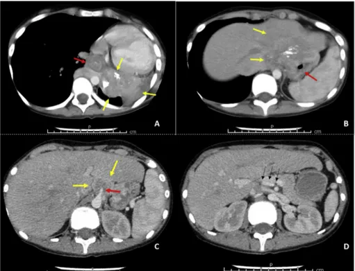
radiotherapy and 3 cycles of actinomycin-D, vincristine and cyclo-phosphamide she was referred to our institute with progressive findings. She was cachectic and decayed. Breathing sounds on left hemithorax were decreased and hemoglobin level was 5.9 g/dL. Computed tomog-raphy showed total atelectasis on left lung due to a 15-cm hilar mass infiltrating left main bronchus and left pulmonary vein (Fig. 1A). Tumor was invading distal esophagus, stomach, left lobe of the liver and extending along retroperitoneum behind the vena cava (Fig. 1B). Celiac trunk was also invaded (Fig. 1C). Angiographic tomography sections showed replaced right hepatic artery from superior mesenteric artery, thereby potential curative surgery with celiac trunk resection was considered (Fig. 1D). Nutritional support, blood transfusions and res-piratory rehabilitation have been provided until the general medical condition of the patient was built up.
Thoracoabdominal incision was performed. The tissues were extremely fibrotic and fragile due to previous radiotherapy and lapa-rotomy, particularly around gastroesophageal junction, that we could not recognize the healthy tissue borders between unattached synchro-nous tumors. Namely, en-block resection for a single giant tumor was presumed. Left lung, distal esophagus, proximal stomach, left lobe of liver, celiac trunk and spleen were resected (Fig. 2). Partial resection of pericardium and left diaphragm were also performed. Reconstruction of the left diaphragm was carried out by primary sutures. During peri-cardiectomy, the patient suffered ventricular fibrillation and corrected to normal sinus rhythm after 5 times of defibrillation. * Corresponding author. Department of General Surgery, Istanbul Medipol University Birlik mah Bahceler cad 5, 34230, Istanbul, Turkey.
E-mail address: naciyearslan@medipol.edu.tr (N.C. Arslan).
Contents lists available at ScienceDirect
Journal of Pediatric Surgery Case Reports
journal homepage: http://www.elsevier.com/locate/epschttps://doi.org/10.1016/j.epsc.2019.101355
Received 1 November 2019; Received in revised form 13 November 2019; Accepted 20 November 2019
Downloaded for Anonymous User (n/a) at Istanbul Medipol University from ClinicalKey.com by Elsevier on April 27, 2020. For personal use only. No other uses without permission. Copyright ©2020. Elsevier Inc. All rights reserved.
Journal of Pediatric Surgery Case Reports 52 (2020) 101355
2
Fig. 1. Preoperative computed tomography sections
of the patient (yellow arrows: tumor). A. Tumor originates from lower lobe of the left lung (red arrow: esophagus) B. Gastric tumor invades distal esophagus and left liver lobe (red arrow: stomach) C. Invasion of celiac trunk (red arrow: celiac trunk) D. Replaced hepatic artery from superior mesenteric artery (black arrows: right hepatic artery). (For interpretation of the references to colour in this figure legend, the reader is referred to the Web version of this article.)
Fig. 2. A. Intraoperative view before esophagogastrostomy (yellow arrow: left portal stump, black arrow: celiac stump) B. Specimen showing en-block resection of
lung, pericardium, diaphragm, liver, esophagus, stomach and spleen (yellow arrow: esophagus). (For interpretation of the references to colour in this figure legend, the reader is referred to the Web version of this article.)
Fig. 3. A. Yellow arrows denote two different tumors originating from lung and stomach B. Destruction of left liver lobe and gastroesophageal junction by the tumor
originating from stomach. (For interpretation of the references to colour in this figure legend, the reader is referred to the Web version of this article.) N.C. Arslan et al.
Downloaded for Anonymous User (n/a) at Istanbul Medipol University from ClinicalKey.com by Elsevier on April 27, 2020. For personal use only. No other uses without permission. Copyright ©2020. Elsevier Inc. All rights reserved.
Journal of Pediatric Surgery Case Reports 52 (2020) 101355
3
Esophagogastrostomy was performed using 26 mm circular stapler. The operative time was 410 min. Blood loss was 300 ml and 2 packs of erythrocyte suspension, 1 fresh frozen plasma and 200 ml human al-bumin 20% were administered intraoperatively. The patient was taken to intensive care unit postoperatively. On postoperative day 1 she was extubated, and enteral nutrition was started. She was discharged to clinic on day 6. Four days later, non-invasive mechanic ventilation was needed due to poor saturations and dyspnea. X-ray revealed pneumonia. Wide spectrum antibiotherapy was started and she returned to intensive care unit. Respiratory parameters were still poor and invasive mechanic ventilation was needed, therefore percutaneous tracheostomy was per-formed. On postoperative day 25, anastomotic leak at esophagogas-trostomy was detected. Intraabdominal and thoracic collections were drained by percutaneous pigtail catheters and leak was managed with silicone covered stenting. Mechanic ventilation was stopped on day 45 and the patient was discharged to the clinic. Four weeks later stent and catheters were removed. The patient was discharged from hospital after closure of tracheostomy on postoperative day 69. Histopathological examination revealed three independent IMTs: Two in lung (7.5 � 5.7 � 4.3 cm and 0.5 � 2x2 cm) and one in proximal stomach (6 � 5.5 � 4.5 cm). Surgical margins were clear (Fig. 3). There was no anaplastic lymphoma kinase (ALK) fusion gene rearrangement. No adjuvant treatment including targeted therapy was considered. The patient has been disease-free without any further problem during follow-up of 33 months.
3. Conclusions
Representing less than 1.5% of lung tumors, IMT is a rare and un-familiar tumor that there are very limited data in the literature to designate the pathogenesis and the optimal treatment [3]. Although it has been considered as a benign or infectious process, current aspect indicates that IMT is a real intermediate-grade neoplasm which carries a non-negligible potential of local recurrence and metastasis [4]. Differ-ential diagnosis is very conflicting due to non-specific symptoms and radiologic findings, besides 30–70% of the patients are asymptomatic. Therefore, a substantial part of the cases is diagnosed postoperatively [1–3]. Given that the most frequent primary lung tumor in pediatric population is IMT, awareness is essential in such cases. Our patient with dysphagia as the major symptom has been treated for achalasia for more than a year. The certain diagnose was obtained after laparotomy and incisional biopsy.
Clinical behavior of IMT varies due to aggressiveness and the loca-tion of the tumor. Different approaches in treatment including resecloca-tion, chemotherapy, radiotherapy, non-steroid anti-inflammatory drugs, and steroids were reported. In a few reports patients had benefit from anti- inflammatory treatment [5,6]. Response to radiotherapy and chemo-therapy is anecdotal, even in adjuvant setting [6,7]. In our case, the tumor has progressed in spite of chemoradiotherapy.
Currently, no standard treatment has been admitted. Resection with clear margins, when possible, is the commonly held treatment, however adjacent organ invasions may preclude surgery. Results of radical sur-gery are satisfying with a 5-year survival rate of 91%. Local recurrence rate has been reported up to 13%, particularly after incomplete re-sections [8]. We did not observe any sign of recurrence after 33 months follow-up.
Our patient had giant tumors invading essential structures. Thus,
surgical risk was extremely high and curative resection possibility was low. Conservative or less invasive treatment options would be cut out for such cases, however other treatment modalities did not provide even palliation and/or regression of IMT in our case. Consequently, we had to push the limits for radical surgery. Nonsurgical treatment modalities may be considered in case of incomplete surgical resection or multi-focal/metastatic disease. Until further studies reveal the optimum management to IMT, radical surgery seemed to remain the most effec-tive treatment [8].
Patient consent
Consent to publish the case report was not obtained. This report does not contain any personal information that could lead to the identifica-tion of the patient.
Funding
No funding or grant support. Authorship
All authors attest that they meet the current ICMJE criteria for Authorship.
Declaration of competing interest
The following authors have no financial disclosures: NCA, SG, OA. Authors declare no grant of support, conflict of interest and financial relationship related to the research or assistance with manuscript preparation.
Appendix A. Supplementary data
Supplementary data to this article can be found online at https://doi. org/10.1016/j.epsc.2019.101355.
References
[1] Gleason BC, Hornick JL. Inflammatory myofibroblastic tumours: where are we now?
J Clin Pathol 2008;61(4):428–37.
[2] Coffin CM, et al. Extrapulmonary inflammatory myofibroblastic tumor
(inflammatory pseudotumor). A clinicopathologic and immunohistochemical study
of 84 cases. Am J Surg Pathol 1995;19(8):859–72.
[3] Takeda S, Onishi Y, Kawamura T, Maeda H. Clinical spectrum of pulmonary
inflammatory myofibroblastic tumor. Interact Cardiovasc Thorac Surg 2008;7. 629:
33.
[4] Cerfolio RJ, Allen MS, Nascimento AG, et al. Inflammatory pseudotumors of the
lung. Ann Thorac Surg 1999;67:933–6.
[5] Dishop MK, Warner BW, Dehner LP, Kriss VM, Greenwood MF, Geil JD, Moscow JA.
Successful treatment of inflammatory myofibroblastic tumor with malignant transformation by surgical resection and chemotherapy. J Pediatr Hematol Oncol
2003;25:153–8.
[6] Diop B, Konate I, Ka S, Sall I, Fall D, Dieng M, Wone Y. Mesenteric myofibroblastic
tumor: NSAID therapy after incomplete resection. J Vis Surg 2011;148:e311–4.
[7] Imperato JP, Folkman J, Sagerman RH, Cassady JR. Treatment of plasma cell
granuloma of the lung with radiation therapy. A report of two cases and review of
the literature. Cancer 1986;57:2127–9.
[8] Fabre D, Fadel E, Singhal S, De Montpreville V, Mussot S, Mercier O, Dartevelle P.
Complete resection of pulmonary inflammatory pseudotumors has excellent long-
term prognosis. J Thorac Cardiovasc Surg 2009;137:435–40.
N.C. Arslan et al.
Downloaded for Anonymous User (n/a) at Istanbul Medipol University from ClinicalKey.com by Elsevier on April 27, 2020. For personal use only. No other uses without permission. Copyright ©2020. Elsevier Inc. All rights reserved.
