Arch Neuropsychiatry 2018;55:146−151 https://doi.org/10.5152/npa.2017.12753 RESEARCH ARTICLE
Blink Reflex in Episodic and Chronic Migraine
Burcu POLAT
1, Fikret AYSAL
1, Musa ÖZTÜRK
2, Belgin MUTLUAY
2, Yavuz ALTUNKAYNAK
2, Nesrin HELVACI YILMAZ
1,
Sevim BAYBAŞ
21Department of Neurology, İstanbul Medipol University, İstanbul, Turkey
2Bakırköy Psychiatric and Neurological Diseases Hospital, 2. Neurology Clinic, Istanbul, Turkey
Introduction: Activation of the trigeminovascular system and sensitization of brainstem trigeminal nuclei play a significant role in the physiopathology of migraine. Our aim was to investigate blink reflex (BR) and its recovery in episodic and chronic migraine patients.
Methods: Twenty-eight chronic migraine patients, thirty-two episodic migraine without aura patients and thirty healthy controls were included in the study. The study was performed using a portable electromyography device with a software specifically prepared for BR. Blink reflex assessments were performed in patients during the pain-free period and in healthy controls using the ‘standard method - double stimulation’ technique in 200 ms, 500 ms, 1000 ms, 2000 ms, and 5000 ms intervals.
Results: Blink reflex recovery was significantly increased in both patient groups as compared to the control group in 200 ms interstimulus interval (ISI) on both sides (p<0.005). Moreover, when it was compared to the control group, recovery was also significantly increased in the chronic migraine group in 2000 ms ISI on the right side and in 5000 ms ISI on the left side as well as in 500 and 1000 ms ISIs on the left side in the migraine
without aura group (p<0.002, p<0.003). R2 recovery curve was noted to be higher in both patient groups as compared to the control group, although could not be demonstrated statistically in all intervals. A statistically significant increase was observed in the migraine group without auras compared with the controls (p <0,037, p <0,011) in the left side at 500 and 1000 ms ISIs. For all intervals in our study, although the increase in recovery was not statistically significant, it was noted that the R2 recovery curve was higher in the patient groups, with respect to the normals. The increase in R2 recovery noted in both patient groups suggested increased sensitization of the trigeminal structures. Significantly increased recovery in low ISI (200 ms) in the two patient groups as compared to the control group raised the thought that the migraine brain goes through two different excitability periods (ictal and interictal).
Conclusion: In conclusion, similar to the previous studies, the findings of this study suggested that there was a reduction in central inhibitory mechanisms during interictal period in migraine patients.
Keywords: Blink reflex, blink reflex recovery, migraine, chronic migraine, trigeminal system
ABSTRACT
Cite this article as: Polat B, Aysal F, Öztürk M, Mutluay M, Altunkaynak Y, Helvacı Yılmaz N, Baybaş S. Blink Reflex in Episodic and Chronic Migraine. Arch Neuropsychiatry 2018;55:146-151. https://doi.org/10.5152/npa.2017.12753
Correspondence Address: Nesrin Helvacı Yılmaz, İstanbul Medipol Üniversitesi, Nöroloji Bölümü, İstanbul,Turkey • E-mail: [email protected] Received: 21.01.2016, Acceptedi: 25.04.2016, Available Online Date: 04.05.2018
©Copyright 2016 by Turkish Association of Neuropsychiatry - Available online at www.noropskiyatriarsivi.com
Activation of the trigeminovascular system and sensitization of brainstem trigeminal nuclei play a significant role in the pathophysiology of migraine. It is thought that cortical and subcortical structures associated with nociceptive and antinociceptive systems become functionally overactive in both during the migraine attack, and interictal period. It has been proposed that there is loss of inhibition in control of pain transmission, and abnormality in cortical control of pain in chronic migraine (1–4). Blink reflex (BR) is an objective electrophysiological method used in the evaluation of the trigeminovascular system, and is obtained by electrical stimulation of the supraorbital nerve (5). The afferent pathway is constituted by the ophthalmic branch of the trigeminal nerve, and its efferent pathway is constituted by the facial nerve (6–8). Blink reflex includes two ipsilateral responses, and one contralateral response (R1, R2, and R2 c, respectively) (9). R1 response indicates the direct synapse between the main sensory nucleus of the trigeminal nerve and ipsilateral facial motor nucleus. All components of the reflex arc are located in
the pons. The neural circuit mediated by the R2 component of the BR involves the trigeminal caudal nucleus, excitatory interneurons of the bulbopontine lateral reticular formation, and pontine facial nuclei innervating orbicularis oculi. R2 response is modulated by segmental and suprasegmental mechanisms. Therefore, it is possible to evaluate the excitability of brainstem reticular formation, and corticoreticular tracts by R2 recordings. While changes in R2 latency result from abnormal synaptic transmission in the brainstem and interneuronal excitability, changes in R2 areas have been mostly associated with changes in the control of higher central structures (7, 10).
Double stimulation technique for BR was first described by Kimura (11). The first response is elicited by a conditioning stimulus, and the second response is the test stimulus. The initial conditioning stimulus induces the reflex response reflecting the input reaching the facial motor neurons. The second test stimulus reflects the capacity of interneurons with altered excitability due to the first stimulus to transmit the second stimulus (8, 10).
INTRODUCTION
Imaging studies conducting on patients with migraine have revealed increased brainstem activity, and suggested that the brainstem might be a migraine generator. BR can be used to investigate the polysynaptic connections in the brainstem of patients with migraine (10, 12). In studies investigating BR in patients with migraine, changes in R2 response area (increase in R2 recovery) by double stimulation technique are important findings suggesting the presence of impairment in central inhibitory mechanisms (13). During migraine attacks, the area-under-the-curve of R2 was found to be increased on the affected side (14). The patients with chronic migraine showed no R2 abnormalities (15), while the patients with frequent attacks displayed shorter R2 latencies (16) compared with the healthy controls. In this study, we aimed to investigate the differences of BR response in patients with episodic and chronic migraine by the double stimulation technique.
METHODS
Sixty patients who were admitted to the outpatient headache clinic, and diagnosed with migraine according to 2004 IHS classification, and 30 healthy controls were also included. Patients were divided into two groups; Group 1 (n=32; 24 females, 8 males) consisted of patients with migraine without aura, and Group 2 (n=28; 25 females and 3 males) consisted of patients with chronic migraine. The healthy controls were designated as Group 3 (n=30; 20 females and 10 males).
The exclusion criteria of the patients included history of more than two attacks in a month, receiving prophylactic treatment within the last three months, history of excessive use of symptomatic drug, having pain within the last 72 hours, having another disease affecting the electrophysiological evaluations or the fifth or seventh cranial nerves, and having abnormal structure of the brainstem by cranial imaging. The tests were performed during the interictal (at least 3-day pain-free) period. The control group consisted of age- and gender-matched subjects. Subjects having a disease affecting electrophysiological evaluations, and having a history of any primary or secondary headache according to IHS classification were excluded. The study was approved by the Ethics Committee of Bakırköy Research and Training Hospital for Psychiatry, Neurology and Neurosurgery. Written and verbal informed consent were obtained from all of the migraine patients and controls included in the study.
Keypoint-portable electromyography (EMG) device with 24 kHz resolution and Keypoint-Net Program (a software specifically prepared for BR) was applied. Patients were asked to refrain from smoking, consuming alcohol, or taking any other medications 3 hours prior to EMG recording. We utilized the conventional method, and used a standard electrode to elicit the BR (2, 5). Blink reflex assessments were performed by using the standard method-double stimulation technique in 200 ms, 500 ms, 1000 ms, 2000 ms, and 5000 ms intervals.
For standard BR assessment, sweep speed of the EMG device was set in 50–800 ms/division interval according to interstimulus interval. Amplitude sensitivity was set in 100–200 µV/division, upper and lower frequency filters were set between 10 Hz and 200 kHz. The patient was in supine position, and relaxed with eyes open. The active electrode was placed inferior and lateral to the orbicularis oculi muscle while the reference electrode was placed on the nose. Ground electrode was placed on the chin. Supraorbital nerve was stimulated over the medial eyebrow by surface subcutaneous Ag-AgCl stimulating electrodes using unilateral electrical shocks. The patients were stimulated first from the right, and then the left side for each time, and each group and the bilateral responses were recorded.
At the beginning of the test, stimulations were performed with 1 mA increases, and the minimum current level of single electrical stimulus
producing a consistent >50 mV R2 response was defined as the R2 threshold intensity. The subjects were asked to identify their first sensation, which was subsequently defined as their individual sensory threshold. The 5–7 times of the individual threshold value was used for the test. In case stimulus intensity (SI) caused discomfort for the patient, the test was continued by reducing SI. For the assessment of BR, each individual’s pain threshold was determined. The value just below the value at which the patient felt discomfort was accepted as the level of stimuli.
Double stimuli consisting of the first conditioning stimulus, and the second test stimulus were randomly performed in 200 ms, 500 ms, 1000 ms, 2000 ms, and 5000 ms intervals. Recordings were performed simultaneously from both orbicularis oculi muscles. In order to prevent habituation, at least a 30 second waiting period was required between interstimulation intervals (ISIs). For every ISI, 6 stimulations were performed, the elicited responses were rectified, and their average was noted. In the conventional method, the classical range (28–42 ms) was used for R2. The onset of latency was measured according to the principle based on a signal ascended over 50% in respect to the basal value. The shortest latency response, “peak to peak” amplitude, and area values were determined. The amplitudes of the EMG responses of the blink reflex show individual variations in different people. Nonetheless, the following values (M±SD) have been suggested as normal: 0.38±0.23 mV for the ipsilateral R1 component, 0.53±0.24 mV for the ipsilateral R2 component, and 0.49±0.24 mV for the contralateral R2 component. The values were obtained both by the first conditioning stimuli (R2 c), and the second (test) stimuli (R2t). The amplitude and the latencies were calculated by measuring the values obtained by the conditioning stimuli. Only R2 area values were calculated using the both values obtained by the conditioning stimuli, and the test stimuli.
Blink reflex recovery curve (BR-RC) is the functional graphic representation of the percent change, in which y-axis indicates the proportion of the area response (mV.ms) elicited by the second stimulus to the area response calculated by the first stimulus (R2t area/R2 c area), and x-axis indicates the ISIs (200 ms, 500 ms, 1000 ms, 2000 ms, 5000 ms).
Statistical Analysis
Comparison of A, B, C, D, E intervals (ISI of 200 ms, 500 ms, 1000 ms, 2000 ms, and 5000 ms, respectively) in each three groups were performed by repeated analysis of variance (ANOVA) test. The cross-sectional comparisons between groups were performed by one-way ANOVA. For post hoc comparisons, Bonferroni was used for repeated ANOVA, and Tukey honestly significant difference test was used for one-way ANOVA. T-test was used for the comparison of R2 and R2 c. The relationship of EMG variables with clinical parameters was assessed by Pearson correlation coefficient. Gender distributions were analyzed by chi-square test. A p value <0.05 was considered as significant.
RESULTS
The mean age was 32±7 years in Group 1, 36±8 years in Group 2, and 34±8 years in controls. There was no significant difference between the three study groups in terms of age and gender. The mean duration of pain was 9.94±8.1 years in the Group 1, and 12.5±8.6 years in the Group 2. The mean pain frequency was 4.3±2.5 days/month in Group 1, and 16.6±4.1 days/month in Group 2. The duration of process of migraine becoming chronic was 2.68±2.6 years in Group 2. The mean visual analog scale (VAS) pain scores were 8.5±1.4 in Group 1, and 7.5±1.4 in Group 2. In the patients with migraine without aura, pain was bilateral in 53% of the patients, and unilateral in 47% of the patients, whereas in the chronic migraine group pain was bilateral in 39% of the patients, and unilateral in 61% of the patients. Neurological examination, routine blood tests, and cranial imaging results were normal in the patient groups.
The mean SI was 24.8±10.37 mA in Group 1, 22.6±4.5 mA in Group 2, and 19.8±6.2 mA in the control group. There was no significant difference between the three groups in terms of right, left, and mean SI values (p=0.33, p=0.70, p=0.36, respectively) (Table 1 and 2).
R2 recovery was found to be significantly increased in 200 ms ISI in both sides (right and left) in both Group 1 and 2, compared to the control group (p=0.005). Compared to the control group, recovery was significantly increased in Group 2 in 2000 ms ISI on the right side, and in 5000 ms ISI on the left side (p=0.002, p=0.003, respectively), as well as in 500 and 1000 ms ISIs on the left side in Group 1 (p=0.037, p=0.011, respectively) (Table 3). However, no significant relationship was found between this finding, and the lateralization of pain.
R2 recovery curve examples from the control group can be seen in Figure 1–4. Although significant differences were noted between patient groups and controls in some intervals, the increase in R2 recovery could not be demonstrated statistically in all intervals. However, R2 recovery curve was noted to be higher in patient groups compared to controls even in these intervals (Figures 5 and 6). When two patient groups were compared, no significant difference was noted with respect to recovery. No significant relationship was noted between recovery values, and clinical parameters (pain duration, pain severity, side of pain, duration of process of pain becoming chronic and VAS scores).
DISCUSSION
The relationship between migraine and the trigeminovascular system has been reported in many studies (17–20). The migraine brains are thought to be different in terms of organization and functional capacity. Regarding the pain mechanism in migraine; electrophysiological studies
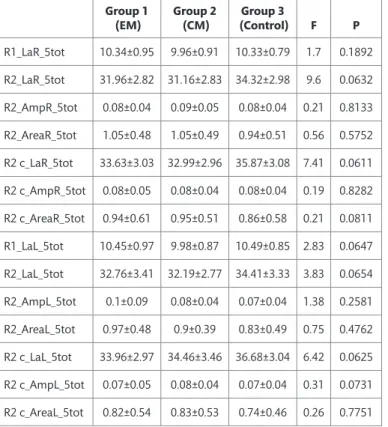
Table 1. Latency, amplitude and area values of R variables elicited
from right and left side in blink reflex examination of the study groups
Group 1
(EM) Group 2 (CM) (Control)Group 3 F P
R1_LaR_5tot 10.34±0.95 9.96±0.91 10.33±0.79 1.7 0.1892 R2_LaR_5tot 31.96±2.82 31.16±2.83 34.32±2.98 9.6 0.0632 R2_AmpR_5tot 0.08±0.04 0.09±0.05 0.08±0.04 0.21 0.8133 R2_AreaR_5tot 1.05±0.48 1.05±0.49 0.94±0.51 0.56 0.5752 R2 c_LaR_5tot 33.63±3.03 32.99±2.96 35.87±3.08 7.41 0.0611 R2 c_AmpR_5tot 0.08±0.05 0.08±0.04 0.08±0.04 0.19 0.8282 R2 c_AreaR_5tot 0.94±0.61 0.95±0.51 0.86±0.58 0.21 0.0811 R1_LaL_5tot 10.45±0.97 9.98±0.87 10.49±0.85 2.83 0.0647 R2_LaL_5tot 32.76±3.41 32.19±2.77 34.41±3.33 3.83 0.0654 R2_AmpL_5tot 0.1±0.09 0.08±0.04 0.07±0.04 1.38 0.2581 R2_AreaL_5tot 0.97±0.48 0.9±0.39 0.83±0.49 0.75 0.4762 R2 c_LaL_5tot 33.96±2.97 34.46±3.46 36.68±3.04 6.42 0.0625 R2 c_AmpL_5tot 0.07±0.05 0.08±0.04 0.07±0.04 0.31 0.0731 R2 c_AreaL_5tot 0.82±0.54 0.83±0.53 0.74±0.46 0.26 0.7751
La, Latency; Amp, Amplitude; R, Right; L, Left; 5tot, The sum of values elicited in all 5 interstimulation intensities; EM, episodic migraine; CM, chronic migraine.
Table 2. Latency, amplitude and area values of the mean R
variables (right+left/2) elicited from right and left side in blink reflex examination of the study groups
Group 1
(EM) Group 2 (CM) (Control)Group 3 F P
R1_LaX_5tot 10.4±0.9 9.97±0.82 10.41±0.76 2.58 0.0817 R2_LaX_5tot 32.36±2.95 31.67±2.68 34.37±2.91 7.07 0.0614 R2_AmpX_5tot 0.09±0.05 0.08±0.04 0.08±0.04 0.57 0.5673 R2_AreaX_5tot 1.01±0.45 0.97±0.38 0.88±0.45 0.74 0.4817 R2 c_LaX_5tot 33.79±2.85 33.72±3.03 36.28±2.72 7.73 0.0678 R2 c_AmpX_5tot 0.07±0.04 0.08±0.04 0.07±0.04 0.28 0.7594 R2 c_AreaX_5tot 0.88±0.53 0.89±0.51 0.8±0.51 0.25 0.7756
La, Latency; Amp, Amplitude; X, The mean value (right+left/2); 5tot, The sum of values elicited in all 5 interstimulation intensities; EM, episodic migraine; CM, chronic migraine.
Table 3. Blink reflex-R2 area recovery percentages (%) in the study
groups for each ISIs
Group 1
(EM) Group 2 (CM) (Control)Group 3 F P
Recovery R_200 ms 23.51±24.86 30.73±37.16 7.93±14.69 5.52 0.0055 Recovery R_500 ms 58.81±29.62 60.39±29.76 44.97±28.13 2.52 0.0864 Recovery R_1000 ms 87.39±26.24 75.37±20.66 73.85±36.25 2.09 0.1297 Recovery R_2000 ms 89.65±25.26 98.96±25.43 77.32±17.27 6.5 0.0023 Recovery R_5000 ms 91.55±25.24 102.97±28.52 88.73±24.87 2.39 0.0976 Recovery L_200 ms 26.39±26.63 34.56±29.86 11.63±16.76 6.34 0.0027 Recovery L_500 ms 69.94±37.68 63.78±26.38 48.57±34.94 3.28 0.0422 Recovery L_1000 ms 85.89±26.1 75.52±24.07 68.01±20.4 4.46 0.0143 Recovery L_2000 ms 98.83±29.18 107.21±38.58 89.8±26.55 2.2 0.1171 Recovery L_5000 ms 93.23±20.53 105.34±31.74 83.66±19.66 5.77 0.0044
R, right; L: left, EM; episodic migraine, CM; chronic migraine.
of trigeminal pathways using BR and corneal reflexes have indicated that trigeminal nociceptors are stimulated during the migraine attack, and continue to be stimulated during the interictal period as well (1, 2). Chronic pain studies have revealed findings suggesting that pain generally results from the dysfunction of diffuse inhibitory control mechanisms (3). Changes in R2 response area by double stimulation technique is an important finding that suggests the presence of impairment in central inhibitory mechanisms (11).
Several studies have been conducted using repetitive stimuli, and BR recovery curve. Shifting of the BR recovery curve to higher levels in low ISIs is suggestive of increased excitability of brainstem interneurons participating in polysynaptic R2 circuit. This can be explained by the loss of inhibition in brainstem interneurons caused by abnormal input coming from suprasegmental structures due to the impairment of central inhibitory mechanisms (7, 21).
In a study using standard BR technique in patients with migraine without aura, R1 latencies were not found to be different from the control group, while R2 latencies were noted to be significantly longer compared to the control group. These findings can be regarded as an objective evidence for the hypothesis that trigeminal afferents or polysynaptic transmission is affected in the brainstem of patients with migraine. However, it was not specified in this study whether or not the measurements were performed during the migraine attacks (22). In contrast, we did not find a significant difference between R2 latencies of the patient groups and controls. This can be attributed to the fact that BR measurements were performed while the patients were in the pain-free period, although R2 is known to be associated with caudal spinal trigeminal nucleus.
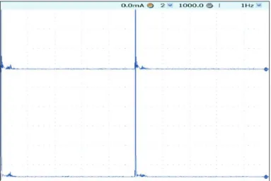
Figure (1–4); Legends (Examples of the Blink Reflex (BR); each curve represents the recordings of BR from a control subject)
Figure 1. Blink reflex responses (rectified) with 6 stimulation in 200 ms interstimulation
interval.
Figure 2. The averaged blink reflex responses of Figure 1.
Figure 3. The rectified and averaged blink reflex responses in 500 ms interstimulation
interval.
Figure 6. Blink reflex recovery curves for each study group for the left side.
(Figure 5–6; x-axis: proportion of the area response (mV. ms) means R2t area/R2 area; y-axis: ISI)
Figure 5. Blink reflex recovery curves for each study group for the right side.
Figure 4. The rectified and averaged blink reflex responses in 1000 ms interstimulation
In a study performed in patients with cervicogenic headache, chronic tension-type headache, and migraine, R2 latency and amplitude values were found to be normal in migraine patients who underwent BR measurements performed using standard electrical stimulation during their interictal period (23). Similarly, in the present study, the patient and control groups were comparable with respect to R2 latency, amplitude, and area values. However, both patient groups were significantly different from the control group with respect to R2 recovery curve obtained using the double stimulation technique in 200 ms interval. On the other hand, there was no significant difference between the two patient groups in this respect.
Aktekin et al. (24) found no significant differences between the migraine patients and control group with respect to the latency of the R1 and R2 components, and the amplitude and size of the R2 component of the BR during the interictal period. Therefore, they concluded that the dysfunction of the trigeminal system that was specific to migraine patients was transient. In the above-mentioned study, the authors found diminished recovery of the R2 component at the 300- and 500-ms interstimulation intervals in the tension-type headache group as compared to the other two groups (migraine without aura and control groups). According to these findings, the authors thought that different underlying mechanisms took a part in the pathogenesis of migraine and tension-type headache. The authors reported short interstimulation interval and small sample size as the limitations of their study (24). According to our results, R2 recovery was significantly increased in 200 ms ISI in both sides (right and left) in both migraine groups as compared to the control group (p<0.005). In the chronic migraine group, a significantly increased recovery was noted in 2000 ms ISI on the right side, and in 5000 ms ISI on the left side (p=0.002 and p=0.003, respectively). In the migraine without aura group, on the other hand, there was a significantly increased recovery in 500 and 1000 ms ISIs on the left side as compared to the control group (p=0.037 and p=0.011, respectively). Unlike Aktekin et al., we observed an increase in R2 recovery in migraine patients. In another study, BR latency and amplitude were studied during attacks in tension-type headache, and migraine patients. Low amplitude values returned normal with sumatriptan injection (25). The authors advocated that a temporary dysfunction occurred only during the attack phase of migraine led to an abnormality in the R2 component, and that this dysfunction returned to normal thereafter. The finding of the present study that there was a significant difference between the patient and control groups with respect to R2 recovery during the interictal period was consistent with the findings of a more recent study, suggesting that there was a dysfunction in inhibitory mechanisms in the pain-free period, and that the migraine brain goes through different excitability periods (26). According to another study, bilateral increases in the cortical and trigeminal excitability were shown in migraine patients with allodynia, and rTMS was effective for reducing clinical allodynia (27).
Methodological differences between studies might create a controversy while comparing the results. On the other hand, there is no recommended single method or gold standard for BR studies in migraine, or other primary headaches.
According to the normal data, R2 responses exhibit greater facilitation at higher ISIs. While R2 responses may completely disappear in ISIs shorter than 200 ms, they may exhibit approximately 40% to 50% increased recovery in 500 ms ISI, and approximately 70% to 90% increased recovery in 1500 ms ISI (8, 12). When we compared the two patient groups with each other, there was no significant difference between the two groups with respect to recovery. However, when the findings were analyzed according to ISI, we found that an increase in R2 recovery in 200 ms ISI,
the lowest ISI, noted during the pain-free period in the chronic migraine group persisted in higher ISIs (in 2000 and 5000 ms ISIs), and concluded that this might indicate increased trigeminal sensitization.
In conclusion, the findings of this study supported that cortical and subcortical structures associated with nociceptive and antinociceptive systems became functionally overactive, and that there was a reduction in inhibitory mechanisms during interictal period in both types of migraine. This study provided electrophysiological evidence that interneuronal brainstem pathways can be altered in patients with episodic and chronic migraine. There is limited number of studies investigating trigeminal sensitization in migraineurs through the study of the BR. For more accurate comparison, studies should evaluate pre-, peri- and post-attack periods. Further studies with larger group of patients are needed to understand the central inhibitory mechanisms of trigeminal sensitization in migraineurs.
Ethics Committee Approval: The study was approved by the Ethics Committee of
Bakırköy Research and Training Hospital for Psychiatry, Neurology and Neurosurgery.
Informed Consent: Written and verbal informed consent were obtained from all of the
migraine patients and controls included in the study.
Author Contributions: Concept - BP, FA, MÖ; Design - BP, FA; Supervision - FA, SB;
Data Collection and/ or Processing - BP, FA; Analysis and/or Interpretation - FA, NHY; Literature Search - BP, MÖ, FA; Writing - BP, SB; Critical Reviews -SB, NHY.
Conflict of Interest: No conflict of interest was declared by the authors.
Financial Disclosure: The authors declared that this study has received no financial
support.
REFERENCES
1. Ambrosini A, Schoenen J. The electrophysiology of migraine. Curr Opin Neurol 2003;16:327–331. [CrossRef]
2. Aguggia M. Neurophysiological test in primary headaches. Neurol Sci 2004;25:S203–S205. [CrossRef]
3. de Tommaso M, Losito L, Difruscolo O, Libro G, Guido M, Livrea P. Changes in cortical processing of pain in chronic migraine. Headache 2005;45:1208– 1218. [CrossRef]
4. Filatova E, Latysheva N, Kurenkov A. Evidence of persistent central sensitization in chronic headaches: a multi-method study. J Headache Pain 2008;9:295–300. [CrossRef]
5. Shahani B. The human blink reflex. J Neurol Neurosurg Psychiatry 1970;33:792–800.
6. Esteban A. A neurophysiological approach to brainstem reflexes. Blink reflex. Neurophysiol Clin 1999;29:7–38. [CrossRef]
7. Kimura J. Studies of the Facial Nerve and the Blink Reflex. In: Kimura J, editor. Electrodiagnosis in Diseases of Nerve and Muscle: Principles and Practice, 4th ed. New York: Oxford University Press Inc; 2001. pp. 409–438.
8. Aramideh M, Ongerboer de Visser BW. Brainstem reflexes: electrodiagnostic techniques, physiology, normative data, and clinical applications. Muscle Nerve 2002;26:14–30. [CrossRef]
9. Berardelli A, Cruccu G, Kimura J, Ongerboer de Visser BW, Valls-Solé J. The orbicularis oculi reflexes. The International Federation of Clinical Neurophysiology. Electroencephalogr Clin Neurophysiol Suppl 1999;52:249– 253.
10. De Marinis M, Pujia A, Colaizzo E, Accornero N. The blink reflex in “chronic migraine”. Clin Neurophysiol 2007;118:457–463. [CrossRef]
11. Kimura J, Harada O. Recovery curves of the blink reflex during wakefulness and sleep. J Neurol 1976;213:189–198.
12. Nardone R, Tezzon F. Brainstem reflexes in migraine patients. In: Clarke LB, editor. Migraine Disorders Research Trends. New York: Nova Science Publishers; 2007. pp. 183–208.
13. Kaube H, Katsarava Z, Przywara S, Drepper J, Ellrich J, Diener HC. Acute migraine headache: possible sensitization of neurons in the spinal trigeminal nucleus? Neurology 2002;58:1234–1238.
14. Brooks JB, Fragoso YD. The blink reflex test does not show abnormalities in a large group of patients with chronic migraine. Arq Neuropsiquiatr 2013;71:862–865. [CrossRef]
15. Zduska A, Cegielska J, Kochanowski J. Variability of the blink reflex in patients with migraine. Neurol Neurochir Pol 2013;47:352–356.
16. Welch KM. Contemporary concepts of migraine pathogenesis. Neurology 2003;61(8 Suppl 4):S2–S8.
17. Silberstein S. Migraine. Lancet 2004;363:381–391. [CrossRef]
18. Moskowitz MA. The neurobiology of vascular head pain. Ann Neurol 1984;16:157–168. https://doi.org/10.1002/ana.410160202
19. Goadsby PJ, Edvinsson L. The trigeminovascular system and migraine: studies characterizing cerebrovascular and neuropeptide changes seen in humans and cats. Ann Neurol 1993;33:48–56. [CrossRef]
20. Lyon LW, Kimura J, McCormick WF. Orbicularis oculi reflex in coma: clinical, electrophysiological, and pathological correlations. J Neurol Neurosurg Psychiatry 1972;35:582–588.
21. Cruccu G, Iannetti GD, Marx JJ, Thoemke F, Truini A, Fitzek S, Galeotti F, Urban PP, Romaniello A, Stoeter P, Manfredi M, Hopf HC. Brainstem reflex circuits revisited. Brain 2005;128:386–394. [CrossRef]
22. Bank J, Bense E, Kiraly C. The blink reflex in migraine. Cephalalgia 1992;12:289–292. [CrossRef]
23. Sand T, Moll-Nilsen B, Zwart JA. Blink reflex R2 amplitudes in cervicogenic headache, chronic tension-type headache and migraine. Cephalalgia 2006;26:1186–1191. [CrossRef]
24. Aktekin B, Yaltkaya K, Ozkaynak S, Oguz Y. Recovery cycle of the blink reflex and exteroceptive suppression of temporalis muscle activity in migraine and tension-type headache. Headache 2001;41:142–149.
25. Avramidis TG, Podikoglou DG, Anastasopoulos IE, Koutroumanidis MA, Papadimitriou AL. Blink reflex in migraine and tension-type headache. Headache 1998;38:69l-696. [CrossRef]
26. Di Clemente L, Coppola G, Magis D, Fumal A, De Pasqua V, Schoenen J. Nociceptive blink reflex and visual evoked potential habituations are correlated in migraine. Headache 2005;45:1388–1393. [CrossRef]
27. Ekizoglu E, Sozer-Topçular N, Baykan B, Oge AE. Assessment of excitability at the brainstem and cortex in primary headaches with allodynia. J Clin Neurophysiol 2015;32:119–129. [CrossRef]