350
Original Ar
ticle
ABSTRACT
Background: Mucins may show aberrant expression, localization, and/
or glycosylation in multiple malignancies. However, information regarding expression of these mucins is mostly unknown in urothelial tumors. Aim: This
study was conducted for examining the expressions of membrane associated and secreted mucin (MUC1) and a secreted gel‑forming mucin (MUC2) in urothelial tumors of the urinary bladder. Subjects and Methods: Archival
transurethral resection materials of 97 urothelial carcinoma cases were reexamined light microscopically and graded according to the 2004 WHO Classification. Pathological stage was given as pTa, pT1, and pT2. Demonstrative sections were recut for immunohistochemistry for MUC1 and MUC2. The results were statistically analyzed, and P < 0.05 was considered statistically
significant. Results: The positivity for MUC1 and MUC2 was 89.7% and 44.3%,
respectively. Independent from pathological stage of the tumor, MUC1 expression showed statistically significant correlation with tumor grade (P < 0.05). We did
not find any correlation between pathological stage and MUC1 and MUC2 expression (P > 0.05). MUC1 staining pattern in papillary urothelial neoplasm of
low malignant potential cases was more commonly apical and superficial (luminal cell layer only). Intermediate cells ± basal cells or isolated cells or islands of tumor cells with cytoplasmic and/or circumferential membrane positivity for MUC1 and MUC2 were more commonly observed in both low‑ and high‑grade carcinomas. The difference between groups in terms of MUC1 and MUC2 staining was statistically significant (P < 0.05). Conclusions: The staining patterns of both
mucins are different between urothelial papillary tumors and may be used to make a differentiation, especially for low‑grade papillary urothelial lesions. This difference may also be important in the carcinomatous transformation of urothelial neoplastic and preneoplastic lesions.
KEY WORDS: Bladder, grading, mucin expression, prognosis, urothelial
carcinomas
Immunohistochemical expression profiles of MUC1 and
MUC2 mucins in urothelial tumors of bladder
Ipek Isik Gonul, Asli Cakir1, Sinan Sozen2
Departments of Pathology and 2Urology, Gazi University Medical School, Ankara, 1Department of Pathology, Istanbul Medipol
University Medical School, Istanbul, Turkey
Address for correspondence:
Dr. Ipek Isik Gonul, Department of Pathology, Gazi University Medical School, Besevler, Ankara, Turkey. E‑mail: [email protected]
INTRODUCTION
Urothelial carcinomas of the bladder constitute the seventh most common cancer in men and the 17th most common cancer in women, and they cause approximately 150,000 deaths
per year worldwide.[1] Although they can range from superficial low‑grade papillary lesions
to muscle‑invasive malignant carcinomas, 70%–80% of cases are usually confined to lamina propria (pT1) or noninvasive (pTa) at the time of diagnosis. However, 10%–30% of these tumors show progression to muscle‑invasive disease (pT2).[2] While superficial tumors are
prone to recurrence, muscle‑invasive tumors show aggressive biological behavior with a poor response to therapy and tendency to early spread and metastasize to distant sites.[3]
Mucins are high‑molecular‑weight glycoproteins with protective role and precisely ordered distribution among epithelia and are categorized into three general types;
membrane‑associated mucins (MUC1, MUC3, MUC4, MUC12, MUC16, and MUC17), gel‑forming‑secreted mucins (MUC2, MUC5A, MUC5B, and MUC6), and soluble‑secreted mucin (MUC7).[4] MUC1 is a transmembrane
glycoprotein that is normally expressed at the basal level in most epithelial cells.[5] MUC2 is a prototype secretory
mucin, which is expressed in the colon, small intestine, and airways. Their gene expressions, rate of synthesis, and extent of their glycosylation may change during malignant transformation of the epithelial tissues.[6] These alterations may have an
impact on the invasive and metastatic capacities of tumors since they can change
How to cite this article: Gonul II, Cakir A,
Sozen S. Immunohistochemical expression profiles of MUC1 and MUC2 mucins in urothelial tumors of bladder. Indian J Pathol Microbiol 2018;61:350-5.
Access this article online Website: www.ijpmonline.org PMID: xxxxxxxxx (when available) DOI: 10.4103/IJPM.IJPM_12_18 Quick Response Code:
This is an open access journal, and articles are distributed under the terms of the Creative Commons Attribution‑NonCommercial‑ShareAlike 4.0 License, which allows others to remix, tweak, and build upon the work non‑commercially, as long as appropriate credit is given and the new creations are licensed under the identical terms.
Gonul, et al.: MUC1 and MUC2 in urothelial tumors
351 the regulation of proliferation and differentiation, immune
recognition, and cellular adhesion mechanisms of cells.[7]
Immunohistochemical studies for mucin expression in various human tumors have demonstrated that the expression of MUC1 is related to invasive proliferation of tumors and/or poor outcome for patients.[8] However, the expression of MUC2 is related to
noninvasive proliferation of tumors and/or favorable outcome for patients.[8]
There are limited data about the expression patterns and significance of mucins in urinary bladder carcinomas.[9‑14]
However, the use of molecular markers, including mucin expression profile, for characterization of bladder urothelial carcinomas may allow more improved and more complete insight in nature of cancer than histologic evaluation alone.[15]
These molecular markers may even serve as valuable tools for the prediction of tumor recurrence and response to therapy and the identification of therapeutic targets.
Our aim in this study was to investigate the relationship between MUC1 and MUC2 expressions and their patterns with the clinicopathological characteristics and biological behavior of urothelial carcinomas of the bladder.
SUBJECTS AND METHODS Patients
Ninety‑seven patients with primary bladder urothelial tumor who underwent transurethral resection with curative intent in our hospital’s Urology Clinic were included in the study. Exclusion criteria included patients with urothelial carcinomas with variant histology.
Histopathological examination
All patients’ biopsies were retrieved from pathology archives, and paraffin blocks were recut for immunostaining. Hematoxylin and eosin‑stained original slides were reexamined and assigned a grade based on the WHO 2004 Grading Scheme as papillary urothelial neoplasm of low malignant potential (PUNLMP), low‑grade carcinoma, or high‑grade carcinoma. Pathological staging was reperformed according to the presence of lamina propria (pT1) or muscularis propria invasion (pT2). Noninvasive tumors were classified as pT0.
Immunohistochemistry
One diagnostic paraffin block from each case was selected, and sections with 4‑µm thickness were cut for immunohistochemical stains of MUC1 (NCL‑MUC1, clone Ma695, 1:100, Novocastra Laboratories, Newcastle‑Upon‑Tyne, UK; NCL) and MUC2 (MUC2, clone Ccp58, 1:100, Novocastra Laboratories, Newcastle‑Upon‑Tyne, UK). Following deparaffinization, antigen retrieval was performed in citrate buffer (pH: 6.0) for both antibodies in a microwave oven for 20 min. Following washing with PBS, sections were incubated with secondary antibody (multispecies ultra streptavidin detection system‑HRP, Zymed, MA, USA) and streptavidin‑biotin complex (Zymed, MA, USA) for 20 min
at room temperature. Diaminobenzidine (diaminobenzidine tetrachloride, Zymed, MA, USA) was used as chromogen, and sections were counterstained using Harris hematoxylin. Both membranous and cytoplasmic staining for MUC1 and MUC2 were assessed. For each antibody, both the degree (percentage of positive cells) and the pattern of staining were recorded. Degree of staining was evaluated semi‑quantitatively for both mucins as follows: 1 = 1%–25% of the neoplastic cells stained; 2 = 26%–50% of the neoplastic cells stained; 3 = 51%–75% of the neoplastic cells stained; and 4 = 76%–100% of the neoplastic cells stained.
Three different patterns of immunostaining were observed; (a) apical membranes of luminal cell layer only, (b) intermediate cells ± basal cell layer, and (c) isolated cells or islands of tumor cells. Single or combined staining pattern was recorded for each case individually.
The results for immunostaining with respect to localization and the percentage of tumor cells stained were compared with tumor grade, pathological stage, and other clinical parameters investigated.
Statistical analysis
All statistical analyses were conducted using the SPSS 10.0 statistical software program (SPSS, Chicago, IL, USA). Pearson’s Chi‑square test and Fisher’s exact test were used for the evaluation of relationship between MUC1 and MUC2 expressions with clinicopathological variables. The results with P < 0.05 were considered to be statistically significant.
RESULTS
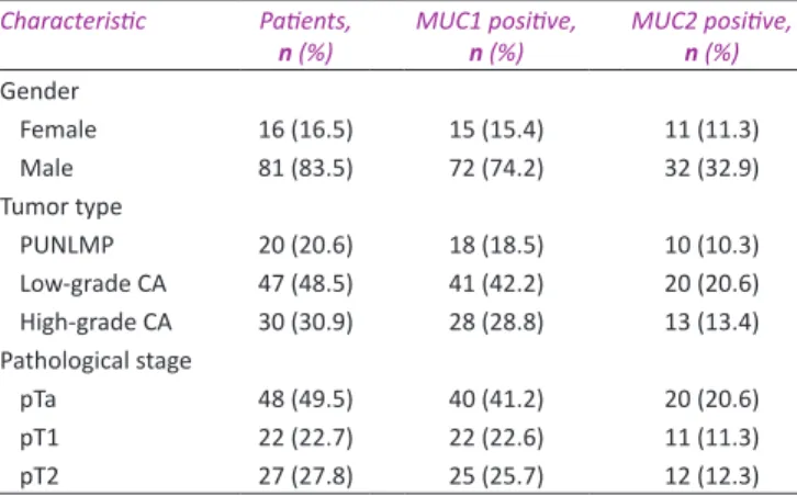
Eighty‑one men (83.5%) and 16 women (16.5%) with potentially curative resection of urothelial tumors were included in the study. The characteristics of the patient population and pathological findings are presented in Table 1. Mean patients’ age was 60.5 years (standard deviation = 12.0; range = 32–90). Of 48 noninvasive tumors, 26 (54.2%) were low‑grade carcinomas and Table 1: The clinicopathological characteristics of 97 cases
Characteristic Patients,
n (%) MUC1 positive, n (%) MUC2 positive, n (%)
Gender Female 16 (16.5) 15 (15.4) 11 (11.3) Male 81 (83.5) 72 (74.2) 32 (32.9) Tumor type PUNLMP 20 (20.6) 18 (18.5) 10 (10.3) Low‑grade CA 47 (48.5) 41 (42.2) 20 (20.6) High‑grade CA 30 (30.9) 28 (28.8) 13 (13.4) Pathological stage pTa 48 (49.5) 40 (41.2) 20 (20.6) pT1 22 (22.7) 22 (22.6) 11 (11.3) pT2 27 (27.8) 25 (25.7) 12 (12.3)
P<0.05, the result is statistically significant. PUNLMP: Papillary urothelial neoplasm of low malignant
352
only two were (4.2%) high‑grade carcinomas. Twenty‑one (44.7%) out of 47 low‑grade carcinomas and one out of 30 high‑grade carcinomas showed lamina propria invasion (pT1). Ninety percent of high‑grade carcinomas (27 out 30 cases) were muscle invasive. Tumor grade was correlated with pathological stage (P < 0.05). Overall, both MUC1 and MUC2 showed variable staining in terms of both reactivity percentage and pattern. Only 10 (10.3%) cases were negative for MUC1 and 54 (55.7%) cases were negative for MUC2. The percentages of cases showing MUC1 and MUC2 expressions in >50% of neoplastic cells were 58.7% (n = 57) and 9% (n = 9), respectively. Independent from pathological stage of the tumor, MUC1 expression showed statistically significant correlation with tumor grade (P < 0.05). Overall, 41 (47.1%) out of 87 MUC1‑positive cases were low‑grade carcinomas, 28 (32%) were high‑grade carcinomas, and 18 (20.6%) were PUNLMP. The highest percentage of staining was observed in the low‑grade carcinoma group (n = 30, 52.6%) when we considered only cases showing MUC1 positivity in >50% of neoplastic cells. Only 6 (10.5%) PUNLMP cases showed MUC1 positivity in >50% of neoplastic cells. Overall, 43 (44.3%) cases were positive for MUC2. Of these cases, 10 (23.2%) were PUNLMP, 20 (46.5%) were low‑grade carcinomas, and 13 (30%) were high‑grade carcinomas. Only 9 (20%) out of 43 cases revealed staining in >50% of neoplastic cells, none of which has been diagnosed as PUNLMP. Twelve (92.3%) out of 13 cases of high‑grade carcinomas showed MUC2 staining in <50% of neoplastic cells. The staining percentage for MUC2 in 20 cases of MUC2‑positive low‑grade carcinomas was highly heterogeneous (1%–25% in 7 cases [35%], 26%–50% in 5 cases [25%], 51%–75% in 4 cases [20%], and 76%–100% in 4 cases [20%]). We did not find a statistical correlation between MUC2 expression and tumor grade independent from pathological stage of the tumor (P > 0.05). Clinicopathological characteristics of MUC1‑ and MUC2‑positive cases are shown in Table 1.
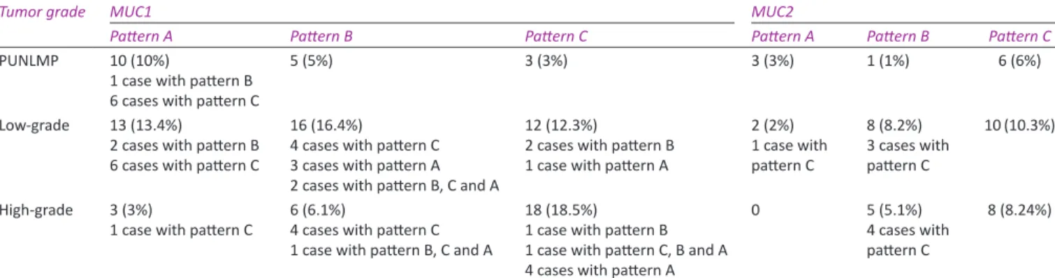
MUC1 and MUC2 staining patterns were heterogeneous between cases, and we have found a statistically significant correlation between MUC1 staining pattern and tumor grade (P < 0.05). MUC1 pattern in PUNLMP cases was more commonly apical and superficial (luminal cell layer only). On the other hand, six out of 10 MUC2‑positive PUNLMP cases showed staining in isolated cells or in islands of tumor cells and only three cases revealed apical and superficial staining. However, intermediate cells ± basal cells, isolated cells, and/or islands of tumor cells with cytoplasmic and/or circumferential membrane positivity for MUC1 and MUC2 were more commonly observed in carcinomas [Table 2]. The difference between groups for MUC1 and MUC2 staining patterns was statistically significant (P < 0.05) [Figures 1 and 2]. We did not find a correlation between pathological stage and either MUC1 expression or MUC2 expression (P > 0.05). Except for the two cases of muscle‑invasive carcinomas, all of the invasive cases (pT1 or pT2) were MUC1 positive. On the other hand, 20 out of 26 noninvasive low‑grade carcinomas and two cases of noninvasive high‑grade carcinomas were also MUC1 positive [Table 3]. MUC2 expression was highly heterogeneous between tumors with different pathological stages. Twenty out of 48 noninvasive tumors, 11 out of 22 pT1 tumors, and 12 out of 27 pT2 tumors were MUC2 positive [Table 3]. Similar to MUC1, we did not find a correlation between pathological stage and MUC2 expression. In addition, there is no correlation between MUC1 and MUC2 expressions of the tumors (P > 0.05). DISCUSSION
MUC1 (epithelial membrane antigen or episialin), which is encoded by a gene on chromosome 1q21, is a transmembrane glycoprotein that is normally expressed at the basal level in most epithelial cells.[5,7,16] While extracellular domain of MUC1 may
be aberrantly glycosylated and leads to putative revelation of Table 2: Staining patterns of MUC1 and MUC2 in different grade groups of the urothelial tumors
Tumor grade MUC1 MUC2
Pattern A Pattern B Pattern C Pattern A Pattern B Pattern C
PUNLMP 10 (10%)
1 case with pattern B 6 cases with pattern C
5 (5%) 3 (3%) 3 (3%) 1 (1%) 6 (6%)
Low‑grade 13 (13.4%)
2 cases with pattern B 6 cases with pattern C
16 (16.4%)
4 cases with pattern C 3 cases with pattern A 2 cases with pattern B, C and A
12 (12.3%)
2 cases with pattern B 1 case with pattern A
2 (2%) 1 case with pattern C 8 (8.2%) 3 cases with pattern C 10 (10.3%) High‑grade 3 (3%)
1 case with pattern C 6 (6.1%)4 cases with pattern C 1 case with pattern B, C and A
18 (18.5%) 1 case with pattern B 1 case with pattern C, B and A 4 cases with pattern A
0 5 (5.1%)
4 cases with pattern C
8 (8.24%)
Pattern A: Apical membranes of luminal cell layer only, Pattern B: Intermediate cells±basal cell layer, Pattern C: Isolated cells or islands of tumor cells. PUNLMP: Papillary urothelial neoplasm of low malignant potential Table 3: Percentage of MUC1 and MUC2 positivity according to the pathological stage of the tumors
Pathological stage MUC1 (%) MUC2 (%)
(‑) 1%‑25% 26%‑50% 51%‑75% >75% (‑) 1%‑25% 26%‑50% 51%‑75% >75%
pTa 8 (8.2) 9 (9.2) 7 (7) 7 (7) 17 (17.5) 28 (28.8) 11 (11.3) 3 (3) 3 (3) 3 (3)
pT1 0 4 (4) 3 (3) 4 (4) 11 (11.3) 11 (11.3) 5 (5) 3 (3) 1 (1) 2 (2)
Figure 2: MUC2 expression patterns in urothelial tumors of the urinary bladder. (a) Isolated small groups of cells positive for MUC2 in PUNLMP (×200). (b) Low‑grade noninvasive papillary urothelial carcinoma showing staining in isolated and small groups of cells, as in the case of PUNLMP (×200). (c) High‑grade invasive urothelial carcinoma showing diffuse and strong staining in neoplastic cells (×200)
Gonul, et al.: MUC1 and MUC2 in urothelial tumors
353 immunogenic epitopes in cancers, cytoplasmic domain functions
as a signal transducer protein.[16] Neoplastic cells of various cancer
types may use MUC1 to increase their proliferation rate, to escape from apoptosis, and to reduce the cell‑to‑cell adhesion to the extracellular matrix by overexpressing it.[7,17] There are several
studies in the literature searching for the role of MUC1 expression in the progression and prognosis of different types of carcinomas of the pancreaticobiliary tract and breast.[6,8,18,19]
Regarding the expression of MUC1 in urinary bladder tumors and in nonneoplastic urinary bladder mucosa, information is limited. Patriarca et al. showed that MUC1 expression was homogeneously confined to apical pole of superficial umbrella cells in nonneoplastic urothelium.[14] Similarly, Kaur et al.
reported that MUC1 expression was restricted to umbrella cells in most of the cases and it sometimes forms a sheath over urothelium in benign bladder mucosa.[13] However, MUC1 did not
show this polarized pattern and co‑localization with E‑cadherin in urothelial carcinoma in situ (CIS).[14] Since our cases did not
include any CIS focus, we could not make any comments about the expression profile of MUC1 in urothelial CIS. However, we observed that MUC1 staining pattern in PUNLMP cases was predominantly apical and superficial (luminal cell layer only), similar to the previously reported staining patterns of nonneoplastic urothelial mucosa and PUNLMP.[9,13,14,20] In
addition to urothelial CIS, MUC1 expression was also reported in bladder carcinomas.[9,12,13,21] A moderate‑to‑strong staining
with MUC1 was observed in luminal as well as in intermediate and basal layer of the urothelium in majority of urothelial papillary carcinomas in a study of Kaur et al.[13] In addition
to the localization of expression, the intensity of staining was also heterogeneous between neoplastic cells in their study.[13]
We have a similar observation that intermediate cells ± basal
cells or isolated cells or islands of tumor cells with cytoplasmic and/or circumferential membrane positivity for MUC1 were more commonly observed in both low‑grade and high‑grade carcinomas when compared to PUNLMP. This difference may be explained by loss of cell polarization in carcinomas. Earlier studies suggested that MUC1 plays a significant role in lumen formation and has an inhibitory role in the cell‑to‑stromal interaction. It was reported that MUC1 expression was predominantly present in the stroma‑facing surface of the cell clusters in invasive micropapillary carcinomas of the breast, pancreas, and gynecologic tract. However, labeling was mostly apical in areas with lumen formation in conventional adenocarcinomas.[22,23] In addition, in vitro and in vivo studies
have shown that MUC1 overexpression and loss of polarization prevents E‑cadherin and integrin‑mediated cell–cell adhesion. As a result, the process of invasion and metastasis becomes easier, especially when coupled with E‑cadherin downregulation.[7,19,24]
In this regard, the presence of MUC1 at the apical cellular site of a urothelial tumor is an indicator of intact MUC pathway. However, aberrant patterns of expression may indicate the defective MUC pathway. In the present study, we showed that higher MUC1 expression levels and altered expression patterns are associated with higher tumor grades. This may reflect differences in biological behavior and aggressiveness between different grades of urothelial tumors, especially for PUNLMP and low‑grade carcinomas. MUC‑1 induced discohesion may even contribute to the well‑known multifocality of bladder urothelial cell CIS.[25]
Elazeez et al. observed that 74% of their cases of papillary urothelial carcinomas were positive for MUC1.[26] They also demonstrated
that MUC1 expression increases as papillary carcinoma grade increases (i.e., 37.5% of grade 1 cases, 75% of grade 2 cases, and
c b a Figure 1: MUC1 expression patterns in urothelial tumors of the urinary bladder. (a) Apical membranous staining in PUNLMP (×200). (b) Low‑grade noninvasive papillary urothelial carcinoma showing basal and suprabasal intermediate cell staining (×200). (c) High‑grade noninvasive papillary urothelial carcinoma showing full thickness strong staining (×200) c b a
354
88.9% of grade 3 cases) (P < 0.01).[26] In the study of Kaur et al.,
expression of MUC1 showed a varied trend, starting from lack of expression (n = 38 or 12%), focal reactivity (n = 12 or 4%, mean H‑score 0.057 ± 0.01), to moderate (n = 147 or 46%, mean H‑score 1.02 ± 0.05) and intense immunoreactivity (n = 117 or 37%, mean H‑score of 2.86 ± 0.02).[13] In our study, overall, 41 out of 87
MUC1‑positive cases (47.1) were low‑grade carcinomas, 28 (32%) were high‑grade carcinomas, and 18 (20.6%) were PUNLMP. The highest percentage of staining was observed in the low‑grade carcinoma group (n = 30, 52.6%) when we considered only cases showing MUC1 expression in >50% of neoplastic cells. We have observed that MUC1 expression showed a statistically significant correlation with tumor grade independent from the pathological stage of the tumor. Interestingly, there is no statistically significant correlation between pathological stage and MUC1 expression of the tumors, and this observation is in agreement with the similar study done by Stojnev et al.[12] However, except for the two cases
of muscle‑invasive carcinomas, all of our invasive cases were MUC1 positive. Although the pattern of staining is different, low‑grade noninvasive carcinomas and even PUNLMP cases also showed MUC1 expression. We suggest that aberrant changes in MUC1 (underglycosylation, sialylation and/or fully glycosylation, overexpression, aberrant surface distribution patterns, etc.) occur early in the carcinomatous transformation and a transformed cell gains ability to invade and/or to make metastasis. In other words, probably, it is not a genetic alteration that occurs with invasion. However, the type of underlying genetic change may influence the degree of anaplasia of cells which is the key factor for grading of an urothelial tumor.
In addition to primary bladder urothelial carcinomas, the incidence and intensity of MUC1 expression were also reported to be high (66%) and strong (mean intensity 2.72 ± 0.45) in metastatic urothelial carcinomas.[13] Furthermore, it was
speculated that the overexpression of MUC1 in bladder tumors might be a good target for radioimmunoscintigraphy and radioimmunotherapy and intended for intravesical radioimmunotherapy of superficial bladder cancer.[27] Although
MUC1 expression was found to be related with prognosis of hepatocellular carcinoma patients, similar association for primary or metastatic urothelial carcinomas of the urinary bladder is not known.[18,23,27] The results of future analyses may lead us to a new
era of MUC1‑based treatment regimen for these patients. MUC2, as a predominant secretory mucin of intestine, has important immunomodulatory effects. It is encoded by a gene located on the chromosome 11p15.5 region as a cluster of mucin–gene complex that also includes MUC5AC, MUC5B, and MUC6 genes.[28] In a study of Stojnev et al., which includes
urothelial bladder cancer tissue samples of 539 patients, MUC2 immunoexpression rate in urothelial bladder cancer was found to be 40.1%.[12] Focal or cluster‑like cytoplasmic staining,
comprising between 10% and 25% of tumor cells, was the most frequent finding in their study and they did not observe MUC2 expression in normal bladder urothelium.[12] These results
were in accordance with the previous study of Walsh et al.[11]
Overall positivity for MUC2 for our cases was 44.3%, of which 23.2% were PUNLMP, 46.5% were low‑grade carcinomas, and 30.3% were high‑grade carcinomas. Importantly, the tumors showing MUC2 expression in >50% of neoplastic cells were all carcinomas. Although low‑grade carcinomas revealed highly heterogeneous staining percentage for MUC2, mostly <50% of neoplastic cells in high‑grade carcinomas was positive for MUC2. Staining pattern of MUC2 was similar to MUC1 for our cases. MUC2 overexpression was reported to be correlated with indolent clinical course and favorable prognosis in pancreaticobiliary neoplasms and in colorectal carcinomas.[6,8,29]
However, the prognostic significance of MUC2 in carcinomas of other sites is controversial.[19,30] Stojnev et al. showed that
MUC2 expression is more frequently observed in low‑grade urothelial carcinomas and significantly correlated with low pathological stage.[12] In addition, MUC2 significantly correlated
with MUC1 expression and increased probability of tumor relapse.[12] Although we did not find a statistical correlation
between MUC2 expression and tumor grade or pathological stage (P > 0.05), it was important for us to observe that none of the high‑grade carcinomas, mostly muscle invasive, expressed MUC2 in >50% of neoplastic cells, in contrast to low‑grade carcinomas. This may contribute to the more favorable outcome of patients with low‑grade carcinomas, showing more significant MUC2 expression compared to high‑grade carcinomas. In the present study, we find no correlation between MUC1 and MUC2 expressions (P > 0.05). This may be related to the small number of cases of high‑grade pT0 and pT1 cases included in our study. CONCLUSIONS
We have examined expressions of MUC1 and MUC2 and their correlation with tumor characteristics in 97 cases of primary urinary bladder urothelial tumors. We observed that MUC1 expression percentage increases as the grade of the papillary urothelial tumor increases. In addition, the pattern of MUC1 expression changes from luminal/superficial staining in PUNLMP to complete membranous and cytoplasmic staining in carcinomas. However, we could not demonstrate any association between MUC1 expression and the pathological stage of the disease. Referring to these observations, we think that aberrant changes in MUC1 occur early in the carcinomatous transformation of urothelial neoplastic and preneoplastic lesions. In addition, MUC1 immunostaining may be used to make a distinction for difficult cases of low‑grade papillary urothelial lesions. MUC2 expression showed no correlation either with the tumor grade or the pathological stage for urothelial tumors in our study. However, we have recognized easy to appreciate staining pattern differences between our cases in terms of MUC1 and MUC2 and this difference was statistically significant.
Financial support and sponsorship Nil.
Conflicts of interest
Gonul, et al.: MUC1 and MUC2 in urothelial tumors
355 REFERENCES
1. Babjuk M, Burger M, Zigeuner R, Shariat SF, van Rhijn BW, Compérat E,
et al. EAU guidelines on non‑muscle‑invasive urothelial carcinoma of
the bladder: Update 2013. Eur Urol 2013;64:639‑53.
2. Jacobs BL, Lee CT, Montie JE. Bladder cancer in 2010: How far have we come? CA Cancer J Clin 2010;60:244‑72.
3. Cheng L, Zhang S, MacLennan GT, Williamson SR, Lopez‑Beltran A, Montironi R, et al. Bladder cancer: Translating molecular genetic insights into clinical practice. Hum Pathol 2011;42:455‑81.
4. Hollingsworth MA, Swanson BJ. Mucins in cancer: Protection and control of the cell surface. Nat Rev Cancer 2004;4:45‑60.
5. Li Y, Liu D, Chen D, Kharbanda S, Kufe D. Human DF3/MUC1 carcinoma‑associated protein functions as an oncogene. Oncogene 2003;22:6107‑10.
6. Yonezawa S, Higashi M, Yamada N, Yokoyama S, Kitamoto S, Kitajima S,
et al. Mucins in human neoplasms: Clinical pathology, gene expression
and diagnostic application. Pathol Int 2011;61:697‑716.
7. Wesseling J, van der Valk SW, Vos HL, Sonnenberg A, Hilkens J. Episialin (MUC1) overexpression inhibits integrin‑mediated cell adhesion to extracellular matrix components. J Cell Biol 1995;129:255‑65.
8. Yonezawa S, Nakamura A, Horinouchi M, Sato E. The expression of several types of mucin is related to the biological behavior of pancreatic neoplasms. J Hepatobiliary Pancreat Surg 2002;9:328‑41.
9. Kaymaz E, Ozer E, Unverdi H, Hucumenoglu S. Evaluation of MUC1 and P53 expressions in noninvasive papillary urothelial neoplasms of bladder, their relationship with tumor grade and role in the differential diagnosis. Indian J Pathol Microbiol 2017;60:510‑4.
10. Scholfield DP, Simms MS, Bishop MC. MUC1 mucin in urological malignancy. BJU Int 2003;91:560‑6.
11. Walsh MD, Hohn BG, Thong W, Devine PL, Gardiner RA, Samaratunga ML,
et al. Mucin expression by transitional cell carcinomas of the bladder.
Br J Urol 1994;73:256‑62.
12. Stojnev S, Ristic‑Petrovic A, Velickovic LJ, Krstic M, Bogdanovic D, Khanh do T, et al. Prognostic significance of mucin expression in urothelial bladder cancer. Int J Clin Exp Pathol 2014;7:4945‑58.
13. Kaur S, Momi N, Chakraborty S, Wagner DG, Horn AJ, Lele SM, et al. Altered expression of transmembrane mucins, MUC1 and MUC4, in bladder cancer: Pathological implications in diagnosis. PLoS One 2014;9:e92742.
14. Patriarca C, Colombo P, Pio Taronna A, Wesseling J, Franchi G, Guddo F,
et al. Cell discohesion and multifocality of carcinoma in situ of the
bladder: New insight from the adhesion molecule profile (e‑cadherin, ep‑CAM, and MUC1). Int J Surg Pathol 2009;17:99‑106.
15. Agrawal R. Immunohistochemical and molecular markers in urothelial carcinoma. Indian J Pathol Microbiol 2017;60:462‑3.
16. Patton S, Gendler SJ, Spicer AP. The epithelial mucin, MUC1, of
milk, mammary gland and other tissues. Biochim Biophys Acta 1995;1241:407‑23.
17. Lau SK, Weiss LM, Chu PG. Differential expression of MUC1, MUC2, and MUC5AC in carcinomas of various sites: An immunohistochemical study. Am J Clin Pathol 2004;122:61‑9.
18. Yuan SF, Li KZ, Wang L, Dou KF, Yan Z, Han W, et al. Expression of MUC1 and its significance in hepatocellular and cholangiocarcinoma tissue. World J Gastroenterol 2005;11:4661‑6.
19. Rakha EA, Boyce RW, Abd El‑Rehim D, Kurien T, Green AR, Paish EC,
et al. Expression of mucins (MUC1, MUC2, MUC3, MUC4, MUC5AC and
MUC6) and their prognostic significance in human breast cancer. Mod Pathol 2005;18:1295‑304.
20. Garbar C, Mascaux C. Expression of MUC1 (Ma695) in noninvasive papillary urothelial neoplasm according to the 2004 world health organization classification of the noninvasive urothelial neoplasm. An immunologic tool for the pathologist? Anal Quant Cytol Histol 2011;33:277‑82.
21. Cardillo MR, Castagna G, Memeo L, De Bernardinis E, Di Silverio F. Epidermal growth factor receptor, MUC‑1 and MUC‑2 in bladder cancer. J Exp Clin Cancer Res 2000;19:225‑33.
22. Levi E, Klimstra DS, Andea A, Basturk O, Adsay NV. MUC1 and MUC2 in pancreatic neoplasia. J Clin Pathol 2004;57:456‑62.
23. Nassar H, Pansare V, Zhang H, Che M, Sakr W, Ali‑Fehmi R, et al. Pathogenesis of invasive micropapillary carcinoma: Role of MUC1 glycoprotein. Mod Pathol 2004;17:1045‑50.
24. Wesseling J, van der Valk SW, Hilkens J. A mechanism for inhibition of E‑cadherin‑mediated cell‑cell adhesion by the membrane‑associated mucin episialin/MUC1. Mol Biol Cell 1996;7:565‑77.
25. Witjes JA. Bladder carcinoma in situ in 2003: State of the art. Eur Urol 2004;45:142‑6.
26. Abd Elazeez TA, El‑Balshy Ael‑L, Khalil MM, El‑Tabye MM, Abdul‑Halim H. Prognostic significance of P27 (Kip 1) and MUC1 in papillary transitional cell carcinoma of the urinary bladder. Urol Ann 2011;3:8‑13. 27. Murray A, Simms MS, Scholfield DP, Vincent RM, Denton G, Bishop MC,
et al. Production and characterization of 188Re‑C595 antibody for
radioimmunotherapy of transitional cell bladder cancer. J Nucl Med 2001;42:726‑32.
28. Pigny P, Guyonnet‑Duperat V, Hill AS, Pratt WS, Galiegue‑Zouitina S, d’Hooge MC, et al. Human mucin genes assigned to 11p15.5: Identification and organization of a cluster of genes. Genomics 1996;38:340‑52.
29. Velcich A, Yang W, Heyer J, Fragale A, Nicholas C, Viani S, et al. Colorectal cancer in mice genetically deficient in the mucin muc2. Science 2002;295:1726‑9.
30. He YF, Zhang MY, Wu X, Sun XJ, Xu T, He QZ, et al. High MUC2 expression in ovarian cancer is inversely associated with the M1/M2 ratio of tumor‑associated macrophages and patient survival time. PLoS One 2013;8:e79769.