Turk J Phys Med Rehab 2021;67(1):122-124 DOI: 10.5606/tftrd.2021.5723
Available online at www.turkishjournalpmr.com
Letter to the Editor
TU RK IS H SO CI ETY OF PH YSICAL MEDICINE A ND R EH AB IL ITA TIO N
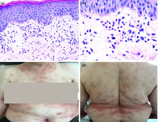
A 65-year-old woman was referred to our clinic with multiple erythematous plaques on the trunk and arms (Figure 1). The eruption occurred three days after starting a 75 mg daily dose of gabapentin prescribed by a physical medicine and rehabilitation specialist for neuropathic pain in her arm. The skin lesions were accompanied by a fever of 38.3°C. The laboratory investigations revealed a white blood count of 18.820/mm3 and neutrophilic leukocytosis with a percentage of 89.4%. Further investigations revealed no infectious condition. A punch biopsy was performed with preliminary diagnoses of Sweet’s syndrome and generalized fixed drug eruption (FDE). Histopathological examination showed a perivascular and interstitial mixed type of inflammatory infiltration with a predominance of neutrophils. A small number of eosinophils was also noted (Figure 1). Gabapentin was immediately discontinued and intramuscular betamethasone dipropionate, topical methylprednisolone, and oral desloratadine were administered; however, the patient was lost to follow-up. Two months later, the patient was readmitted with similar lesions on the same sites and she reported that the lesions appeared again two days ago, after taking a new drug prescribed by another physical medicine and rehabilitation specialist for the intense pain in her arm (Figure 2). The new drug was switched to gabapentin with a different brand name, as the patient did not inform the physician regarding the reaction to the previous
drug. A fever of 38.0°C once again accompanied the rash, and the blood tests revealed a white blood count of 16.230/mm3 with 88.2% neutrophils. A diagnosis of generalized neutrophilic FDE was established based on the clinical and pathological findings. Gabapentin was discontinued, and the lesions resolved within one week following the administration of oral desloratadine and a short course of topical and oral methylprednisolone. A written informed consent was obtained from the patient.
Fixed drug eruption is a cutaneous drug reaction with unknown mechanism characterized by recurrent erythematous and bullous skin lesions. Sulfonamides, trimethoprim, tetracyclines, and anticonvulsants are well-known culprits.[1] Gabapentin- induced oral classical FDE has been previously reported.[2] However, to the best of our knowledge, this is the first report of neutrophilic FDE related to the gabapentin use.
Neutrophilic FDE is a rare type of FDE which was first described by Agnew and Oliver[3] in association with amoxicillin-clavulanic acid. Subsequently, the entity has been reported in association with naproxen, trimethoprim-sulfamethoxazole, fluconazole, metronidazole, and dasatinib.[4-6] In the present case, given the accompanying fever and the peripheral neutrophilic leukocytosis, Sweet’s syndrome was the main provisional diagnosis. There was a typical, dense neutrophilic infiltration of Sweet’s syndrome and leukocytoclasis, but no lesions recurred at the same
Generalized neutrophilic fixed drug eruption induced by gabapentin
Emine Müge Acar1, Serpil Çorum Şirin2, Asuman Kilitci3, Ömer Faruk Elmas1, Funda Kemeriz2, Ümran Günay4 1Department of Dermatology, Kırşehir Ahi Evran University Faculty of Medicine, Kırşehir, Turkey
2Department of Dermatology, Aksaray University Faculty of Medicine, Aksaray, Turkey 3Department of Pathology, Kırşehir Ahi Evran University Faculty of Medicine, Kırşehir, Turkey
4Department of Dermatology, University of Health Sciences, Kartal Dr. Lütfi Kırdar Training and Research Hospital, Istanbul, Turkey Received: December 13, 2018 Accepted: February 17, 2020 Published online: March 04, 2021
Corresponding author: Asuman Kilitci, MD. Kırşehir Ahi Evran Üniversitesi Tıp Fakültesi Patoloji Anabilim Dalı, 40200 Kırşehir, Türkiye.
e-mail: [email protected]
Cite this article as:
Acar EM, Çorum Şirin S, Kilitci A, Elmas ÖF, Kemeriz F, Günay Ü. Generalized neutrophilic fixed drug eruption induced by gabapentin. Turk J Phys Med Rehab 2021;67(1):122-124.
This is an open access article under the terms of the Creative Commons Attribution-NonCommercial License, which permits use, distribution and reproduction in any medium, provided the original work is properly cited and is not used for commercial purposes (http://creativecommons.org/licenses/by-nc/4.0/).
123 Gabapentin induced neutrophilic fixed drug eruption
sites, strongly supporting the diagnosis of FDE.[7] Peripheral neutrophilic leukocytosis, which is not an expected finding in classical FDE, was evident for both episodes of the eruption, indicating a possible systemic component of generalized neutrophilic FDE.
Histopathological features of classical FDE include hydropic degeneration of the epidermal basal cell layer, dyskeratotic cells in the epidermis, and a lymphocytic infiltrate in the upper dermis.[8] The histopathology of the present case showed a mixed type of inflammatory Figure 1. (a, b) Perivascular and interstitial mixed inflammatory infiltration with neutrophil
predominance and neutrophil exocytosis in the epidermis (H-E ¥100; H-E ¥400). (c, d) Widespread erythematous-nummular plaques on the trunk and arms after the first gabapentin use.
(a)
(c)
(b)
(d)
Figure 2. (a, b) Erythematous plaques occurring on the same localizations after the second use of gabapentin.
Turk J Phys Med Rehab 124
infiltration with a predominance of neutrophils, instead. Controversy exists as to whether neutrophilic FDE is a separate entity as it has been suggested that it may be only a stage of the histopathological evolution of FDE.[9] Van Voorhees et al.[10] reported thickening of the epidermis, a few scattered necrotic keratinocytes, edema in the upper dermis and a mixed inflammatory infiltrate predominantly consisting of neutrophilic polymorphonuclear leucocytes in the first day histopathology of trimethoprim/sulfamethoxazole -induced FDE, while the examination on Day 5 showed the classical features of FDE. In the present case, the punch biopsy was performed on Day 7 of the eruption and did not reveal necrotic keratinocytes, spongiosis, lichenoid dermatitis, vacuolar degeneration in the basal cell layer, extracellular melanin or melanophages, which are the characteristics of the classical FDE. On the other hand, neutrophilic predominance was still notable.
In the first reported case of neutrophilic FDE which was presented by Agnew and Oliver,[3] classical epidermal changes of FDE were not observed and, similar to that seen in our case, neutrophilic infiltration was the predominant histological feature. The authors did not prefer the diagnosis of drug-induced Sweet’s syndrome due to the recurrent nature of the eruption in the same place and the absence of leukocytoclasis on the histopathological examination. Although fever and peripheral neutrophilic leukocytosis were observed in our patient, we also did not prefer the diagnosis of Sweet’s syndrome, as the lesions had the same distribution in both attacks, and the dermal infiltration was not as intensive as expected in Sweet’s syndrome. Suzuki et al.[4] also used the term “neutrophilic FDE” in a case report, in which histological examination showed the histological features of classical FDE accompanying with neutrophilic infiltration. In another case of neutrophilic fixed reaction which was presented by Bergman et al.,[6] the epidermis was also unaffected and the authors used the term "neutrophilic fixed dermatosis" instead of "FDE”. On the contrary, Ozkaya et al.[5] preferred the term "neutrophilic FDE", although the eruption did not reveal the classical
epidermal involvement of FDE. All these data show that there is no consensus among researchers regarding the description of the entity, which has been reported only in a small number of cases. We suggest that the terms “neutrophilic FDE” and “drug-related fixed neutrophilic dermatosis” can be used interchangeably to describe the eruption.
Declaration of conflicting interests
The authors declared no conflicts of interest with respect to the authorship and/or publication of this article.
Funding
The authors received no financial support for the research and/or authorship of this article.
REFERENCES
1. Dalla Costa R, Yang CY, Stout M, Kroshinsky D, Kourosh AS. Multiple fixed drug eruption to minocycline at sites of healed burn and zoster: An interesting case of locus minoris resistentiae. JAAD Case Rep 2017;3:392-4.
2. Gupta S, Gupta S, Mittal A, David S. Oral fixed drug eruption caused by gabapentin. J Eur Acad Dermatol Venereol 2009;23:1207-8.
3. Agnew KL, Oliver GF. Neutrophilic fixed drug eruption. Australas J Dermatol 2001;42:200-2.
4. Suzuki S, Ho J, Rosenbaum M, Bhawan J. Neutrophilic fixed drug eruption: a mimic of neutrophilic dermatoses. Clin Exp Dermatol 2019;44:236-8.
5. Ozkaya E, Büyükbabani N. Neutrophilic fixed drug eruption caused by naproxen: a real entity or a stage in the histopathologic evolution of the disease? J Am Acad Dermatol 2005;53:178-9.
6. Bergman JC, Ly TY, Keating MM, Hull PR. Recurrent and Fixed Neutrophilic Dermatosis Associated With Dasatinib. J Cutan Med Surg 2018;22:621-3.
7. Cohen PR. Sweet's syndrome--a comprehensive review of an acute febrile neutrophilic dermatosis. Orphanet J Rare Dis 2007;2:34.
8. Weyers W, Metze D. Histopathology of drug eruptions - general criteria, common patterns, and differential diagnosis. Dermatol Pract Concept 2011;1:33-47.
9. Waldman L, Reddy SB, Kassim A, Dettloff J, Reddy VB. Neutrophilic Fixed Drug Eruption. Am J Dermatopathol 2015;37:574-6.
10. Van Voorhees A, Stenn KS. Histological phases of Bactrim-induced fixed drug eruption. The report of one case. Am J Dermatopathol 1987;9:528-32.