Research Article
Effects on Lower Extremity Neuromuscular Control Exercises on
Knee Proprioception, Muscle Strength, and Functional Level in
Patients with ACL Reconstruction
Defne Kaya ,
1,2,3Hande Guney-Deniz,
4Cetin Sayaca,
1Mahmut Calik,
1and Mahmut Nedim Doral
51Department of Physical Therapy and Rehabilitation, Faculty of Health Sciences, Uskudar University, ˙Istanbul, Turkey
2Department of Physical Therapy and Rehabilitation, Faculty of Health Sciences, Istinye University, ˙Istanbul, Turkey
3Department of Physical Therapy and Rehabilitation, Faculty of Health Sciences, Istanbul Okan University, ˙Istanbul, Turkey
4Department of Physical Therapy and Rehabilitation, Faculty of Health Sciences, Hacettepe University, Ankara, Turkey
5Department of Orthopaedics & Traumatology, Faculty of Medicine, Ufuk University, Ankara, Turkey
Correspondence should be addressed to Defne Kaya; [email protected] Received 1 October 2019; Accepted 31 October 2019; Published 15 November 2019 Academic Editor: Nihal Apaydin
Copyright © 2019 Defne Kaya et al. This is an open access article distributed under the Creative Commons Attribution License, which permits unrestricted use, distribution, and reproduction in any medium, provided the original work is properly cited.
Objective. The purpose of this study was to determine the effects of lower extremity motor control exercises on knee
pro-prioception, muscle strength, and functional level in patients with anterior cruciate ligament reconstruction (ACL-R). Materials
and Methods. Thirty-two of the 57 patients with ACL-R using tibialis anterior allografts were divided into two groups. Group I:
lower extremity motor control exercises were added to the standard rehabilitation program. Group II: standard rehabilitation program was applied. Effects of lower extremity motor control exercises on quadriceps and hamstring muscles strength, knee joint position sense, and hop test were evaluated. Results. There were no differences in muscle strength and endurance of the quadriceps and hamstring between the operative and nonoperative sides in Group I (p > 0.05) while there were significant differences in strength of the quadriceps and hamstring between the operative and nonoperative sides in Group II (p < 0.05). There were significant differences in the endurance of the quadriceps and hamstring and knee joint position sense at 15°, 45°, and 75°between
the operative sides of the patients in both groups (p < 0.05). Conclusions. The neuromuscular control exercises program was found to be more effective in reducing the difference in strength while the standard program was found to be more effective in reducing the difference in endurance between the operated knee and the other knee. This study revealed that neuromuscular control exercises should also be used to improve knee proprioception sense following ACL-R.
1. Introduction
A successful rehabilitation program should benefit the pa-tients to return to an active lifestyle and preinjury levels. Outcomes of the numerous rehabilitation programs such as standard, aggressive, accelerated, home-based, supervised, and intensive programs in the literature have been discussed [1–7].
Rehabilitation programs for anterior cruciate ligament reconstruction (ACL-R) include a lot of parameters such as regaining the early full passive knee extension, immediate
range of motion (ROM), proprioception, quadriceps and hamstring strengthening, and fast return to normal daily living activities and sports [1–4].
Asymmetries in physical performance such as hopping, jumping, landing, loading, and movement pattern between the reconstructed and uninjured (another side) legs were described in the patient with ACL-R [8, 9]. This asymmetry can be seen for several years after ACL-R surgery [9, 10], and it must be treated to protect the joints from abnormal loading, to prevent secondary injuries, and to develop
correct movement patterns. Neuromuscular control
Volume 2019, Article ID 1694695, 7 pages https://doi.org/10.1155/2019/1694695
exercises such as squat, stairs descending and ascending, and landing should be added in a rehabilitation program to enhance bilateral symmetry and motor control of the leg.
To the best of our knowledge, there have been no studies evaluating the mentioned outcomes of the neuromuscular motor control exercise programs after ACL-R with tibialis anterior tendon allograft. Therefore, the purpose of this study was to assess the effects of motor control exercises on muscle strength, proprioception, and functional level after ACL-R with tibialis anterior tendon allograft. The hypothesis of the present study was that nonathletic patients who had undergone a standard rehabilitation program with lower extremity motor control exercises had better outcomes compared to patients who had undergone a standard re-habilitation program.
2. Materials and Methods
2.1. Study Design. This randomized-controlled study with two-year follow-up was designed to evaluate the effects of the standard rehabilitation program with motor control exer-cises on muscle strength, proprioception, and functional level in patients with ACL-R.
2.2. Participants. Patients were recruited between January 2016 and December 2018. Fifty-seven patients who had undergone primary ACL-R using tibialis anterior tendon allograft at Hacettepe University were invited to participate in the present study. Patients were eligible for the study if they (a) were from 14 to 55 years of age, (b) were male sex, (c) had ACL-R surgery at least before two years, and (d) had no previous history of knee surgery. Patients who underwent meniscal repair/meniscectomy/chondral surgery were also excluded from the study. The present study protocol was reviewed and approved by the Institutional Review Board of Yedikule Chest Diseases and Thoracic Surgery Training and Research Hospital Institutional Review Board (29012015/ 617). The written informed consent form was signed by all patients before the study, which was conducted according to the ethics guidelines and principles of the Declaration of Helsinki.
Patients were divided into two groups using the software
program (Random Allocation Software
®
) by the fourthauthor (MC) who was blinded to the assessment and group details. Group I: lower extremity neuromuscular motor control exercises were applied into the standard re-habilitation program. Group II: standard rere-habilitation program was applied.
2.3. Blinding. The assessor of muscle strength, pro-prioception, hop distance, and functional scores was also blinded to the patient’s identity and rehabilitation groups/ protocols. The first author who designed and carried out the rehabilitation program and followed up the patients was blinded to tests. The third author who carried out the re-habilitation program and followed up the patients was also blinded to tests. All tests were performed by the second author (HGD) who was blinded to the groups.
2.4. Interventions. The standard rehabilitation program was applied to all patients of both groups. The standard re-habilitation protocol was performed in light of the accepted orthopedic rehabilitation textbook [11]. Neuromuscular control exercises were started at the third week after ACL-R surgery for patients in Group I [12, 13].
All patients underwent a standard rehabilitation pro-gram from the first day to the end of the second week after ACL-R surgery. All patients performed exercises in their home. Follow-up of the rehabilitation program was per-formed by the first author (DK) and the third author (CS) at the third week, sixth week, third month, sixth months, a year, and two years following surgery. The rehabilitation progress was determined by the functional level of patients. Follow-up checklist of the exercises was used.
On the first day after surgery, all patients underwent a standard rehabilitation program and tolerated weight-bearing mobilization with crutches. The movement of the
knee was set to the extension/flexion of 0°–90°from the first
days after surgery to the third week. The restrictions such as active knee terminal extension exercises were lifted after the
6th week after surgery. Running was recommended at the
13thweek and agility and sports training at the 18th week.
Plyometrics and agility exercises were started from the 20th
week to the 24th week. Return to sports was not allowed
before nine months after surgery.
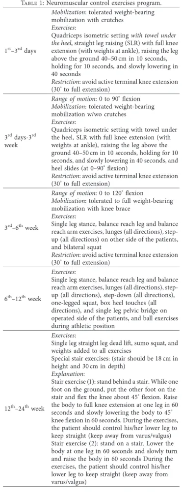
Neuromuscular control exercises were added to the rehabilitation program for patients in Group I (see Table 1). Single leg stance, balance reach leg and balance reach arm exercises, lunges (all directions), step-up (all directions) on the other side of the patients, and bilateral squat were started at 3 to 4 weeks after surgery. Single leg stance, balance reach leg and balance reach arm exercises, lunges (all directions), step-up (all directions), step-down (all directions), one-legged squat, box heel touches (all directions), and single leg pelvic bridge on the operated side of the patients, and ball exercises during athletic position were started at 5 to 8 weeks after surgery. Single leg straight leg dead lift, sumo squat, and weights added to all exercises were performed at 9 to 12 weeks.
2.5. Outcome Measures. All tests were performed at two years after surgery. Subjective tests, knee muscle strength tests, knee joint position sense tests, and one-legged hop test were done by the second author (HGD).
All patients executed 5-minute warm-up and then learned to exert peak torque in a practice session before proper data collection. The warm-up consisted of four submaximal contractions at each speed test.
2.5.1. Subjective Parameters. The pivot shift, anterior drawer, and valgus stress tests were applied to control the laxity of the ligaments by fully trained surgeons (MND). 2.5.2. Knee Muscle Strength. Two-minute rest interval was given between warm-up and testing. Before isokinetic measurement, all patients participated in a general body
warm-up. The warm-up consisted of 5 minutes of
sub-maximal exercise for lower extremity on a Biodex
®
SystemPro3 (Biodex Corp. Shirley NY, USA) at 60°/sec. The same
measurement protocol was reproduced for each of the testing sessions.
The isokinetic concentric peak torque of the knee muscles was determined bilaterally at an angular velocity
30°/s and 60°/s used for slow speeds, 180°/s for medium
speed, and 330°/s used for a high speed by using a Biodex
®
System Pro3 isokinetic dynamometer. Tests were performed
at 90°to 0°flexion for the knee. The order of the evaluation
between the right and left sides of the knee was determined randomly. The monitor was placed in such a way to provide visual feedback to the patients. Three maximal repetitions at all velocity were performed with 5-minute rest between the tests. Standardized verbal instructions and encouragement were given.
The isokinetic test for the knee muscles was performed
with the patients seated at 70°hip flexion (from the supine
position) and the knee angle at 90°flexion [14]. The knee
joint articulation axis was aligned to the dynamometer mechanic arm lever axis. The length of the dynamometer arm, which was fastened to the distal portion of the tibia by a Velcro strap, was altered according to the length of the leg of the patient.
2.5.3. Knee Joint Position Sense. Proprioceptive status was assessed by joint position sense (JPS) using an active angle reproduction (AAR) technique with eyes closed [15], using
the Biodex
®
System Pro 3 isokinetic dynamometer. Duringthe AAR detection, patients were eyes closed and had their head turned away, looking at the opposite side of the dy-namometer. The actual angle achieved by the patient and its difference from the target angle was recorded from the on-screen goniometer. When patients felt they had reached the target angle, they pressed the stop button and the angle was recorded; they were not permitted to correct the angle. This process was repeated six times for each target angle. The midrange angles were selected in an attempt to maximize sensory input from muscle proprioceptors. We decided that
if a patient was 5°or more away from their target angle, the
proprioceptive deficit was accepted [16].
The knee was moved from a 90°flexion starting position
passively to each of the target angles of 75°, 45°, and 15°(with
15°close to full extension). The leg was held there for 10 s for
the patient to memorize the position and then returned to
90°knee flexion. After a pause of 5 s, the patient moved the
lower limb by active contraction at an angular velocity
approximating 2°/s and stopped when the patient perceived
that the target angle had been reached.
The mean of the six trials was taken for each patient at each angle and used to calculate the difference between the actual angles achieved and the target angles [17]. The solute error of the total of six readings was taken. The ab-solute error is the difference between the actual angles relative to the target angle; this has no directional bias. The reliability for AAR testing was examined in our facility on 11
Table 1: Neuromuscular control exercises program.
1st–3rddays
Mobilization: tolerated weight-bearing
mobilization with crutches
Exercises:
Quadriceps isometric setting with towel under
the heel, straight leg raising (SLR) with full knee
extension (with weights at ankle), raising the leg above the ground 40–50 cm in 10 seconds, holding for 10 seconds, and slowly lowering in 40 seconds
Restriction: avoid active terminal knee extension
(30°to full extension)
3rddays-3rd week
Range of motion: 0 to 90°flexion
Mobilization: tolerated weight-bearing
mobilization w/wo crutches
Exercises:
Quadriceps isometric setting with towel under the heel, SLR with full knee extension (with weights at ankle), raising the leg above the ground 40–50 cm in 10 seconds, holding for 10 seconds, and slowly lowering in 40 seconds, and heel slides (at 0–90°flexion)
Restriction: avoid active terminal knee extension
(30°to full extension)
3rd–6thweek
Range of motion: 0 to 120°flexion
Mobilization: tolerated to full weight-bearing
mobilization with knee brace
Exercises:
Single leg stance, balance reach leg and balance reach arm exercises, lunges (all directions), step-up (all directions) on other side of the patients, and bilateral squat
Restriction: avoid active terminal knee extension
(30°to full extension)
6th–12thweek
Exercises:
Single leg stance, balance reach leg and balance reach arm exercises, lunges (all directions), step-up (all directions), step-down (all directions), one-legged squat, box heel touches (all directions), and single leg pelvic bridge on operated side of the patients, and ball exercises during athletic position
12th–24thweek
Exercises:
Single leg straight leg dead lift, sumo squat, and weights added to all exercises
Special stair exercises: (stair should be 18 cm in height and 30 cm in depth)
Explanation:
Stair exercise (1): stand behind a stair. While one foot on the ground, put the other foot on the stair and flex the knee about 45°flexion. Raise
the body to full knee extension at one leg in 60 seconds and slowly lowering the body to 45°
knee flexion in 60 seconds. During the exercises, the patient should control his/her lower leg to keep straight (keep away from varus/valgus) Stair exercise (2): stand on a stair. Lower the body at one leg in 60 seconds and slowly turn and raise the body in 60 seconds During the exercises, the patient should control his/her lower leg to keep straight (keep away from varus/valgus)
healthy controls having ICC2,1of 0.716 (SEM 4.5) with eyes
open and ICC2,10.404 (SEM 3.87) with eyes closed.
2.5.4. Functional Level. One-legged hop test was used to determine the functional level of patients. The one-legged hop test has good reliability and its intraclass correlation co-efficients range from 0.97 to 0.99 [18]. Patients were asked to hop as far as possible from a predetermined line and to land on the same leg. The three trials were collected from both sides. The normal limb was tested first, followed by the reconstructed limb [19].
2.6. Statistical Analysis. The normality of the distribution of the data was investigated by Kolmogorov–Smirnov testing with alpha set at 0.05. This testing confirmed that the data were normally distributed and that further statistical ana-lyses using the parametric testing would be appropriate. All data were analyzed with the Statistical Package for the Social
Sciences (SPSS
®
) version 21.0 (SPSS Inc, Chicago, Illinois,USA). A comparison between the groups was performed using an independent t-test for age, body weight, and body height to determine whether the groups presented similar demographics. The paired samples t-test was used to compare operative and nonoperative sides of the patients. The independent samples t-test was used to compare for the data of muscle strength, joint position sense, and the single-legged hop test between the operative sides of the patients in groups. Statistical significance was set for all testing at
p< 0.05.
3. Results
3.1. Recruitment and Sample Size. Thirty-two of the 57 pa-tients completed the original study. The Consolidated Standards of Reporting Trials (CONSORT) table depicts patient flow throughout the study (see Table 2). The dis-tributions of all numerical data were normal. There were no significantly differences in age (F � 0.220, p � 0.64), height (F � 0.002, p � 0.97), and body weight (F � 0.000, p � 0.98) between groups.
The mean age was 29.35 ± 9.71 years (range of 16–53 years), the mean height was 174.47 ± 8.26 cm (range of 162–192 cm), and the mean weight was 75.29 ± 12.21 kg (range of 53–95 kg) of the patients in Group I. The mean age was 31.60 ± 8.45 years (range of 23–53 years), the mean height was 177.07 ± 7.24 cm (range of 168–188 cm), and the mean weight was 81.27 ± 12.31 kg (range of 63–105 kg) of the patients in Group II.
3.2. Outcomes of Subjective Parameters. The pivot shift, anterior drawer, and valgus stress tests of the patients were normal in both groups. Additionally, there was no lack of the knee flexion and extension range of motion of the patients. 3.3. Changes in Muscle Strength, Proprioception, and Hop Test Length. There were significant differences in muscle
strength of the quadriceps and hamstring at 30°/s angular
speed and in hop test between the operative and non-operative sides in both groups (p < 0.05) (see Table 3). There were no differences in muscle strength of the quadriceps and
hamstring at 60°/s, 180°/s, and 330°/s between the operative
and nonoperative sides in Group I (p > 0.05). There were significant differences in muscle strength of the quadriceps
(p < 0.001) and hamstring (p � 0.01) at 60°/s between the
operative and nonoperative sides in Group II (see Table 3). There was no difference in the knee joint position sense at
15°, 45°, and 75° between the operative and nonoperative
sides in both groups (p > 0.05) (see Table 3). There was a difference in the hop test length between the operative and nonoperative sides in both groups (p < 0.05) (see Table 3). There were significant differences in muscle strength of
the quadriceps (p � 0.01) and hamstring (p � 0.01) at 330°/s
and joint position sense at 75°, 45°, and 15° between the
operative sides of the patients (p < 0.05) in Group I and Group II (see Table 4). There was no difference in the hop test length between the operative sides of the patients in both groups (p > 0.05) (see Table 4).
4. Discussion
The most important finding of the present study was that
joint position sense at 15°, 45°, and 75°knee flexion of the
patients in neuromuscular control exercises group was better than that in the standard rehabilitation group. Secondly, there was no difference in muscle strength between the
groups at the lower and medium angular speeds (at 30°/s,
60°/s, and 180°/s). At the higher angular speed (at 330°/s),
there was a difference between groups. Additionally, the pivot shift, anterior drawer, and valgus stress tests of the groups were similar. This is the first study to compare the neuromuscular control exercises program and standard
Table 2: Consolidated Standards of Reporting Trials (CONSORT) table.
1. Assessed for eligibility n � 57
2. Enrolment n � 57 ∗Exclusions: n � 17 Meniscal repair/ meniscectomy/chondral repair: n � 13 Female patients: n � 4 Group I Group II 3. Randomized n � 20 n � 20 4. Lost to follow-up n � 3 n � 5
5. 6 months after surgery n � 17 n � 15
6. 24-month follow-up n � 17 n � 15
Group I: neuromuscular control exercises were performed; Group II: standard rehabilitation program was performed; n: number of patients.
Table 1: Continued.
To 9th month
(3) running program (started at the 13thweek) (4) jumping (multidirectional) (started at the 18thweek)
(5) plyometrics and agility exercises (started at the 20thto 24thweek)
Restriction: return to sports is not allowed before
rehabilitation program for patients with ACL-R using tibialis anterior tendon graft.
The autografts are commonly used for good functional outcomes [20] while donor muscle deficits of the hamstring and patellar tendon autografts are also well known [21]. Although allografts have potential advantages such as the absence of donor site morbidity, shorter operative times, improved cosmetics, and easier rehabilitation over auto-grafts in ACL-R [22–24], functional scores of the ACL-R with a single loop freeze-dried irradiated tibialis anterior allograft were found similarly in four-strand hamstring autograft in nonathletic patients [25].
There is limited evidence on functional outcomes of the ACL-R with tibialis anterior allograft in the literature [22–25]. To our knowledge, limited number of studies evaluated only functional and subjective outcomes (IKDC and knee laxity) after the ACL-R using tibialis anterior allograft without the
measurement of the muscle strength, hop distance, and proprioception. In the present study, clinical assessments, quadriceps and hamstring muscle strength, hop test, and proprioception of the knee joint were assessed. Additionally, outcomes of the neuromuscular control exercises and stan-dard rehabilitation protocols were compared.
To the best of our literature research, we could not identify any previous study that evaluated the strength of the knee muscles, hop distance, and proprioception in patients with ACL-R using tibialis anterior allograft. Previous studies showed that intensive/accelerated rehabilitation program should improve quadriceps strength after ACL re-construction using autografts, but it may not fully recover to preinjury level [6, 7, 26, 27]. Although muscle strength of the operative side of the patients was still lower than that of the nonoperative side in all groups, there was no considerable difference in the quadriceps and hamstring muscle strength
Table 3: Descriptive and compared values of muscle strength, joint position sense, and hop tests obtained for the operated side and the other side of patients. Group I (n � 17) Group II (n � 15) Operated side (mean ± SD) Other side (mean ± SD) p Operated side (mean ± SD) Other side (mean ± SD) p Quadriceps muscle strength (Nm) 30°/s 138.71 ± 35.7 162.24 ± 49.12 0.01∗ 138.67 ± 34.05 160.27 ± 39.05 0.001∗ 60°/s 128.06 ± 33.47 140.94 ± 30.53 0.19 120.13 ± 36.86 147.00 ± 37.16 0.001∗ 180°/s 88.82 ± 24.09 93.53 ± 24.77 0.39 94.93 ± 40.88 98.73 ± 26.61 0.60 330°/s 57.00 ± 9.50 58.82 ± 18.47 0.64 62.80 ± 25.36 68.20 ± 26.15 0.28 Hamstring muscle strength (Nm) 30°/s 98.88 ± 25.66 112.94 ± 28.60 0.01∗ 96.73 ± 23.64 114.20 ± 29.88 0.02∗ 60°/s 99.00 ± 20.46 101.18 ± 20.93 0.62 92.13 ± 25.00 109.20 ± 26.07 0.01∗ 180°/s 79.41 ± 16.62 82.00 ± 18.37 0.62 79.07 ± 27.59 85.20 ± 23.12 0.45 330°/s 73.24 ± 11.24 74.41 ± 11.78 0.77 76.13 ± 20.49 77.67 ± 24.73 0.74 JPS (°) 75° 74.55 ± 1.54 74.61 ± 2.98 0.93 75.85 ± 3.77 76.92 ± 3.42 0.22 45° 45.63 ± 2.94 45.19 ± 4.11 0.64 47.32 ± 6.05 47.18 ± 5.84 0.92 15° 15.44 ± 1.74 15.78 ± 2.40 0.64 16.95 ± 3.13 17.07 ± 2.49 0.93 Hop test (cm) 141.37 ± 34.35 156.12 ± 24.04 0.01∗ 146.53 ± 19.56 157.31 ± 19.53 <0.001∗
∗Paired sample t-test. JPS: joint position sense; Group I: neuromuscular control exercises were performed; Group II: standard rehabilitation program was performed; SD: standard deviation; n: number of patients.
Table 4: Descriptive and compared values of muscle strength, joint position sense, and hop tests obtained for the operated side of the patients in Group I and the operated side of patients in Group II.
Group I (n � 17) Group II (n � 15) F p Operated side (mean ± SD) Other side (mean ± SD) Operated side (mean ± SD) Other side (mean ± SD) Quadriceps muscle strength (Nm) 30°/s 138.71 ± 35.7 162.24 ± 49.12 138.67 ± 34.05 160.27 ± 39.05 0.362 0.55 60°/s 128.06 ± 33.47 140.94 ± 30.53 120.13 ± 36.86 147.00 ± 37.16 0.037 0.85 180°/s 88.82 ± 24.09 93.53 ± 24.77 94.93 ± 40.88 98.73 ± 26.61 2.389 0.13 330°/s 57.00 ± 9.50 58.82 ± 18.47 62.80 ± 25.36 68.20 ± 26.15 10.138 0.01∗ Hamstring muscle strength (Nm) 30°/s 98.88 ± 25.66 112.94 ± 28.60 96.73 ± 23.64 114.20 ± 29.88 0.371 0.55 60°/s 99.00 ± 20.46 101.18 ± 20.93 92.13 ± 25.00 109.20 ± 26.07 0.371 0.55 180°/s 79.41 ± 16.62 82.00 ± 18.37 79.07 ± 27.59 85.20 ± 23.12 1.565 0.22 330°/s 73.24 ± 11.24 74.41 ± 11.78 76.13 ± 20.49 77.67 ± 24.73 7.102 0.01∗ JPS (°) 75° 74.55 ± 1.54 74.61 ± 2.98 75.85 ± 3.77 76.92 ± 3.42 28.990 0.001∗ 45° 45.63 ± 2.94 45.19 ± 4.11 47.32 ± 6.05 47.18 ± 5.84 7.899 0.01∗ 15° 15.44 ± 1.74 15.78 ± 2.40 16.95 ± 3.13 17.07 ± 2.49 9.014 0.01∗ Hop test (cm) 141.37 ± 34.35 156.12 ± 24.04 146.53 ± 19.56 157.31 ± 19.53 2.323 0.14
∗Independent sample t-test. JPS: joint position sense; Group I: neuromuscular control exercises were performed; Group II: standard rehabilitation program was performed; SD: standard deviation; n: number of patients.
between the groups at the lower and medium angular speeds. The standard program was found to be more effective in reducing the difference in endurance while the neuromus-cular control program was found to be more effective in reducing the difference in strength between the operated and other knees. All patients included in the present study were sedentary. Positive effects on physical performance, muscle strength, functional level, and proprioception in sedentary patients who underwent neuromuscular control exercises program may be less than those in professional athletes.
It is emphasized that weight-bearing and postural/pro-prioceptive exercises would help to improve joint stability and proprioception following treatment in patients with ACL-R
[27]. In the present study, decided that if a patient was 5°or
more away from their target angle, the proprioceptive deficit was accepted. There were no proprioception deficits in
pa-tients. Additionally, joint position sense at 75°, 45°, and 15°of
the operative side of the patients in Group I was found better than that of the operative side of the patients in Group II. Improving the proprioceptive sense is critical to prevent secondary injuries. Results of the proprioceptive sense could be explained by the specific exercises in the program with neuromuscular control exercises, which were designed to control lower extremity alignment and to help weight-bearing during the functional activities.
The primary limitation of the present study was that nonathletic patients who underwent ACL-R using tibialis anterior tendon allograft were included. The neuromuscular control exercises program should be performed in different grafts and professional athletes. Second limitation was that biomechanical and radiological evaluation was not per-formed. Biomechanical and radiological studies are required to determine the effects of the rehabilitation program on tunnel enlargement, gait and movement pattern, and graft healing. The third limitation of the present study was the 2-year follow-up. Long-term results of the program should be needed. Lastly, although the patients were frequently fol-lowed up, all patients were folfol-lowed up with a home exercise program. Home exercises follow-up schedule was used while there is a need for studies where the exercises will be fol-lowed under the observation by physiotherapists.
Patient age and activity level, graft types, and re-habilitation protocols may contribute significantly to the outcome. The reason why these results are similar can be explained by the fact that the patients were sedentary and the exercises were followed by home exercises program.
Considering our results from muscle strength, pro-prioceptive sense, and functional test, the program with neuromuscular control exercises seems to be a reasonable and preferable option for nonathletic patients who undergo pri-mary ACL-R with tibialis anterior allograft. Clinical relevance of the present study is that the program with neuromuscular control should be used to improve muscle strength and pro-prioceptive sense after ACL-R with tibialis anterior allograft.
5. Conclusion
The neuromuscular control exercises program was found to be more effective in reducing the difference in strength,
while the standard program was found to be more effective in reducing the difference in endurance between the op-erated and other knees. This study revealed that neuro-muscular control exercises should also be used to improve knee proprioception sense following ACL-R. Future studies might evaluate and compare the effects of the neuromus-cular control exercises rehabilitation program for different grafts and professional athletes.
Data Availability
The data used to support the findings of this study are available from the corresponding author upon request.
Conflicts of Interest
All authors have no conflicts of interest with respect to the data collected and procedures used within this study.
Authors’ Contributions
Kaya D performed conceptualization. Guney-Deniz H was responsible for data curation. Sayaca C and Calik M carried out formal analysis. Kaya D, Guney-Deniz H, and Doral MN devised the methodology. Kaya D and Sayaca C provided software. Kaya D, Guney-Deniz H, Sayaca C, and Calik M investigated the study. Doral MN supervised the study. Kaya D, Guney-Deniz H, and Sayaca C were involved in the writing of the original draft.
References
[1] K. Przybylak, M. Sibi´nski, M. Dom˙zalski, A. Kwapisz, A. M. Momaya, and M. Zieli´nska, “Supervised physiotherapy leads to a better return to physical activity after anterior cruciate ligament reconstruction,” The Journal of Sports
Medicine and Physical Fitness, vol. 59, no. 9, 2018.
[2] D. Chrzan, D. Kusz, W. Bołtu´c, A. Bryła, and B. Kusz, “Subjective assessment of rehabilitation protocol by patients after ACL reconstruction—preliminary report,” Ortopedia
Traumatologia Rehabilitacja, vol. 15, no. 3, pp. 215–225, 2013.
[3] A. Gokeler, M. Bisschop, A. Benjaminse, G. D. Myer, P. Eppinga, and E. Otten, “Quadriceps function following ACL reconstruction and rehabilitation: implications for op-timisation of current practices,” Knee Surgery, Sports
Trau-matology, Arthroscopy, vol. 22, no. 5, pp. 1163–1174, 2014.
[4] J. C. Christensen, L. R. Goldfine, and H. S. West, “The effects of early aggressive rehabilitation on outcomes after anterior cruciate ligament reconstruction using autologous hamstring tendon: a randomized clinical trial,” Journal of Sport
Re-habilitation, vol. 22, no. 3, pp. 191–201, 2013.
[5] G. Harput, H. E. Kilinc, H. Ozer, G. Baltaci, and C. G. Mattacola, “Quadriceps and hamstring strength re-covery during early neuromuscular rehabilitation after ACL hamstring-tendon autograft reconstruction,” Journal of Sport
Rehabilitation, vol. 24, no. 4, pp. 398–404, 2015.
[6] J.-M. Lim, J.-J. Cho, T.-Y. Kim, and B.-C. Yoon, “Isokinetic knee strength and proprioception before and after anterior cruciate ligament reconstruction: a comparison between home-based and supervised rehabilitation,” Journal of Back
and Musculoskeletal Rehabilitation, vol. 32, no. 3, pp. 421–429,
[7] K. E. Webster and J. A. Feller, “A research update on the state of play for return to sport after anterior cruciate ligament reconstruction,” Journal of Orthopaedics and Traumatology, vol. 20, no. 1, p. 10, 2019.
[8] E. S. Gardinier, K. Manal, T. S. Buchanan, and L. Snyder-Mackler, “Gait and neuromuscular asymmetries after acute anterior cruciate ligament rupture,” Medicine & Science in
Sports & Exercise, vol. 44, no. 8, pp. 1490–1496, 2012.
[9] R. Castanharo, B. S. da Luz, M. Duarte, A. C. Bitar, C. O. D’Elia, and W. Castropil, “Males still have limb asymmetries in multijoint movement tasks more than 2 years following anterior cruciate ligament reconstruction,” Journal
of Orthopaedic Science, vol. 16, no. 5, pp. 531–535, 2011.
[10] S. A. Xergia, E. Pappas, F. Zampeli, S. Georgiou, and A. D. Georgoulis, “Asymmetries in functional hop tests, lower extremity kinematics, and isokinetic strength persist 6 to 9 months following anterior cruciate ligament reconstruction,”
Journal of Orthopaedic & Sports Physical Therapy, vol. 43,
no. 3, pp. 154–162, 2013.
[11] M. D’Amato and B. R. Bach, “Knee Injuries” in Clinical
Or-thopaedic Rehabilitation, pp. 251–370, Mosby, Maryland
Heights, MI, USA, 2003.
[12] M. A. Risberg, M. Mørk, H. K. Jenssen, and I. Holm, “Design and implementation of a neuromuscular training program following anterior cruciate ligament reconstruction,” Journal
of Orthopaedic & Sports Physical Therapy, vol. 31, no. 11,
pp. 620–631, 2001.
[13] G. D. Myer, M. V. Paterno, K. R. Ford, and T. E. Hewett, “Neuromuscular training techniques to target deficits before return to sport after anterior cruciate ligament re-construction,” Journal of Strength and Conditioning Research, vol. 22, no. 3, pp. 987–1014, 2008.
[14] D. R. Keskula, J. S. Dowling, V. L. Davis, P. W. Finley, and D. L. Dell’omo, “Interrater reliability of isokinetic measures of knee flexion and extension,” Journal of Athletic Training, vol. 30, pp. 167–170, 1995.
[15] T. Frid´en, D. Roberts, R. Z¨atterstr¨om, A. Lindstrand, and U. Moritz, “Proprioception in the nearly extended knee,”
Knee Surgery, Sports Traumatology, Arthroscopy, vol. 4, no. 4,
pp. 217–224, 1996.
[16] D. Kaya, M. Calik, M. J. Callaghan, B. Yosmaoglu, and M. N. Doral, “Proprioception after knee injury/surgery/re-habilitation,,” in Proprioception Orthopaedics, Sports Medicine
and Rehabilitation, pp. 123–142, Springer, International
Publishing, Berlin, Germany, 2018.
[17] J. Selfe, M. Callaghan, A. McHenry, J. Richards, and J. Oldham, “An investigation into the effect of number of trials during proprioceptive testing in patients with patellofemoral pain syndrome,” Journal of Orthopaedic Research, vol. 24, no. 6, pp. 1218–1224, 2006.
[18] L. D. Booher, K. M. Hench, T. W. Worrell, and J. Stikeleather, “Reliability of three single-leg hop tests,” Journal of Sport
Rehabilitation, vol. 2, no. 3, pp. 165–170, 1993.
[19] M. A. Risberg, I. Holm, G. Myklebust, and L. Engebretsen, “Neuromuscular training versus strength training during first 6 months after anterior cruciate ligament reconstruction: a randomized clinical trial,” Physical Therapy, vol. 87, no. 6, pp. 737–750, 2007.
[20] J. T. Cavanaugh and M. Powers, “ACL rehabilitation pro-gression: where are we now?,” Current Reviews in
Musculo-skeletal Medicine, vol. 10, no. 3, pp. 289–296, 2017.
[21] A. Heijne and S. Werner, “A 2-year follow-up of rehabilitation after ACL reconstruction using patellar tendon or hamstring tendon grafts: a prospective randomised outcome study,”
Knee Surgery, Sports Traumatology, Arthroscopy, vol. 18, no. 6,
pp. 805–813, 2010.
[22] T. B. Shybut, B. Pahk, G. Hall et al., “Functional outcomes of anterior cruciate ligament reconstruction with tibialis anterior allograft,” Bulletin of the Hospital for Joint Diseases, vol. 71, pp. 138–143, 2013.
[23] S.-J. Kim, J.-H. Bae, and H.-c. Lim, “Comparison of Achilles and tibialis anterior tendon allografts after anterior cruciate ligament reconstruction,” Knee Surgery, Sports Traumatology,
Arthroscopy, vol. 22, no. 1, pp. 135–141, 2014.
[24] K. W. Lawhorn, S. M. Howell, S. M. Traina, J. E. Gottlieb, T. D. Meade, and H. I. Freedberg, “The effect of graft tissue on anterior cruciate ligament outcomes: a multicenter, pro-spective, randomized controlled trial comparing autograft hamstrings with fresh-frozen anterior tibialis allograft,”
Ar-throscopy: The Journal of Arthroscopic & Related Surgery,
vol. 28, no. 8, pp. 1079–1086, 2012.
[25] A. Issin, A. Oner, H. Sofu, and H. Yurten, “Comparison of freeze-dried tibialis anterior allograft and four-strand ham-string autograft in anterior cruciate ligament reconstruction,”
Acta Orthopaedica et Traumatologica turcica, vol. 53,
pp. 45–49, 2019.
[26] R. M. Palmieri-Smith, A. C. Thomas, and E. M. Wojtys, “Maximizing quadriceps strength after ACL reconstruction,”
Clinics in Sports Medicine, vol. 27, no. 3, pp. 405–424, 2008.
[27] L. M. Kruse, B. Gray, and R. W. Wright, “Rehabilitation after anterior cruciate ligament reconstruction,” The Journal of
Stem Cells
International
Hindawi www.hindawi.com Volume 2018 Hindawi www.hindawi.com Volume 2018 INFLAMMATIONEndocrinology
International Journal ofHindawi www.hindawi.com Volume 2018 Hindawi www.hindawi.com Volume 2018
Disease Markers
Hindawi www.hindawi.com Volume 2018 BioMed Research InternationalOncology
Journal of Hindawi www.hindawi.com Volume 2013 Hindawi www.hindawi.com Volume 2018Oxidative Medicine and Cellular Longevity
Hindawi
www.hindawi.com Volume 2018
PPAR Research
Hindawi Publishing Corporation
http://www.hindawi.com Volume 2013 Hindawi www.hindawi.com
The Scientific
World Journal
Volume 2018 Immunology Research Hindawi www.hindawi.com Volume 2018 Journal ofObesity
Journal of Hindawi www.hindawi.com Volume 2018 Hindawi www.hindawi.com Volume 2018 Computational and Mathematical Methods in Medicine Hindawi www.hindawi.com Volume 2018Behavioural
Neurology
Ophthalmology
Journal of Hindawi www.hindawi.com Volume 2018Diabetes Research
Journal ofHindawi
www.hindawi.com Volume 2018
Hindawi
www.hindawi.com Volume 2018 Research and Treatment
AIDS
Hindawi
www.hindawi.com Volume 2018
Gastroenterology Research and Practice
Hindawi www.hindawi.com Volume 2018