Available online at www.medicinescience.org
ORIGINAL RESEARCH
Medicine Science 2019;8(4):800-6
Elevated blood basophil count may has a role in etiopathogenesis of isolated
coronary artery ectasia
Mucahid Yilmaz1, Hidayet Kayancicek2, Nevzat Gozel3, Yusuf Cekici4, Mehmet Nail Bilen1, Guney Sarioglu1, Fikret Keles1, Hasan Korkmaz5
1Elazig Education and Research Hospital, Department of Cardiology, Elazig, Turkey 2Elazig Medical Park Hospital Affiliated to Istinye University , Department of Cardiology, Elazig, Turkey
3Dr. Ersin Arslan Education and Research Hospital, Department of Cardiology, Elazig, Turkey 4Malatya Education and Research Hospital, Department of Cardiology, Malatya, Turkey
5Firat University Faculty of Medicine, Department of Cardiology, Elazig, Turkey
Received 17 May 2019; Accepted 23 September 2019 Available online 23.10.2019 with doi:10.5455/medscience.2019.08.9090
Copyright © 2019 by authors and Medicine Science Publishing Inc. Abstract
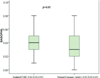
The pathophysiology of isolated coronary artery ectasia (CAE) takes in inflammation and atherosclerosis. It is clear that basophils have a critical role in endothelial dysfunction, atherosclerosis and inflammation. In this study, it was aimed to examine the association between isolated CAE and basophilia. All cases who underwent coronary angiography between January 2013 and April 2018 evaluated retrospectively. Of 10985 cases, 173 (107 males) with isolated CAE and 220 with normal coronary angiography (NCA) that age and gender matched subjects (119 males) were recorded. Hospital’s database was used to derive the biochemical and hematological test results, and baseline characteristics. White blood cell (WBC) count and basophil count were significantly elevated for the cases that have angiographic isolated CAE when compared to the subjects with NCA [ 7.87 (6.83-9.42)109/ L vs 7.48 (6.27-8.67) 109/ L, p=0.01; 0.04 (0.03-0.05) 109/ L vs 0.03 (0.02-0.05) 109/ L, p=0.03, respectively].
According to receiver operating characteristics curve analyses (ROC); the specificity of a basophil value > 0.037 109/ L (measured prior to coronary angiography) in
predicting isolated CAE was 57,3% and the sensitivity was 57.2% (area under the curve [AUC] 0.562, 95% CI 0.505, 0.618; p=0.03). Patients with isolated CAE have higher blood basophil count. Elevated blood basophil count may have a substantial mission in the pathogenesis of isolated CAE.
Keywords: Isolated coronary artery ectasia, blood basophil levels, inflammation
Medicine Science International Medical Journal
Introduction
Being a congenital or acquired coronary anomaly, CAE is described as the local or wide extension of a partial or entire epicedial coronary artery which is larger 1.5 times more than the adjacent normal coronary artery’s diameter [1-5]. CAE aetiology has been attributed to atherosclerosis (50%), congenital malformations (20– 30%) and connective tissue or inflammatory diseases (10–20%) [6]. It is generally considered to be a variant of atherosclerotic cardiac disease. However, many studies have revealed a presence of more intense inflammation in CAE than obstructive coronary artery disease [7,8].
Basophils are the cells with the lowest proportion in circulation with important immunomodulatory characteristics [9]. There are
many studies that demonstrate basophils have effect on coronary artery disease. Mochizuki et al., [10] have determined a connection between atherosclerotic risk factors and basophil count. According to Toyama et al., [11] for the patients with coronary artery disease, basophil count decrease by statin treatment and this causes decrease in arterial wall stiffness. There are also studies showing an association between histamine that released from basophils and atherosclerosis [12,13].
Is it possible to associate basophils with isolated CAE of which inflammation and atherosclerosis play a role in ethiopathogenesis? We aimed to examine whether there is an association between plasma basophil count and presence and severity of isolated CAE to answer this question.
Material and Methods Study Population
Angiography records of 10985 Turkish patients who underwent coronary angiography because of chest pain or objective signs
*Coresponding Author: Mucahid Yilmaz, Elazig Education and Research
Hospital, Department of Cardiology, Elazig, Turkey E-mail: [email protected]
of ischemia (with treadmill exercise or myocardial perfusion SPECT [single photon emission computed tomography]) were retrospectively evaluated for the presence of isolated CAE. Angiography records belonged to coronary angiography procedures performed between January 2013 and April 2018 at Elazığ Training and Research Hospital. The study included 173 subjects who have isolated CAE and 220 age and gender matched subjects who have normal coronary anatomy (NCA). Routine clinical and laboratory values (complete blood count and total biochemistry values, etc.) of the subjects were obtained from their files (Figure 1).
The study was carried out in conformity with Helsinki principles and ethics approval obtained from the Presidency of University Ethics Committee. Because the used data was anonymous in nature, no written informed consent obtained.
Coronary angiography and assessment of isolated CAE
A “Siemens Axiom Artis FC diagnostic device (Siemens Healthcare GmbH, Forchheim, Germany)”, was utilized to perform coronary angiography and Judkins technique was applied during operation [14].
Coronary angiography records were obtained from right and left anterior oblique cranial, AP cranial, right anterior oblique, caudal, and horizontal positions. As an opaque ingredient during the coronary angiogram, iohexol 350mgI / ml (Amersham Health, Co. Cork, Ireland) were used. 6 ml of opaque ingredient was injected into coronaries at each position during the procedure. Angiography was recorded digitally with the frame rate at 25 frames/millisecond. Coronary artery diameters were determined by computerized quantitative angiography. These measurements were obtained by analysing the digital data obtained from coronary angiographies using the Scientific Quantification Coronary Analysis software (Siemens Healthcare Gmbh, Forcheim, Germany). A calibration was applied to evaluate the coronary artery lumen’s real diameter, with the catheter diameter. To identify the artery segment as ectatic, measurements were taken at the proximal, mid and distal segments of the coronary arteries in patients who were considered to have an ectatic coronary segment. The CAE was defined as 1.5 times or more enlargement of the coronary artery compared to the adjacent coronary artery. Isolated CAE defined as regional or widespread expansion without significant coronary artery stenosis. Angiographic stenosis of more than 50% of the coronary artery was considered as significant stenosis. Patients without significant coronary artery stenosis and have ectatic segment were included in the isolated CAE group. The characteristics of the CAE were categorized as diffuse or discrete ectasia for classifying the severity of the CAE. The fusiform dilatations of the coronary arteries are identified as diffuse ectasia. On the other hand, localized/focal vesicular or spheroidal dilatation of the coronary arteries are identified as discrete ectasia (Figure 2-5) [6].
The classification by Markis et al,. [4] was used to detect the distribution of coronary artery ectasia. The diffuseness of ectasia is the basis of this classification. Accordingly, patients who have isolated CAE was classified in four groups: Type I; diffuse ectasia in two or three vessels, Type II; diffuse ectasia in only one vessel and focal ectasia in the another vessel, Type III; diffuse ectasia in only one vessel, Type IV; focal ectasia.
Study Exclusion Criteria
Subjects with acute coronary syndrome, significant coronary artery stenosis (angiographic stenosis>50%), isolated coronary slow flow, anaemia (Htc<%30), cardiac failure, thyroid dysfunction, malignancy, chronic renal deficiency (Glomerular Filtration Rate [GFR] <60mL/min/1.73 m2), chronic liver failure, chronic obstructive pulmonary disease and/or bronchial asthma in addition to subjects who were found to be using immunosuppressive therapy or steroid medication or BMI> 30 kg / m2 were excluded from the study.
Also subjects had a recent history of an acute infection or and high body temperature > 37.2 °C, an inflammatory or allergic disease were not included in the analysis.
Statistical Evaluation
The “SPSS version 16.0 (SPSS Inc., Chicago, IL, USA)” was employed for statistical analyses. Ability of continuous variables to normal distribution, was analyzed utilizing the Kolmogorov-Smirnov test. Continuous variables were presented as means with standard deviations or medians with 25th–75th percentiles. Categorical variables were represented as numbers with percentages. Continuous data were analyzed by Student’s t-test for normally distributed variables and Mann–Whitney U test for non-normally distributed variables. With the exception potassium and calcium, all continuous values had not normal distribution and Mann-Whitney U test was utilized to compare these parameters. Categorical data were analyzed using chi-square test. Bonferroni test was used to validate one-way ANOVA analysis for comparison between groups (among subjects with diffuse ectasia, focal ectasia and NCA). ROC test was used to evaluate the specificity and sensitivity of basophil and its optimum cut-off value. Spearman’s correlation test was executed for correlation analyses. p<0.05 were considered to indicate statistical significance.
Results
The data about 10985 subjects who went through coronary angiography were screened retroprospectively. 173 subjects (107 males) with isolated CAE and 220 age and gender matched subjects (119 males) with NCA were enrolled into the study. White blood cell (WBC) count and basophil count for isolated CAE group were significantly higher when they were compared to NCA group [ 7.87 (6.83-9.42) 109/ L vs 7.48 (6.27-8.67) 109/ L, p= 0.01; 0.04
(0.03-0.05) 109/ L vs 0.03 (0.02-0.05) 109/ L, p=0.03, respectively] (Table
I, Figure 6). On the other hand, levels of high density lipoprotein cholesterol (HDL-C) for the NCA group were significantly higher when compared to the other group (46.95 (40.0-56.0) mg/dL vs 41.0 (35.8-50.0) mg/dL, p<0.0001, respectively) (Table I). In the one-way ANOVA analysis among subjects with diffuse ectasia, focal ectasia and NCA , basophil levels were higher in subjects with diffuse ectasia than in subjects with NCA, on the other hand no significant difference was observed between subjects with focal ectasia and NCA and between subjects with diffuse ectasia and focal ectasia (Table II, Figure 7).
In correlation analysis, there was no significant relationship between any Markis classification and basophil count (p=0.49, r =-0.052) or diffuse or focal ectasia (p=0.44, r =0.059) (Table III).
doi: 10.5455/medscience.2019.08.9090 Med Science 2019;8(4):796-9
According to the ROC curve analysis, in guessing isolated CAE, the specificity of an basophil level > 0.037 109/ L (measured prior
to coronary angiography) was 57,3% and the sensitivity was 57.2% (area under the curve [AUC] 0.562, 95% CI 0.505, 0.618;
p=0.03) (Figure 8).
No meaningful difference was detected within the groups with regard to other investigated laboratory parameters (Table I).
Table 1. Inter-group comparison of demographical and laboratory data
CAE (173) NCA (220) P value
Sex, n (male/ female) 107/66 119/101 0.12
Hypertension, n (%) 58/173 (33.5) 59/220 (26.8) 0.15 Hyperlipidemia, n (%) 71/173 (41.0) 82/220 (37.3) 0.44 Diabetes mellitus, n (%) 39/173 (22.5) 45/220 (20.5) 0.61 Smoking, n (%) 66/173 (38.2) 71/220 (32.3) 0.22 Age,( year) 56.0 (52.5-61.0) 55.0 (51.0-59.0) 0.06 Platelet,( 109/ L) 260.0 (227.5-298.0) 255.0 (214.25-300.0) 0.19 Glucose, ( mg/dl) 99.0 (90.0-110.50) 96.0 (89.0-108.0) 0.14 Triglycerides(mg/dL) 150.0 (108.50-218.0) 137.45 (115.0-155.0) 0.09
Low density lipoprotein cholesterol, ( mg/dl) 115.0 (92.5-139.0) 112.0 (90.0-125.0) 0.05
Total cholesterol, ( mg/dl) 190.0 (161.5-213.5) 188.20 (166.15-200.0) 0.28
HDL cholesterol, (mg/dL) 41.0 (35.8-50.0) 46.95 (40.0-56.0) <0.0001
Basophil,( 109/ L) 0.04 (0.03-0.05) 0.03 (0.02-0.05) 0.03
Hemoglobin,(g/dl) 14.40 (13.5-15.15) 14.0 (13.5-15.0) 0.08
Hematocrit, (%) 42.90 (40.35-45.60) 42.0 (40.5-45.0) 0.13
White blood cell,( 109/ L) 7.87 (6.83-9.42) 7.48 (6.27-8.67) 0.01
Urea, ( mg/dl) 30.0 (24.0-36.0) 29.85 (23.7- 35.0) 0.24
Creatinine, ( mg/dl) 0.69 (0.51-0.80) 0.67 (0.57-0.78) 0.72
Sodium, (mmol/L) 140 (138-142) 140.0 (137.0-142.0) 0.29
Potassium, (meq/L) 4.35± 0.40 4.42± 0.51 0.15#
Calcium, ( mg/dl) 9.24± 0.46 9.28± 0.48 0.38#
#Normality of the distribution was evaluated by the Kolmogorov-Smirnov test and the Mann–Whitney U test applied to compare for continous variables except from white blood cell, potassium and calcium.
Table 2. Comparison of the basophil count among diffuse ectasia (Markis class
1, 2, 3), focal ectasia (Markis class 4) and NCA
Basophil counts P value Normal coronary anatomi (n=220) 0.035±0.018109/ L NS, 0.04#
Focal ectasia (n=81) 0.037±0.017109/ L NS
Diffuse ectasia (n=92) 0.041±0.024109/ L 0.04#, NS #: P value between diffuse ectasia and NCA= 0.04 All other p values >0.5
Table 3. Spearman’s correlation analysis between extension of the CAE and
basophil count-Markis classification and the basophil count.
r p
Extension of the isolated CAE (as diffuse) 0.059 0.44
Markis classification -0.052 0.49
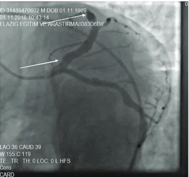
Figure 2. Demonstration of a fusiform ectasia image in the LAD (Left anterior
descending) artery, in the region between the white arrows Figure 3. Demonstration of a fusiform ectasia image in the CX (Circumflex) artery, in the region between the white arrows
Figure 4. Demonstration of a fusiform ectasia image in the RCA (Right
Figure 8. Receiver operator characteristic curve (ROC) analysis showing specificity and sensitivity of blood basophil count in predicting isolated CAE. AUC; area under the curve, CI; Confidence interval.
Discussion
We demonstrated significant difference regarding basophil count between two groups (Table I, Figure 6). In isolated CAE, elevated basophil count may reflect inflammation that may be seen in coronary arteries. The study, therefore, provided another evidence for the role of inflammation in isolated CAE. Last, higher basophil count may portend the presence of isolated CAE.
Isolated CAE was described as a diffuse or localized non-obstructive lesion of the epicardial coronary arteries. It was defined as a luminal dilation 1.5-times more compared to the normal adjacent segment [1,5].
Coronary artery ectasia can be congenital or acquired. Most ectasias (50%) occurs as a result of coronary atherosclerosis [15]. In various publications, it has been noted that Kawasaki syndrome, inflammatory diseases, Ehlers-Danlos syndrome, infections, connective tissue disorders, scleroderma, polyarthritis
nodoza, systemic lupus erythematosus, Marfan syndrome and Takayasu’s disease is related to CAE [7,16-21]. Despite the fact that it is generally considered as a different form of atherosclerotic cardiovascular disease, in some studies the different findings obtained and addressing the existence of denser inflammation than occlusive coronary artery disease [22,23].
The association between inflammation and CAE has been demonstrated utilizing well-recognized inflammatory markers such as WBC count, neutrophil count, lymphocyte count, neutrophil to lymphocyte ratio, interleukin-6, INF-γ, TNF-α, IL-1β, IL-8 and C-reactive protein (CRP) [8,24-26].
Basophils may have an substantial mission in the progress of chronic inflammation [27]. Increased risk of cardiovascular diseases amplified by systemic inflammation via destabilizing plaques, causing arterial stiffness, impairing endothelial function, or accelerating atherosclerosis [28,29]. Mochizuki et al. have found a link between atherosclerotic risk factors and peripheral basophil counts [10]. Toyama et al., [11] have indicated that an improvement detected in the arterial wall inflexibility, following the statin treatment of coronary artery disease, was associated with a decline in basophil count. Moreover, Sasaguri et al., [12] stated that histamine secreted by basophils has relation with atherosclerosis. Further argument for the role of basophils in the progression of inflammation, atherosclerosis, impairing endothelial disfunction, arterial stiffness, or destabilize plaques, is the Kounis syndrome. The clinically acute coronary syndrome that has been seen in this disease is believed to be generated by the vasospasm which is stemmed from degranulation of eosinophils, mast cells, and the basophils that can lead to rupture and/or erosion of plaque [30,31].
Nitricoxide (NO) has an important role in flow-mediated vasodilation. That is mainly released from the endothelium by the effect of endothelial shear stress. A pharmacological inhibition or genetic deficiency of endothelial NO synthase (eNOS) has an effect on decreasing endothelium-dependent vasodilation, which leads to an increase in vascular resistance [32]. When it
Figure 6. Comparison of basophil count between Isolated CAE and
NCA. The heavy black horizontal lines for each group represent the means, the extremities of the box are the 25th and 75th percentiles, the error bars are the minimum and maximum outliers.
Figure 7. Comparison of blood basophil count among diffuse ectasia,
focal ectasia and normal coronary artery. The heavy black horizontal lines for each group represent the means, the extremities of the box are the 25th and 75th percentiles, the error bars are the minimum and maximum outliers.
comes to vascular actions of NO, it regulates the vascular tone, as well as suppresses vascular smooth muscle proliferation. NO, blocks leukocyte-endothelial cell interaction, and inhibits platelet adhesion to the vascular endothelium [33]. Decreased NO activity causes abnormal vasoconstriction in the process of vascular pathology [34]. Gurlek et al. reported that NO levels decreased in CAE patients and this finding indicates elevated arterial stiffness in CAE [35].
High-density lipoprotein (HDL) seems to have a role being an innate part of the immune system, which partially uses an enhanced oxidative phase as a nonspecific way of protection against various pathogens. HDL is anti-inflammatory in humans, rabbits, and mice in the lack of chronic or acute inflammation [36]. Several studies have previously reported that HDL cholesterol levels decrease in the presence of systemic inflammation. Systemic and vascular inflammation also impair the structure of HDL cholesterol, disrupt its function, and reduce its protective effect on vascular endothelium [36-38]. In this research, it was observed that for the group with isolated CAE, HDL-C was low contrast to the other group with NCA (Table I). This finding may be reflective of the systemic and vascular inflammation, consistent with previous studies. Moreover, low HDL-cholesterol levels observed in the isolated CAE group may be considered as one of the mechanisms responsible for endothelial dysfunction, vascular destruction and arterial stiffness.
In our study, we observed that basophil count is increased in isolated CAE patients compared with the control group with NCA (Table I, Figure 6). Furthermore, in the analysis of one-way ANOVA among cases with diffuse ectasia, focal ectasia and NCA, basophil levels in cases with diffuse ectasia were significantly higher than cases with NCA, but there was no significant difference in basophil levels between cases with focal ectasia and NCA. However, no significant correlation was found between blood basophil levels and Markis classification nor between blood basophil levels and diffuse ectasia (Table II, Table III, Figure 7). In CAE cases, the Markis classification is performed after the ectatic segment is defined roughly as fusiform ectasia or saccular / focal ectasia. The diameter of the ectatic segment or the length of the fusiform lesion is not considered. This situation may have caused the correlation analysis in the ectasia group to be insignificant, although high basophil levels were found in the diffuse ectasia group compared to focal ectasia and NCA group. Another reason may be the systemic effect of inflammation which is thought to cause CAE.
Conclusions
The research results may contribute to etiopathogenesis of isolated CAE. These findings, also, may help to elucidate pathophysiological mechanisms of elevated prevalence of cardiovascular morbidity in isolated CAE patients. Elevated basophil levels may explain unstable plaques, endothelial dysfunction, vascular destruction, and arterial stiffness that may be seen in these patients.
Limitation of the study
Although there is an atherosclerotic plaque over a wide area, the vessel lumen could be observed as normal. This condition was demonstrated by autopsy and intravascular ultrasound studies conducted in cases of atherosclerotic heart disease [39,40]. Intravascular ultrasound (IVUS) was not applied for diagnosis of
isolated CAE and NCA in the recent study. Thus, the limitation of the present study was the lack of IVUS application.
Conflict of interest
The authors declare that there are no conflicts of interest. Financial Disclosure
All authors declare no financial support. Ethical approval
The study was approved by Presidency of T.C. Firat University Ethics Committee. Mucahid Yilmaz ORCID: 0000-0003-1458-8637
Hidayet Kayancicek ORCID: 0000-0001-6493-5591 Nevzat Gozel ORCID: 0000-0001-7326-6860 Yusuf Cekici ORCID: 0000-0002-4585-3707 Mehmet Nail Bilen ORCID: 0000-0003-1468-2930 Guney Sarioglu ORCID: 0000-0003-1458-8637 Fikret Keles ORCID: 0000-0003-1012-3875 Hasan Korkmaz ORCID: 0000-0001-8455-6724 References
1. Yetkin E, Waltenberger J. Novel insights into an old controversy: is coronary artery ectasia a variant of coronary atherosclerosis? Clin Res Cardiol. 2007;96:331-9.
2. Swaye PS, Fisher LD, Litwin P, et al. Aneurysmal coronary artery disease. Circulation. 1983;67:134-8.
3. Falsetti HL, Carroll RJ. Coronary artery aneurysm. A review of the literature with a report of 11 new cases. Chest. 1976;69:630-6.
4. Markis JE, Joffe CD, Cohn PF, et al. Clinical significance of coronary arterial ectasia. Am J Cardiol. 1976;37:217-22.
5. Maehara A, Mintz GS, Ahmed JM, et al. An intravascular ultrasound classification of angiographic coronary artery aneurysms. Am J Cardiol. 2001;88:365-70.
6. Türkmen M, Bitigen A, Esen AM. Coronary artery ectasia. J Med Sci. 2006;26:68-72.
7. Adiloglu AK, Can R, Nazli C, et al. Ectasia and severe atherosclerosis: relationships with chlamydia pneumoniae, helicobacterpylori, and inflammatory markers. Tex Heart Inst J. 2005;32:21-7.
8. Aydin M, Tekin IO, Dogan SM, et al. The levels of tumor necrosis factor-alpha and interleukin-6 in patients with isolated coronary artery ectasia. Mediators Inflamm. 2009: 106-145.
9. Karasuyama H, Mukai K, Obata K, et al. Nonredundant roles of basophils in immunity. Annu Rev Immunol. 2011;29:45-69.
10. Mochizuki K, Miyauchi R, Misaki Y, et al. Associations between leukocyte counts and cardiovascular disease risk factors in apparently healthy Japanese men. J Nutr Sci Vitaminol Tokyo. 2012;58:181-6.
11. Toyama K, Sugiyama S, Oka H, et al. Combination treatment of rosuvastatin or atorvastatin, with regular exercise improves arterial wall stiffness in patients with coronary artery disease. Plos One. 2012;7: 413-69.
12. Sasaguri Y, Wang KY, Tanimoto A, et al. Role of histamine produced by bone marrow-derived vascular cells in pathogenesis of atherosclerosis. Circ Res. 2005;96:974-81.
13. Clejan S, Japa S, Clemetson C, et al. Blood histamine is associated with coronary artery disease, cardiac events and severity of inflammation and atherosclerosis. J Cell Mol Med. 2002; 6:583- 92.
14. Kern MN, Seto AH. Cardiac catheterization, cardiac angiography and coronary blood flow and pressure measurements. In: Fuster V, Walsh R, Harrington R (eds) Hurst’s the Heart. 12th ed. McGraw-Hill Companies, New York. 2009; 471–4.
15. Yilmaz H, Sayar N, Yilmaz M, et al. Coronary artery ectasia: clinical and angiographical evaluation. Turk Kardiyol Dern Ars. 2009;36:530-5.
16. Papathanasiou AI, Katsouras CS, Goudevenos IA, et al. Rare association of diffused coronary ectasia and anomalous origin of left circumflex coronary artery in a man with heterozygous familial hypercholesterolemia: a case report. Angiology. 2005;56:343-5.
17. Dieter RS, Murtaugh T, Black J, et al. Coronary arteriomegaly in a patient with ehlers-danlos syndrome and multiple aneurysms-a case report. Angiology. 2003;54:733-6.
18. Crook BR, Raftery EB, Oram S. Mycotic aneurysms of coronary arteries. Br Heart J. 1973;35:107-9.
19. Chaithiraphan S, Goldberg E, O’Reilly M, et al. Multiple aneurysms of coronary artery in sclerodermal heart disease. Angiology. 1973;24:86-93. 20. Sumino H, Kanda T, Sasaki T, et al. Myocardial infarction secondary to
coronary aneurysm in systemic lupus erythematosus. An autopsy case. Angiology. 1995;46:527-30.
21. Suzuki H, Daida H, Tanaka M, et al. Giant aneurysm of the left main coronary artery in Takayasu aortitis. Heart. 1999;81:214-7.
22. Turhan H, Erbay AR, Yasar AS, et al. Plasma soluble adhesion molecules; intercellular adhesion molecule-1, vascular cell adhesion molecule-1 and E-selectin levels in patients with isolated coronary artery ectasia. Coron Artery Dis. 2005;16:45-50.
23. Yilmaz H, Tayyareci G, Sayar N, et al. Plasma soluble adhesion molecule levels in coronary artery ectasia. Cardiology. 2006;105:176-81.
24. Ayhan SS, Ozturk S, Erdem A, et al. Relation of neutrophil/lymphocyte ratio with the presence and severity of coronary artery ectasia. Turk Kardiyol Dern Ars. 2013;41:185–90.
25. Turhan H, Erbay AR, Yaşar Ast al. Comparison of C-reactive protein levels in patients with coronary artery ectasia versus patients with obstructive coronary artery disease. Am J Cardiol. 2004;94:1303–6.
26. Boles U, Johansson A, Wiklund U, et al. Cytokine disturbances in coronary artery ectasia do not support atherosclerosis pathogenesis. Int J Mol Sci. 2018;19.
27. Yamanishi Y, Karasuyama H. Basophils and mast cells in immunity and inflammation. Semin Immunopathol. 2016;38:5:535-7.
28. Dregan A. Arterial stiffness association with chronic inflammatory disorders in the UK Biobank study. Heart. 2018;104:1257-62.
29. Sattar N, McCarey DW, Capell Het al. Explaining how “high-grade” systemic inflammation accelerates vascular risk in rheumatoid arthritis. Circulation. 2003;108: 2957–63.
30. Yilmaz M, Korkmaz H. Kounis syndrome: a paradoxal non-ST elevation myocardial infarction case after triamcinolone treatment for dermatitis. Turk Kardiyol Dern Ars. 2018;46:223-7.
31. Hoshi T, Sato A, Akiyama Det al. Kounis syndrome manifesting as coronary aneurysm and very late coronary stent thrombosis. JACC Cardiovasc Interv. 2014;7:173-6.
32. Cooke JP, Dzau VJ. Nitric oxide synthase: role in the genesis of vascular disease. Annu Rev Med. 1997;48:489–509.
33. Garg UC, Hassid A. Nitric oxide-generating vasodilators and 8-bromocyclic guanosine monophosphate inhibit mitogenesis and proliferation of cultured rat vascular smooth muscle cells. J Clin Invest. 1989;83:1774–77.
34. Nabel EG, Selwyn AP, Ganz P. Large coronary arteries in humans are responsive to changing blood flow: an endothelium-dependent mechanism that fails in patients with atherosclerosis. J Am Coll Cardiol. 1990;16 349–56. 35. Gürlek A, Esenboğa K, Özcan ÖU, et al. Serum nitric oxide levels in patients
with coronary artery ectasia. Anatol J Cardiol. 2016;16:947-52.
36. Navab M, Anantharamaiah GM, Fogelman AM. The role of high-density lipoprotein in inflammation. Trends Cardiovasc Med. 2005;15:158-61. 37. Yılmaz M, Kayançiçek H. A New ınflammatory marker: elevated monocyte
to HDL cholesterol ratio associated with smoking. J Clin Med. 2018;7:76. 38. Feingold KR, Grunfeld C. Effect of inflammation on HDL structure and
function. Curr Opin Lipidol. 2016;27:521-30.
39. Nakatani S, Yamagishi M, Tamai J, et al. Assessment of coronary artery distensibility by intravascular ultrasound. Application of simultaneous measurements of luminal area and pressure. Circulation. 1995;91:2904–10. 40. Tuzcu EM, Kapadia SR, Tutar E, et al. High prevalence of coronary
atherosclerosis in asymptomatic teenagers and young adults: evidence from intravascular ultrasound. Circulation. 2001;103:2705–10.