Full Terms & Conditions of access and use can be found at
https://www.tandfonline.com/action/journalInformation?journalCode=ynns20
Nutritional Neuroscience
An International Journal on Nutrition, Diet and Nervous System
ISSN: 1028-415X (Print) 1476-8305 (Online) Journal homepage: https://www.tandfonline.com/loi/ynns20
The effects of walnut supplementation on
hippocampal NMDA receptor subunits NR2A and
NR2B of rats
Hicran Hicyilmaz, Huseyin Vural, Namik Delibas, Recep Sutcu, Fatih Gultekin
& Nigar Yilmaz
To cite this article: Hicran Hicyilmaz, Huseyin Vural, Namik Delibas, Recep Sutcu, Fatih Gultekin & Nigar Yilmaz (2017) The effects of walnut supplementation on hippocampal NMDA receptor subunits NR2A and NR2B of rats, Nutritional Neuroscience, 20:3, 203-208, DOI: 10.1179/1476830514Y.0000000166
To link to this article: https://doi.org/10.1179/1476830514Y.0000000166
Published online: 17 Mar 2017.
Submit your article to this journal
Article views: 162
View related articles
View Crossmark data
Citing articles: 5 View citing articles
D
Taylor & Francis~ Tll~lorf,J,;t1,caC:rlklj1 Nutritional I~ . I -•~" •C:J••--~
-
---CrossMarkThe effects of walnut supplementation on
hippocampal NMDA receptor subunits
NR2A and NR2B of rats
Hicran Hicyilmaz
1, Huseyin Vural
2, Namik Delibas
3, Recep Sutcu
4,
Fatih Gultekin
2, Nigar Yilmaz
51
Burdur State Hospital, Turkey,2Department of Biochemistry, Medical Faculty, Suleyman Demirel University, Isparta, Turkey,3Department of Biochemistry, Medical Faculty, Hacettepe University, Ankara, Turkey,
4
Department of Biochemistry, Medical Faculty, Izmir Kâtip Celebi University, Turkey,5Department of Biochemistry, Medical Faculty, Mugla Sitki Kocman University, Turkey
Objectives: Walnuts contain numerous selected dietary factors that have an impact on brain functions, especially learning and memory formation in the hippocampus. Hippocampal N-methyl D-aspartate
receptors (NMDARs) are involved in the formation of cognitive functions. In this study, we aimed to investigate the molecular effects of walnut supplementation on the hippocampal expressions of NMDARs involved in cognitive functions and lipid peroxidation levels in rats.
Methods: The male Sprague-Dawley rats (6 months old, n = 24) were fed with a walnut-supplemented diet (6% walnut diet, n = 12) and a control diet (rat food, n = 12) as ad libitum for 8 weeks. At the end of this period, NMDAR subunits NR2A and NR2B in the hippocampi were assayed by western blotting. Lipid peroxidation levels were measured using the thiobarbituric acid.
Results: The expression of NR2A and NR2B was elevated in the walnut-supplemented rats compared with the control group (P < 0.05). In addition, the levels of lipid peroxidation in the walnut-supplemented group were significantly decreased compared with the control group.
Discussion: We suggested that walnut supplementation may have protective effects against the decline of cognitive functions by regulating NMDAR and lipid peroxidation levels in the hippocampus. The study provides evidence that selected dietary factors (polyunsaturated fatty acids, melatonin, vitamin E, and flavonoids) within walnut may help to trigger hippocampal neuronal signal transduction for the formation of learning and memory.
Keywords: Walnut diet, NMDAR, Lipid peroxidation, Hippocampus
Introduction
Certain dietary factors in food are important as they affect the central nervous system in health and disease.1Walnuts are valuable food sources, because they contain various dietary factors, such as polyunsa-turated fatty acids (PUFAs), melatonin, vitamin E, and flavonoids. These factors have both antioxidant and anti-inflammatory properties and protect brain tissue from oxidative damage.2 Several studies have shown preventive and therapeutic effects of walnut on age-related motor and cognitive deficits. Those effects were assessed with behavioral experiments such as Morris water maze.3Walnuts contain signifi-cant amounts of n-6 and n-3 PUFA, linoleic acid (LA) (18:2n-6), and alpha-linolenic acid (ALA)
(18:3n-3).3,4 Researches have shown that essential fatty acids regulate a number of cellular processes, including learning and memory in the brain.5 Growing epidemiological studies suggest that dietary deficiency of n-3 PUFA is a candidate risk factor for the development of Alzheimer’s disease (AD).6,7 The amount of dietary n-3 PUFA must be sufficient for optimal cognitive performance in several animal species and in an animal model of AD.7Several studies using rat models have focused on the ability of dietary alterations or supplementation to ameliorate age-related loss of cognitive function.8–11Healthy diets consisting of high amounts of n-3 fatty acids can stimulate molecular systems involved in neuronal func-tions and plasticity in the brain.1
N-methyl D-aspartate receptor
(NMDAR)-depen-dent long-term potentiation (LTP) is the major cellular mechanism thought to underlie spatial learning and
Correspondence to: R Sutcu, Department of Biochemistry, Faculty of Medicine, Izmir Katip Celebi University, 32260 Izmir, Turkey.
memory in the hippocampus.8 NMDAR subunits, especially NR2A and NR2B, are essential for LTP induction and maintenance and are required for hip-pocampal synaptic plasticity. NMDARs contain various combinations of NR1 and NR2 subunits (A–D). The NR2 subunit regulates the duration of Ca2+ influx through the NMDA ion channel.12 Molecular alterations in NMDAR subunits can affect physiological and pathological processes in the hippocampus. NMDARs are involved in numerous physiological processes, including basic neuronal com-munication, axonal pathfinding, mood regulation, and memory formation.13Hyperactivity of NMDARs has been implicated in a variety of neurodegenerative dis-orders, such as Parkinson’s disease and AD.13 Excessive or deficiency of certain components in foods can result in the overactivation or inhibition of the func-tions of NMDAR subunits. It has been reported that dietary n-3 fatty acid depletion leads to a significant NR2B decrease while n-3 fatty acids enrichment results in NR2B increase.6,7Keleshian et al.14evaluated the effects of n-3 PUFA deficiency and supplementation of ALA on chronic NMDA-induced changes in rat brain, and found that n-3 PUFA deficiency worsened NMDA-induced changes, whereas n-3 supplementation did not affect NMDA-induced responses.
Although the beneficial cognitive effects of a walnut-supplemented diet in rats have been documen-ted, the molecular changes contributing to its effects are not fully understood. The aim of this study was to investigate the effect of dietary intake of walnut on NMDAR subunit expression and the level of mal-ondialdehyde (MDA) as a marker of lipid peroxi-dation in rat hippocampus.
Materials and methods Animals
Sprague-Dawley male rats (Suleyman Demirel University, Medical Faculty, Animal Experimental Laboratories, Isparta, Turkey), 6–7 months old, weighing between 200 and 240 g, were housed indivi-dually to monitor their daily food intake. They were maintained in environmentally controlled rooms (22–24°C, 50–55% humidity) with a 12-hour light/ dark cycle. The rats were randomly divided into two groups of 12 animals: one group received a control diet (control group, n = 12), and the other group received a diet enriched with walnuts (walnut group, n = 12) for 8 weeks. The diets were provided ad libitum during the experimental period. The animals’ weights were recorded throughout the study, and their food intake during a 24-hour period was assessed. At the end of 8 weeks, each rat was anesthetized separately by injecting intraperitoneal 2% xylazine (10 mg/kg) and then 10% ketamin (80 mg/kg). This anesthesia gave us 1-hour time window to sacrifice the animals.
The experimental protocol of the study was approved by the ethical committee of the Medical Faculty of Suleyman Demirel University. The animals involved in the procedure were maintained and used in accordance with the Animal Welfare Act and the Guide for the Care and Use of Laboratory Animals prepared by the Suleyman Demirel University Animal Ethical Committee (the number of the document: 04.13.01.06.2006).
Tissue preparation
Rats were decapitated and the brain was rapidly removed. The hippocampi were carefully dissected (within 5 minutes) on an apparatus, which was icy and wetted with phosphate buffer (50 mM) and frozen in the eppendorfs, which were filled with phosphate buffer (50 mM). Samples were stored at−80°C until assayed.
Diets
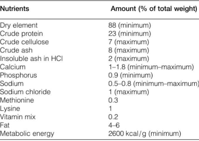
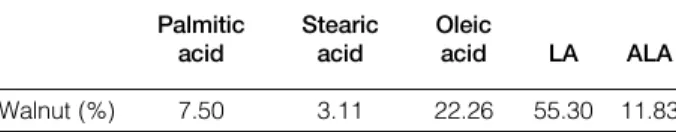
Walnuts were obtained from Burdur, a city in the Mediterranean region of Turkey. The walnuts were combined with the control diet, and the amount of maize in the control diet was adjusted to compensate for the added volume of walnuts. Control group rats were fed with ordinary food, whereas walnut group were fed with walnut 6% of intake food during the experimental procedure.3 The standard diet in this study contains 4–6% fat. The metabolizable energy of this diet was 2600 kcal/g for adult rats. The minimum amounts of the required nutrients ( protein, amino acid, fat, vitamins, and some minerals) for adult rats are listed in Table 1. The fatty acid compo-sitions of the walnut diets are shown in Table 2. The main PUFAs present in the walnuts were LA (55.30%, omega-6) and ALA (11.83%, omega-3). It is crucial to realize the ideal n-6/n-3 ratio for optimal cognitive performance in animal species.3 The ideal ratio was 4:1 in the study. The fatty
Table 1 The chemical constituents of control rat diet
Nutrients Amount (% of total weight)
Dry element 88 (minimum)
Crude protein 23 (minimum)
Crude cellulose 7 (maximum)
Crude ash 8 (maximum)
Insoluble ash in HCl 2 (maximum)
Calcium 1–1.8 (minimum–maximum)
Phosphorus 0.9 (minimum)
Sodium 0.5–0.8 (minimum–maximum)
Sodium chloride 1 (maximum)
Methionine 0.3
Lysine 1
Vitamin mix 0.2
Fat 4–6
Metabolic energy 2600 kcal/g (minimum)
The nutritive requirements for the rat are listed in the table. The content of each nutrient is expressed as g/100 g dry weight. Control rat diet was purchased from Korkutelim Feed Industry, Antalya, Turkey.
Hicyilmaz et al. Effects of walnut on NMDA receptor subunits 2A and 2B levels
Nutritional Neuroscience 2017 VOL.20 NO.3
acid profile of the walnuts was measured by gas chromatography/mass spectrophotometer (GC/MS) (Schimadzu QP5050, Japan) at the Suleyman Demirel University’s Central Laboratory.
Antibodies and chemicals
Anti-glutamate receptor NR2A, anti-glutamate recep-tor NR2B, monoclonal anti-rabbit IgG alkaline phos-phatase conjugate, beta-actin, a pre-stained molecular weight marker kit, nitroblue tetrazolium /5-bromo-4-chloro-3-indolyl phosphate (NBT/BCIP), leupeptin, aprotinin, benzamidine, and ethylene glycol-bis (beta-aminoethyl ether)-N,N,N′,N′-tetraacetic acid (EGTA) were all purchased from Sigma (St Louis, MO, USA). All reagents were of an analytical grade or the highest grade available.
Lipid peroxidation assay
MDA, an end product of lipid peroxidation, was assayed by the method of Drapper and Hadley.15 The principal of the method is spectrophotometric measurement of the color produced during the reac-tion of thiobarbituric acid (TBA) with MDA. One hip-pocampus was homogenized (1/5 w/v) in a glass Teflon homogenizer in ice-cold buffer (0.05 M Na2PO4/KH2PO4 buffer, pH 7.4). The homogenate
was centrifuged at 10 000× g for 15 minutes at 4°C and used for determining the MDA concentration. For this purpose, 1.25 ml of 20% trichloroacetic acid solution was added to 250μl of homogenate in a centrifuge tube and placed in a boiling water bath for 15 minutes. After being cooled in tap water, the mixture was centrifuged at 1000× g for 10 minutes, and 1 ml of the supernatant was added to 0.5 ml of 0.67% TBA solution in a test tube and placed in a boiling water bath for 15 minutes. The solution was then cooled in tap water, and its absorbance was measured spectrophotometrically (Shimadzu UV-1601, Kyoto, Japan) at 532 nm. The concentration of MDA was calculated using the value of the MDA–TBA complex of 1.56 × 10⁵/cm/mol and was expressed as nanomole MDA per milligram protein.
Protein determination
Protein in hippocampus homogenate was assayed by Lowry et al.’s (1951) method.16 To 1.0 ml of super-natant from above, 5.0 ml of alkaline copper sulfate
reagent is added and thoroughly mixed. Allow to stand for 10 minutes and then add 0.5 ml of Folin’s reagent. To develop color, this is kept standing for 30 minutes. This was followed by recording absorbance in spectrophotometer at 660 nm, against a blank. The blank is prepared by taking 1.0 ml of 0.5 M NaOH in place of sample in a cuvette. Bovine serum albumin is used to draw a standard curve, and the amounts of proteins in different samples are estimated.
Western blot analyses
The other hippocampi were homogenized (1/5 w/v), using a handheld homogenizer in ice-cold buffer (50 mM Tris–HCl (pH 7.5), 0.15 M NaCl, 1% Triton X-100, 1 mM EDTA, 1 mM EGTA, 25μg/ml leupeptin, 25μg/ml aprotinin, and 10 μM benzami-dine). The homogenate was centrifuged at 10 000× g, 15 minutes, at 4°C, and aliquot was taken for protein determination using Lowry et al.’s method.20 Equal amounts of protein from each sample (20μg of protein per lane) were separated by SDS–PAGE on 7.5% minigels, blotted electrophoretically to PVDF membrane (Immobilon P), and incubated in Tris-buffered saline with Tween 20 (TBST) (50 mM Tris–HCl (pH 7.5), 150 mM NaCl, and 0.1% Tween 20 containing 3% bovine serum albumin for 30 minutes). Blots were incubated overnight with anti-NR2A (1:3000) or anti-NR2B (1:5000) in 1% BSA. The blots were then subjected to three additional 10-minute washings in TBST. The blots were incubated with alkaline phosphatase-conjugated monoclonal anti-rabbit IgG (1:10 000) in 1% BSA for 1 hour at room temperature, and three additional 10-minute washes were carried out with TBST. The membrane was incu-bated in 20 ml of fresh reagent solution (BCIP/NBT) until color development. Immunoblotting for beta-actin (1:5000) was used as an internal standard to confirm equal protein loading and sample transferring. Images of the immunoblots were analyzed with a com-puterized image analysis system (Kodak Image MM Station, Ultra-Violet Products Ltd, Cambridge, UK). SDS–PAGE and western blot analyses were performed for three independent hippocampus preparations (two to three animals/group).
Statistical analysis
A statistical analysis of the data was performed using SPSS Version 13.0 (Chicago, IL, USA), and the Mann–Whitney U test was used for comparison of the control and walnut groups. The significance level was set at P < 0.05.
Results
The western blot analyses of the NR2A and beta-actin bands and the graphic of NR2A protein expression are shown in Fig. 1A and B, respectively. The western blot
Table 2 Fatty acid composition of the walnuts Palmitic acid Stearic acid Oleic acid LA ALA Walnut (%) 7.50 3.11 22.26 55.30 11.83
The fatty acid composition of the walnuts was measured by GC/MS (Schimadzu QP5050, Japan). The principal fatty acids were LA (linoleic acid) and ALA (alpha-linolenic acid). It is the effective n-6/n-3 ratio: 1/4 for cognitive functions in the walnut.
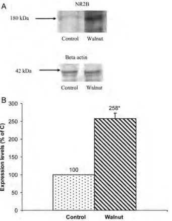
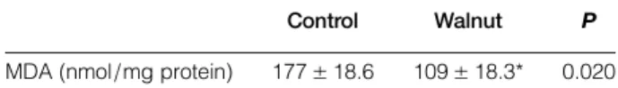
bands of NR2B and beta-actin and the graphic of NR2B protein expression are shown in Fig. 2A and B, respectively. The protein concentrations of NR2A and NR2B were higher (112 and 158%) in the walnut supplementation group compared with the control group (P < 0.05). In addition, the levels of MDA were significantly lower in the walnut group compared with the control group (P < 0.05). The mean levels of MDA are shown in Table 3.
Discussion
The study aimed to shed light on the molecular mech-anisms underlying the effect of walnut supplemen-tation on hippocampal cognitive functions, such as learning and memory formation, at the experimental level. The primary findings of the study are that walnut consumption has a positive effect on the
expression of hippocampal NMDAR subtypes, especially NR2A and NR2B. In addition, we found that a walnut-supplemented diet decreased lipid per-oxidation in the hippocampus.
Increasing evidence indicates that dietary factors have an influence on cognitive functions in aging and neurodegenerative disorders, such as AD.1,5,7 A number of dietary factors, such as saturated fatty acids, higher calorie intake, and excessive alcohol, have been reported to increase the risk of dementia and AD.17 In contrast, antioxidants, fish, methion-ine-rich proteins, and vitamins were shown to be pro-tective against the disease. Several cross-sectional studies suggested a relationship between particular nutrients and the presence of cognitive changes.17–18
NMDARs are required for hippocampal-dependent learning and memory.19The activation of NMDARs can be either toxic to neurons or promote their survi-val and plasticity.20 Aberrant NMDAR activity can
Figure 1 Protein expression of NR2A. (A) Western blotting samples of NR2A and beta-actin. (B) Protein expressions of NR2A. Representative western blotting NR2A bands of all groups from hippocampi. Blotting of beta-actin was used as an internal standard to confirm equal protein loading and sample transferring. Expression of NR2A protein was normalized against that of beta-actin. The density of protein band in the control group was accepted 100% and the walnut group was calculated as a percentage of the control value. Experiments were done on three independent hippocampus preparations (two to three animals/group). Size marker is indicated on the left (alpha-2-macroglobulin, 180 kDa). Walnut supplementation significantly increased NR2A level in the hippocampus. The asterisk indicates significant changes compared with the control group (P < 0.05).
Figure 2 Protein expression of NR2B. (A) Western blotting samples of NR2B and beta-actin. (B) Protein expressions of NR2B. Representative western blotting NR2B bands of all groups from hippocampi. Blotting of beta-actin was used as an internal standard to confirm equal protein loading and sample transferring. Expression of NR2B protein was normalized against that of beta-actin. The density of protein band in the control group was accepted 100% and the walnut group was calculated as a percentage of the control value. Experiments were done on three independent hippocampus preparations (two to three animals/group). Size marker is indicated on the left (alpha-2-macroglobulin, 180 kDa). Walnut supplementation significantly increased NR2B level in the hippocampus. The asterisk indicates significant changes compared with the control group (P < 0.05).
Hicyilmaz et al. Effects of walnut on NMDA receptor subunits 2A and 2B levels
Nutritional Neuroscience 2017 VOL.20 NO.3
206 A 180 k.Da 42 k.Da B 300
~
250 0-
0 ~ " 200 1/) ~ > -9:! 150 C: .2 1/) Cl) e! 100 C. ) ( w 50 0 NR2A Control Walnut Beta acrio Control Walnut 100 ... . . . . ~ . ' . . .... ~ .. .. .. .. .. . . . ..
..
. . . . ..
. . ' ..
.
'..
. . . . . . . . . . . . . . . . ' . ' . . . .. . ... -... -. Control 212· Walnut A 180kDa -42k.Do -_ 250 0 0c
200 !fl. Q> > ,S! 150 e .2 1/).,
! Q. >< 100 w 50 0 NR2B Conttol WolnUL Beta actin Control W11lnut25s•
cause excitotoxicity, resulting in uncontrolled Ca entry via NMDAR channels.20 On the other hand, the activity of synaptic NMDARs is crucial for the survi-val of neurons.20 The degree of activation of NMDARs is important for hippocampal-dependent cognitive functions. Sufficient expression of NMDARs is needed for LTP induction in the hippo-campus. Excess activation of NMDARs causes gener-ation of reactive oxygen species (ROS), and increased ROS can alter NMDAR-dependent calcium influx, ultimately resulting in oxidative damage in the hippo-campus.21Long-term oxidative stress led to a decrease in LTP formation, and the supplementation of antiox-idant-rich dietary factors was shown to reverse the impaired LTP response.8,21,22 In the study, the ben-eficial effects of the walnuts were dependent on several factors, one of which was the ratio of ALA and LA in the walnuts. The walnuts studied in the study were found to contain LA and ALA at a ratio of 1:4.7 (55.0 and 11.83%, respectively). This finding was also concordant with the previously suggested optimal ratio of ALA and LA (4:1), which improved cognitive performance.3 PUFAs can activate mem-brane-bound receptors, such as the NMDAR subtypes, NR2A and NR2B, which are involved in cognitive functions. The flexibility of the membrane is crucial for the function of membrane-embedded receptors, such as NR2B, and signal transduction.23In addition, PUFAs can contribute to synaptic membrane fluidity, reduce oxidative stress, and modulate signaling mech-anisms. Several studies reported that enriching the diet of old rats with PUFAs reverses age-related impair-ments in LTP in the hippocampus.24,25Dyall et al.24 showed that dietary supplementation with n-3 PUFA reversed age-related decreases in GluR2 and NR2B subunits in the prefrontal cortex, striatum, and hippo-campus. It has been shown that the extracts of flavo-noid-rich plant or specific molecules, such as blueberry,7grape,8,26and baicalein27, can prevent cog-nitive dysfunctions and can achieve NMDAR-mediated LTP response by enhancing in the rat hippo-campus. Results showed that another dietary factor in walnut is melatonin, which has had the effects on the expressions of hippocampal NMDARs for cognitive functions. Our previous study demonstrated that mela-tonin regulated the hippocampal expressions of NR2A and NR2B in a dose-dependent manner without
causing lipid peroxidation.28 Delibas et al.29 showed that NR2A and NR2B concentrations in hippocampus of rats maintained in dark showed significant increases compared with the control and functional pinealect-omy groups. There was no significant increase in lipid peroxidation while the NMDAR concentration increased significantly.29These results suggest that mel-atonin regulates on the hippocampal expressions of NMDARs. Melatonin and flavonoids in walnut can contribute to modulate the expressions of NMDARs. These effects may vary depending on the levels of the components in walnut.
It has been suggested that the oxidative stress plays a major role in the pathogenesis of neurodegenerative disease and aging.30 MDA, a marker of oxidative tissue damage, is by-product of lipid peroxidation induced by free radicals.8 Dietary antioxidants help to protect against free radical-induced damage in the brain. Walnuts contain a variety of important dietary factors with strong antioxidative properties, such as melatonin, vitamin E, and flavanoids.3,4 In our study, we used walnuts obtained from Burdur city in the Mediterranean area of Turkey. We ana-lyzed the percent content of fatty acids in the walnuts. According to the results of the analysis, the walnuts were rich in essential fatty acids, especially omega-6 vs. omega-3 PUFA. A limitation of the study is that we did not conduct a detailed analysis of the content of other dietary components in the walnuts. In the study, the hippocampal levels of MDA decreased significantly in the walnut sup-plementation group compared with the control group. This result demonstrated that the production of the lipid peroxidation in the hippocampus is pre-vented by dietary factors in walnut. Dietary factors in walnuts protect against oxidative stress-induced damage by reducing the generation of free radicals and inhibiting membrane lipid peroxidation. Essential fatty acids (n-3 PUFA) in walnuts may prevent lipid peroxidation by changing the compo-sition of membrane lipids and membrane fluidity. Another protective factor, flavonoids can affect tran-scriptional upregulation of antioxidant enzymes, such as glutathione synthesizing enzymes.18 Walnut supplementation increases the total antioxidant capacity of blood by increasing melatonin levels.31 Other nutrients in walnuts, such as vitamin E, melato-nin, and polyphenols, may prevent NMDAR-mediated excitotoxicity by increasing antioxidant capacity and decreasing lipid peroxidation, thereby contributing to the effects of walnut’s antioxidant. These components can also cause inhibition of stress signals and activation of protective signals. Walnut polyphenols include ellagitannins, which have been shown to inhibit oxidative stress and to modulate cell-signaling cascades.23
Table 3 Levels of MDA in the hippocampus in walnut (n:12) and control groups (n:12)
Control Walnut P
MDA (nmol/mg protein) 177± 18.6 109± 18.3* 0.020 The results are expressed as mean± SD.
*Significant change compared with the control group. Walnut diet significantly decreased the levels of MDA compared with the control groups in the hippocampus.
In the study, in addition to inducing the expression of hippocampal NMDARs, walnut supplementation decreased lipid peroxidation in the rat hippocampus. Considering some dietary factors within walnut, the mechanism of effect on hippocampal-dependent func-tions may be associated with the cumulative effects of PUFAs, and above-mentioned factors can help to trigger the mechanism.
Disclaimer statements
Contributors H.H.: the planning of the study, tissue preparation, western blotting, and imaging. H.V.: the planning of the study, statistical analysis, and interpret-ation of the results. N.D.: western blotting and interpretation of results. R.S.: imaging and evaluation of the bands. F.G.: the determination of the protein and evaluation of the results. N.Y.: tissue preparation and the determination of the lipid peroxidation. Funding None.
Conflicts of interest None.
Ethics approval I have read and have abided by the statement of ethical standards for manuscripts sub-mitted to the nutritional neuroscience. The animals involved in the procedure were maintained and used in accordance with the Animal Welfare Act and the Guide for the Care and Use Laboratory Animals pre-pared by the Süleyman Demirel University Ethical Committee. The study was approved by the Ethics Committee for Animal Experiments, Süleyman Demirel University’s Medical Faculty.
References
1 Gomez-Pinilla F, Gomez AG. The influence of dietary factors in central nervous system plasticity and injury recovery. PM R 2011;3(6 Suppl. 1):S111–6.
2 Muthaiyah B, Essa MM, Lee M, Chauhan V, Kaur K, Chauhan A. Dietary supplementation of walnuts improves memory deficits and learning skills in transgenic mouse model of Alzheimer’s disease. J Alzheimers Dis 2014;42(4):1397–405. 3 Willis LM, Shukitt-Hale B, Cheng V, Joseph JA.
Dose-depen-dent effects of walnuts on motor and cognitive function in aged rats. Br J Nutr 2009;101(8):1140–44.
4 Muthaiyah B, Essa MM, Chauhan V, Chauhan A. Protective effects of walnut extract against amyloid beta peptide-induced cell death and oxidative stress in PC12 cells. Neurochem Res 2011;36(11):2096–103.
5 Willis LM, Bielinski DF, Fisher DR, Matthan NR, Joseph JA. Walnut extract inhibits LPS-induced activation of BV-2 micro-glia via internalization of TLR4: possible involvement of phos-pholipase D2. Inflammation 2010;33(5):325–33.
6 Lim GP, Calon F, Morihara T, Yang F, Teter B, Ubeda O, et al.. A diet enriched with the omega-3 fatty acid docosahexaenoic acid reduces amyloid burden in an aged Alzheimer mouse model. Neuroscience 2005;25(12):3032–40.
7 Calon F, Lim GP, Morihara T, Yang F, Ubeda O, Salem N, Jr, et al.. Cole GM. Dietary n-3 polyunsaturated fatty acid depletion activates caspases and decreases NMDA receptors in the brain of a transgenic mouse model of Alzheimer’s disease. Eur J Neurosci 2005;22(3):617–26.
8 Coultrap SJ, Bickford PC, Browning MD. Blueberry-enriched diet ameliorates age-related declines in NMDA receptor-depen-dent LTP. Age 2008;30(4):263–72.
9 Joseph JA, Shukitt-Hale B, Denisova NA, Bielinski D, Martin A, McEwen JJ, et al.. Reversals of age-related declines in
neuronal signal transduction, cognitive, and motor behavioral deficits with blueberry, spinach, or strawberry dietary sup-plementation. J Neurosci 1999;19(18):8114–21.
10 Yilmaz N, Vural H, Yilmaz M, Sutcu R, Sirmali R, Hicyilmaz H, et al.. Calorie restriction modulates hippocampal NMDA receptors in diet-induced obese rats. J Recept Signal Transduct Res 2011;31(3):214–9.
11 Delibas N, Altuntas I, Sutcu R, Yonden Z, Koylu H. Effects of dietary long chain PUFAs on hippocampal lipid peroxidat-ion and NMDA receptor subunits A and B concentratperoxidat-ion in streptozotocin-diabetic rats. Int J Neurosci 2004;114(10): 1353–64.
12 Shapiro M. Plasticity, hippocampal place cells and cognitive maps. Arch Neurol 2001;58:874–81.
13 Hovelsø N, Sotty F, Montezinho LP, Pinheiro PS, Herrik KF, Mørk A. Therapeutic potential of metabotropic glutamate recep-tor modularecep-tors. Curr Neuropharmacol 2012;10(1):12–48. 14 Keleshian VL, Kellom M, Kim HW, Taha AY, Cheon Y,
Igarashi M, et al.. Neuropathological responses to chronic NMDA in rats are worsened by dietary n-3 PUFA deprivation but are not ameliorated by fish oil supplementation. PLoS One 2014;9(5):e95318.
15 Drapper HH, Hadley M. Malondialdehyde determination as index of lipid peroxidation. Methods Enzymol 1990;186:421–531. 16 Lowry O, Rosenbrough N, Farr L. Protein measurement with
the folin phenol reagent. J Biol Chem 1951;193:265–75. 17 Rosenzweig ES, Barnes CA. Impact of aging on hippocampal
function: plasticity, network dynamics, and cognition. Prog Neurobiol 2003;69(3):143–79.
18 Ramesh BN, Rao TS, Prakasam A, Sambamurti K, Rao KS. Neuronutrition and Alzheimer’s disease. J Alzheimers Dis 2010;19(4):1123–39.
19 Chytrova G, Ying Z, Gomez-Pnilla F. Exercise contributes to the effects of DHA dietary supplementation by acting on mem-brane-related synaptic systems. Brain Res 2010;23(1341C):32–40. 20 Shi L, Adams MM, Linville MC, Newton IG, Forbes ME, Long AB, et al.. Caloric restriction eliminates the aging-related decline in NMDA and AMPA receptor subunits in the rat hippocampus and induces homeostasis. Exp Neurol 2007;206(1):70–9. 21 Zhou Q, Sheng M. NMDA receptors in nervous system diseases.
Neuropharmacology 2013;74:69–75.
22 Farr SA, Yamada KA, Butterfield DA, Abdul HM, Xu L, Miller NE, et al.. Obesity and hypertriglyceridemia produce cognitive impairment. Endocrinology 2008;149(5):2628–36.
23 Gomez-Pnilla F. Collaborative effects of diet and exercise on cognitive enhancement. Nutr Health 2011;20(3–4):165–69. 24 Dyall SC, Michael GJ, Whelpton R, Scott AG, Michael-Titus
AT. Dietary enrichment with omega-3 polyunsaturated fatty acids reverses age-related decreases in the GluR2 and NR2B glu-tamate receptor subunits in rat forebrain. Neurobiol Aging 2007; 28(3):424–39.
25 Kelly L, Grehan B, Chiesa AD, O’Mara SM, Downer E, Sahyoun G, et al.. The polyunsaturated fatty acids, EPA and DPA exert a protective effect in the hippocampus of the aged rat. Neurobiol Aging 2011;32(12):p2318.e1–15.
26 Sarkaki A, Rafieirad M, Hossini SE, Farbood Y, Motamedi F, Mansouri SM, et al.. Improvement in memory and brain long-term potentiation deficits due to permanent hypoperfusion/ ischemia by grape seed extract in rats. Iran J Basic Med Sci 2013;16(9):1004–10.
27 Wang W, Wang F, Yang YJ, Hu ZL, Long LH, Fu H, et al.. The flavonoid baicalein promotes NMDA receptor-dependent long-term potentiation and enhances memory. Br J Pharmacol 2011;162(6):1364–79.
28 Sutcu R, Yonden Z, Yilmaz A, Delibas N. Melatonin increases NMDA receptor subunits 2A and 2B concentrations in rat hip-pocampus. Mol Cell Biochem 2006;283(1–2):101–5.
29 Delibas N, Tuzmen N, Yonden Z, Altuntas I. Effect of func-tional pinealectomy on hippocampal lipid peroxidation, antiox-idant enzymes and N-methyl-D-aspartate receptor subunits 2A and 2B in young and old rats. Neuro Endocrinol Lett 2002; 23(4):345–50.
30 Darvesh AS, Carroll RT, Bishayee A, Geldenhuys WJ, Van der Schyf CJ. Oxidative stress and Alzheimer’s disease: dietary poly-phenols as potential therapeutic agents. Expert Rev Neurother 2010;10(5):729–45.
31 McKay DL, Chen CY, Yeum KJ, Matthan NR, Lichtenstein AH, Blumberg JB. Chronic and acute effects of walnuts on anti-oxidant capacity and nutritional status in humans: a random-ized, cross-over pilot study. Nutr J 2010;12:9–21.
Hicyilmaz et al. Effects of walnut on NMDA receptor subunits 2A and 2B levels
Nutritional Neuroscience 2017 VOL.20 NO.3