ORIGINAL ARTICLE
Investigation of anti-DFS70 antibody in patients with systemic
autoimmune rheumatic diseases
Bilal Olcay Peker1
&Aslı Gamze Şener1&Emine Figen Tarhan2&Selçuk Kaya3
Received: 7 June 2019 / Revised: 5 July 2019 / Accepted: 30 July 2019 # International League of Associations for Rheumatology (ILAR) 2019 Abstract
Introduction Dense fine-speckled 70 (DFS70) antibody is defined as an antinuclear antibody (ANA) pattern in indirect immu-nofluorescence (IIF). The presence of anti-DFS70 antibody has been shown as a potential marker for the exclusion of systemic autoimmune rheumatic diseases (SARD) (without any other SARD-associated autoantibodies). We aimed to investigate the frequency of anti-DFS70 antibodies in patients with SARD and in the blood bank donors (BD).
Materials and methods The study group consists of 418 rheumatoid arthritis (RA), 101 systemic lupus erythematosus (SLE), 71 Sjogren’s syndrome (SS), 43 ankylosing spondylitis (AS), 36 systemic sclerosis-scleroderma (SSc), 2555 undifferentiated connective tissue disease (UCTD), and 507 BD. All samples were tested on the HEp-2 IIF-ANA assay. Samples that showed DFS70 pattern in IIF were confirmed by a specific DFS70 antibody enzyme-linked immunosorbent assay (ELISA).
Results The DFS70 pattern was detected in 43 (1.33%) in SARD and four (0.78%) in BD. The anti-DFS70 antibody was detected in three (0.59%) in BD, six (1.43%) in RA, three (2.97%) in SLE, one (1.40%) in SS, and 25 (0.97%) in UCTD, however, it was not detected in AS and SSc by ELISA. There was no significant difference between BD and SARD (p = 0.28). Distinctly, the frequency of anti-DFS70 was significantly different for SLE in SARD (p = 0.02). Conclusion Anti-DFS70 antibody was more prevalent in the subsets of SARD than BD. This result may be related to the demographic formation of study groups and individual immunological status. More comprehensive studies are needed to investigate the importance of the anti-DFS70 antibody for SARD.
Key Points
• This study draws attention to the importance of anti-DFS70 antibodies in the diagnostic algorithm in systemic autoimmune rheumatic diseases. • This study emphasizes the further investigation of anti-DFS70 antibodies in undifferentiated connective tissue diseases.
• This study emphasizes the need to verify the DFS70 pattern detected in IIF-ANA test for definitive diagnosis with additional confirmation methods. Keywords Anti-DFS70 antinuclear antibody . Indirect immunofluorescence . Systemic autoimmune rheumatic disease . Systemic lupus erythematosus
Introduction
Autoimmune diseases consist of a number of systemic diseases with multiple organ involvement, as well as organ-specific. Systemic autoimmune rheumatic diseases (SARD) are heteroge-neous disorders associated with significant morbidity and mor-tality. SARD consists of rheumatoid arthritis (RA), systemic lu-pus erythematosus (SLE), Sjogren’s syndrome (SS), systemic sclerosis-scleroderma (SSc), ankylosing spondylitis (AS), idio-pathic inflammatory myositis, and systemic vasculitis. Although the classic symptoms facilitate diagnosis, multiple organ involve-ment makes the diagnosis difficult [1]. Antinuclear antibody (ANA) is a serological indicator used for the diagnosis of * Bilal Olcay Peker
Department of Medical Microbiology, Izmir Katip Çelebi University Atatürk Training and Research Hospital, Karabağlar,
35360 Izmir, Turkey
2 Department of Rheumatology, Muğla Sıtkı Koçman University Research and Training Hospital, Muğla, Turkey
3
Department of Medical Microbiology, Izmir Katip Çelebi University Medical Faculty, Izmir, Turkey
SARD and described by indirect immunofluorescence (IIF) as-say on HEp-2 cells [2]. The IIF assay on HEp-2 cells has a great advantage for the diagnosis of SARD economically [3]. American College of Rheumatology (ACR) recommends the IIF-ANA test on HEp-2 cells as a screening assay and remains the gold standard test for ANA detection [4]. It is very important to interpret ANA positivity as it can be positive in healthy indi-viduals (HI) up to 20%. These positive results might belong to autoantibodies that target the dense fine-speckled 70 (DFS70) antigen also known as lens epithelium-derived growth factor (LEDGF). However, the DFS70 IIF pattern was reported in up to 33% of positive HI serum, but not reported in ANA-positive SARD serum [3]. Although anti-DFS70 antibody was firstly shown in the case of interstitial cystitis, either that was shown in clinical cases such as chronic inflammatory conditions, cancer, and atopic dermatitis in HI [5].
The International Consensus on ANA Patterns (ICAP) committee recently classified the DFS pattern as an AC-2 competency pattern level which was identified as a stained pattern uniformly distributed throughout the interphase nucle-us and mostly localized on metaphase chromosomes in IIF assay [6]. When defining the pattern on HEp-2 substrates on IIF assay, it might be confused with other nuclear patterns, depending on the evaluation and therefore additional tests are needed to verify [7]. In order to prevent misinterpretations, the diagnostic algorithm was developed by ICAP and the pseudo-DFS pattern was defined. This pattern refers to a nu-clear speckled pattern with nu-clear staining of the metaphase plate but without the typical features of the DFS (AC-02) pattern [8]. It is emphasized that distinguishing pseudo-DFS from the classical DFS model is important to increase the accuracy of IIF-ANA test interpretation [9]. Anti-DFS70 an-tibody has been shown as around 10% in HI but has been reported as 2–3% in SARD patients. The presence of this antibody, along with clinical studies, has been shown as a potential marker for the exclusion of SARD (without any oth-er SARD-associated autoantibodies) [5].
In the routine laboratory of our hospital, we often observe this pattern in the IIF test. According to these observations, we aimed to investigate the frequency of anti-DFS70 antibodies in patients with SARD and in the blood bank donors (BD).
Materials and methods
Patients
The study consists of blood bank donors (BD) and pa-tients who were admitted to the rheumatology outpatient clinic in Izmir Katip Çelebi University Atatürk Training and Research Hospital between January 2013 and September 2014. The control group was designed for healthy BD who were admitted to Izmir Katip Çelebi
University Atatürk Training and Research Hospital Blood Bank Laboratory. The patient group was selected from with and without ANA-associated rheumatic dis-eases (AARDs) as a SARD group. Undifferentiated con-nective tissue disease (UCTD) patients were included in the SARD group. The study group consisted of 507 BD as a control group and 3224 patients. A single serum sample was accepted from each patient for the study. Serum sam-ples were collected after ethical approval and stored at – 40 °C with 1 ml volumes. The study was designed on adult (aged ≥ 18 years) male and female individuals. The patient group consisted of confirmed diseases with clini-cal and laboratory findings diagnosed by a rheumatolo-gist. Diagnostic groups were defined 418 as RA, 101 as SLE, 71 as SS, 43 as AS, 36 as SSc, and 2555 as UCTD patients.
Procedures
Anti-DFS70 analysis by IIF-ANA and ELISA
ANAs were evaluated by a standard IIF technique using HEp-20-10/liver biochip (Monkey) (Euroimmun AG, Lübeck, Germany) conjugated with a specific anti-human IgG (Euroimmun AG). Samples were considered as positive for ANAs if immunofluorescent staining was observed at a serum dilution of 1:100 according to the manufacturer’s protocol. The fluorescence intensity was performed with a light-emitting diode light source in a microscope (Eurostar III plus, Lübeck, Germany). During the interpretation of the IIF, pat-terns were evaluated as a semi-quantitatively 1+ to 4+ accord-ing to the intensity of the positive control. Samples with DFS70 positive patterns were confirmed by Human PC4 and SFRS1 interactive protein 1 (PSIP1) enzyme-linked immuno-sorbent assay (ELISA) kit (CusaBio Biotech, Wuhan, China). The serum concentrations were measured by absorbance read-er and obtained optical densities wread-ere calculated by four-parameter logistic curve-alignment calculation formula with the manufacturer’s recommendations.
Extractable nuclear antigen analysis by line immunoassay Samples with DFS70 positive patterns in IIF were tested for association with additional patterns by LIA. The line immunoassay (LIA) was performed using the EUROLINE kit (Euroimmun AG, Lübeck, Germany) for extractable nuclear antigen (ENA). Each strip consisted of nRNP/Sm (U1-nRNP), Sm, SS-A, Recombinant Ro-52 (Ro-52, 52 kDa), SS-B, DNA topoisomerase I (Scl-70), PM-Scl, histidyl-tRNA synthetase (Jo-1), centromere protein B (CENP B), dsDNA, nucleosome, histones, and pyruvate dehydrogenase complex antigens and studied with the manufacturer’s protocol.
Ethical approval
This study was approved by the Katip Celebi University ethics committee of clinical research (April 9, 2014, Approval Number 60) and performed in accordance with the ethical standards as laid down in the 1964 Declaration of Helsinki.
Statistical analysis
Pearson’s chi-square test was used to compare the median age value between the SARD and BD group. The average age was compared among the SARD groups using the Mann-Whitney U test. There was not a normal distribution of at least one disease group. The Person’s chi-square test was used to com-pare the qualitative results obtained in the categorical vari-ables. The Kruskal-Wallis H test was used to compare anti-DFS70 positivity among the SARD groups. Statistical analy-ses were performed using IBM SPSS statistics 21 (SPSS Inc., Chicago, IL, USA). GraphPad Prism version 7.00 (GraphPad Software, La Jolla, CA, USA) was used for drawing chart. Data are shown as mean ± standard deviation (SD), number (n), and percentage (%). The p value < 0.05 was considered statistically significant.
Results
The mean age was 47.95 ± 14.25 (range 18–98 years) for con-trol and patient groups. There was no normal distribution for the SARD and BD groups in terms of age. The median age values among the groups were statistically significant (p < 0.001, Mann-Whitney U). The mean age of the BD group was 36.64 ± 9.6 (range 20–65 years). The BD group consisted of younger age and male sex majority compared to individuals with SARD. The mean age of the SARD group was 49.72 ± 14.44 (range 18–98 years). Age ranges of the individuals in the SARD group did not fit the normal distribution. The age distribution was normal in SS and SSc. The Mann-Whitney U test was used for the control and patient groups except for the SS and SSc groups.
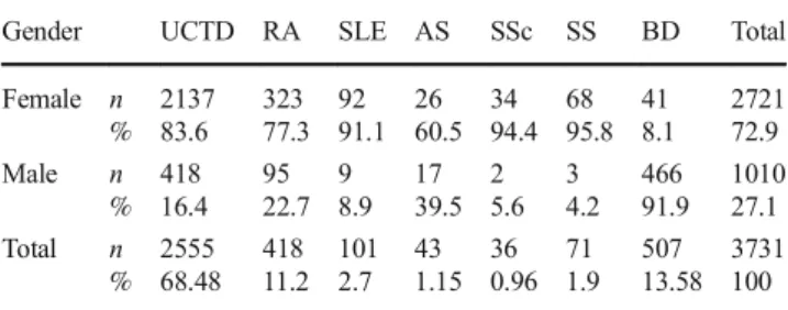
The gender distribution of the volunteers included in our study were 72.9% (2731/3731) female and 27.1% male (1010/3731). The gender distributions in the study groups were not equal and are shown in Table1in terms of number and percentage. The female gender was dominant in the SARD group and the male gender was dominant in the BD group.
The positivity rates of ANA pattern in the IIF test were 10.28% (43/418) in RA, 48.51% (49/101) in SLE, 4.65% (2/43) in AS, 22.53% (16/71) in SS, 58.33% (21/36) in SSc, 9.27% (237/2555) in UCTD, and 0.78% (4/507) in BD. The ANA positivity was statistically significant be-tween the SARD and BD group (p < 0.001, Pearson’s
chi-square). The distribution of IIF-ANA positivity rates in study groups was shown in Table 2.
The DFS70 pattern was detected in the HEp-2 cells by the IIF assay. It was seen as a fine speckle, uniformly distributed over the interphase nuclei and on the meta-phase chromatin. This type of staining should be differen-tiated from homogeneous and dense fine-speckled stain-ing. The fluorescence intensity of positive samples was considered 3+ and 4+ and shown in Fig.1. Samples with nuclear fine-speckled (NFS) pattern defined according to ICAP criteria in IIF-ANA test were not included in the study. This pattern is described as negative staining of mitotic chromatin with tiny fine speckles [10].
We detected the DFS70 pattern in four of 507 samples in the BD group. The DFS70 pattern was identified in six RA, three SLE, one SS, and 33 UCTD patients by the IIF assay. The positivity rate of DFS70 pattern was 1.35% (5/368) in the ANA-positive SARD group. The ANA positivity rate in the BD group was 0.78% (4/507) and none of the ANA-positive Table 1 The distribution of gender in study groups
Gender UCTD RA SLE AS SSc SS BD Total Female n % 2137 83.6 323 77.3 92 91.1 26 60.5 34 94.4 68 95.8 41 8.1 2721 72.9 Male n % 418 16.4 95 22.7 9 8.9 17 39.5 2 5.6 3 4.2 466 91.9 1010 27.1 Total n % 2555 68.48 418 11.2 101 2.7 43 1.15 36 0.96 71 1.9 507 13.58 3731 100 AS ankylosing spondylitis, BD blood bank donor, ELISA enzyme-linked immunosorbent assay, RA rheumatoid arthritis, SLE systemic lupus ery-thematosus, SS Sjogren’s syndrome, SSc systemic sclerosis-scleroderma, UCTD undifferentiated connective tissue disease, and % value for within gender
Table 2 The distribution of IIF-ANA and anti-DFS70 positivity in study groups
Study groups IIF-ANA n (%) Anti-DFS70 n (%)* Total n UCTD 237 (9.27) 3 (1.26) 2555 RA 43 (10.28) 0 418 SLE 49 (48.51) 2 (4.08) 101 AS 2 (4.65) 0 43 SSc 21 (58.33) 0 36 SS 16 (22.53) 0 71 BD 4 (0.78) 0 507
ANA antinuclear antibody, AS ankylosing spondylitis, BD blood bank donor, DFS70 dense fine speckled, IIF indirect immunofluorescence, RA rheumatoid arthritis, SLE systemic lupus erythematosus, SS Sjogren’s syndrome, SSc systemic sclerosis-scleroderma, UCTD undif-ferentiated connective tissue disease, % value for within groups, and * value is indicated that within ANA-associated rheumatic diseases
samples were detected DFS70 pattern positive (0/4). The dis-tribution of anti-DFS70 and IIF-ANA positivity rates were shown in Table2for the study groups.
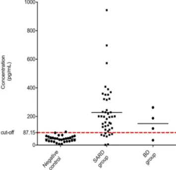
Differences in the techniques may lead to differences in evaluating results between laboratories. Thus, we performed a specific ELISA test to confirm anti-DFS70 antibodies for samples with the DFS70 staining pattern in IIF-ANA. A total of 43 samples in the SARD group, four samples in the BD group, seven standard samples, and 32 negative control serum samples were studied. The cut-off value was calculated for anti-DFS70 positive serum and negative control serums. The calculated cut-off value was 87.15 pg/mL (mean + 2SD) and the concentration of the anti-DFS70 antibody was shown in Fig.2.
In the ELISA test, 80.85% (38/47) of the DFS70 positive samples that were confirmed anti-DFS70 positive. Only one negative control serum was calculated above the cut-off value. The anti-DFS70 positivity between study groups were six for RA, three for SLE, one for SS, 25 for UCTD, and three for BD. The anti-DFS70 positivity was 32 for female and three for male in the SARD group. All positive samples belonged to the male gender in the BD group.
There was no statistically significant difference in the SARD group except for SLE (p = 0.02, Kruskal-Wallis H test). Anti-DFS70 positivity rates for RA, SLE, SS, and UCTD were 1.43% (6/418), 2.97% (3/101), 1.40% (1/71), and 0.97% (25/2555), respectively. Anti-DFS70 positivity rates were 0.59% (3/507) and 1.08% (35/3224) for BD and SARD. The prevalence of anti-DFS70 antibody in patients with AARDs was 1.35% (5/368). There was no significant difference between the BD and the SARD group (p = 0.28, Mann-Whitney U test). The prevalence in the disease cohorts varied between 0.0% and 2.97%. The distribution of anti-DFS70 positivity between IIF and ELISA was shown in Table3for study groups.
Anti-DFS70 positive samples were tested by LIA for the presence of additional ANA. Anti-dsDNA was found positive in only one of the six SLE patients who were detected anti-DFS70 positive. Anti-Ro52 was found Fig. 2 The anti-DFS70 cut-off concentration chart. Enzyme-linked im-munosorbent assay (ELISA) analysis of anti-dense fine-speckled 70 (anti-DFS70) antibody levels in DFS70-positive samples in blood bank donor (BD), systemic autoimmune rheumatic diseases (SARD) group, and neg-ative control serums. The anti-DFS70 antibody level was expressed as a measured concentration from the optical density reads on a positive stan-dard sample chosen for reference. Each calculated value for the samples was shown in both groups. The dashed line indicates the cut-off level (87.15 pg/mL). Thirty five SARD, three BD, and one control serum were calculated above the cut-off level
Table 3 The distribution of anti-DFS70 positivity between IIF and ELISA test
Study groups IIF-ANA** n (%) ELISA n (%) Total n p UCTD 33 (1.29) 25 (0.97) 2555 0.36 RA 6 (1.43) 6 (1.43) 418 0.19 SLE 3 (2.97) 3 (2.97) 101 0.02 AS 0 0 43 0.61 SSc 0 0 36 0.64 SS 1 (1.40) 1 (1.40) 71 0.43 BD 4 (0.78) 3 (0.59) 507 0.28*
ANA antinuclear antibody, AS ankylosing spondylitis, ELISA enzyme-linked immunosorbent assay, BD blood bank donor, IIF indirect immu-nofluorescence, RA rheumatoid arthritis, SLE systemic lupus erythemato-sus, SS Sjogren’s syndrome, SSc systemic sclerosis-scleroderma, UCTD undifferentiated connective tissue disease, and %, value for within groups, p value is indicated for among systemic autoimmune rheumatic diseases (SARD), * p value for analyses between SARD and BD, ** dense fine-speckled 70 pattern in IIF-ANA test
Fig. 1 The DFS70 antibody pattern by IIF. The DFS70 pattern is seen as dense fine speckled and uniformly distributed over the interphase nuclei and on the metaphase chromatin in the HEp-2 cells by IIF
positive in two patients with RA and SS. Two serum sam-ples were found positive for centromere B and anti-Scl70 in BD.
Discussion
The IIF method is the most frequently used and gold standard test for routine ANA detection [4]. We aimed to determine the incidence of anti-DFS70 antibody in BD and SARD (AS, RA, SLE, SS, SSc, UCTD) using the IIF test. The typical DFS staining pattern is considered as the fine speckle pattern uni-formly distributed throughout the interphase stage of the nu-cleus and in the metaphase chromatin [11]. The evaluation of IIF-ANA test in our laboratory is carried out by an experi-enced specialist for 15 years. When defining a low-level DFS pattern, it can be confused with other classical patterns and emphasizes that anti-DFS70 needs to be confirmed by specific immunoassays [9]. Recently, this pattern has been included as a“pseudo-DFS” pattern in the ICAP classification algorithm [8]. The typical patterns that were confused with the DFS70 pattern (pseudo-DFS) were excluded in the study. In the following, results were confirmed by ELISA test. It is recommended to confirm the presence of anti-DFS70 antibod-ies with a DFS70-specific immunoassay to reach a definite conclusion [12]. Based on this inference, DFS70-positive samples were confirmed by a specific ELISA test.
In our study, the anti-DFS70 antibody rate was 0.59% (3/507) in BD. Watanabe et al. [13] found that anti-DFS70 antibodies were positive in 11% of whole individuals and 54% of ANA-positive individuals on 597 hospital workers. Their study showed that anti-DFS70 antibodies could be used in the exclusion of systemic autoimmune diseases in ANA-positive individuals. In contrast to this study, we did not detect anti-DFS70 in ANA-positive BD. Ayaki et al. [14] found that anti-LEDGF antibodies were positive in 5.4% individuals on 650 BD by ELISA test and there was no significant difference between age and anti-LEDGF antibody reactivity.
The immunogenetic background could influence the pro-duction of anti-DFS70 antibodies among individuals with var-ious clinical manifestations [15]. The demographic data of BD could not be obtained in detail. It is thought that the low rate of anti-DFS70 positivity might be related to individual immuno-logical conditions. The level of development of the society, environmental factors, and stress can cause the production of autoantibody in the individual, and autoantibodies may in-crease in autoimmune diseases [2]. The BD group samples have male gender dominance, whereas SARD are generally observed in the female population. Due to the inequality of gender distribution between the SARD and BD group in our study, it is difficult to interpret on the distribution of anti-DFS70 antibodies between males and females. The main lim-itation of our study was the inability to adapt the data to the
general population due to the disproportionate gender distri-bution in the selection of the control group.
The age distribution of anti-DFS70 antibody-positive pa-tients in the SARD group was 66.6% in papa-tients for over 40 years old. The mean age was 44.5 (range 25–69 years) in females and 56.6 (range 44–69 years) in males. All anti-DFS70 positive individuals in BD were over 50 years old and male. Mahler et al. [16] found that the mean age of fe-males with an anti-DFS70 antibody was 47.5 (range 14– 80 years), and the mean age of males was 45.3 (range 25– 63 years). There was no difference between DFS70 anti-body reactivity and the mean age of individuals. The preva-lence of anti-DFS70 antibody pattern in the IIF test was 1.62% (n, 3263). In the study, DFS70 pattern–positive samples were tested for anti-DFS70 by a novel chemiluminescence immu-noassay (CLIA) and ELISA test. The prevalence of anti-DFS70 antibodies was significantly higher in healthy individ-uals (8.9%) compared to patients with SARD (2.8%, p < 0.001).
Bizarro et al. [17] found that 172 (0.8%) patients had a positive anti-DFS70 antibody in the study which consisted of 21,516 consecutive samples by the IIF assay. In our study, the frequency was 1.33% (43/3224) in the SARD group by the IIF assay.
In our current study, 80.85% (38/47) of the DFS70-positive samples that were accepted by the IIF-ANA test were found anti-DFS70 positive by ELISA. Mutlu et al. [18] found an 83.8% correlation between a commercial ELISA kit and IIF-ANA test. LIA test was used as a validation test in another study and an 84% correlation was found with the IIF-ANA test [19]. Kang et al. [20] found a 75.3% correlation between an anti-DFS70 antibody–specific western blot (WB) assay and IIF-ANA. In a study using multiple complementary anal-ysis methods (ELISA, CLIA, and WB), strong concordance was achieved in the detection of anti-DFS70 [21]. These re-sults showed that confirmation tests for the detection of anti-DFS70 antibodies should be included in diagnostic algo-rithms. However, this classic double-step approach leads to prolonged result time and additional cost when presenting the laboratory report. With this in mind, a recent study using HEp-2 DFS70-Ko cell lines has shown that ANA patterns can be interpreted correctly in one step, even in the presence of mixed patterns [22].
In our study, anti-DFS70 positivity in SLE patients was 2.97% and this observation rate was similar to previous stud-ies [11, 17]. We have found a statistically significant anti-DFS70 positivity rate in SLE among the SARD group (p = 0.02). Since our study groups did not comply with the normal distribution, we could not make a general assumption.
The anti-DFS70 antibody positivity rate was 1.35% in pa-tients with AARDs and this rate was closed to another study (1.9%) [23]. Muro et al. [11] found that the anti-DFS70 pos-itivity rate for SLE and SS was 6% and 11%. However, the
anti-DFS70 positivity rate was 2.97% and 1.40% in our study. There is a wide variation in the anti-DFS70 antibody range (8– 40%) for UCTD [24]. We found that the frequency of anti-DFS70 antibody was 0.97% in UCTD. This range is probably due to differences in UCTD patient populations, demographic characteristics, and test methods for the detection of the anti-DFS70 antibody as indicated in the previous study [24].
There was no subtype association in the SLE group. However, the anti-Ro antibody was detected in two patients by LIA. One of these patients belonged to the RA group and the other to the SS group. It is difficult to distinguish the types of nuclear stained pattern by the IIF method. Therefore, the LIA method is rapid and has the advantage of detecting sev-eral different antibodies on one strip at the same time. However, there are disadvantages such as giving false-positive results depending on the technical errors and the stor-age condition of the solutions. Anti-dsDNA was found posi-tive in only one of the six anti-DFS70-posiposi-tive SLE patients. Muro et al. [11] found SSA/Ro52 antibody in four, Sm antibody in six, and dsDNA in two of seven anti-DFS70-positive SLE patients. In our study, the anti-DFS70 antibody was rarely seen simultaneously with SARD-related autoantibodies.
Kang et al. [25] investigated the relationship of the DFS70 pattern between HI and various diseases. In the IIF test, the anti-DFS70 ratio was found to be 13.3% in 2654 patients. The frequency of DFS70 pattern was 14.3% in seborrheic derma-titis, 16.9% in RA, 15.4% in SLE, and 14.3% in SS. In con-trast to previous studies, it was stated that there is a need to identify the relationship of DFS70 pattern with other diseases instead of the association with autoimmune diseases.
Şener and Afşar [26] reported that the anti-DFS70 antibody rate was 1.2% in IIF-ANA test. They emphasized that it was a rare autoantibody in SARD patients in accordance with pre-vious studies. That was a preliminary study and our study was carried out in the same demographic region.
There is still a gap to understand the meaning of DFS70 antibody occurrence in SARD patients. The anti-DFS70 antibody was more prevalent in the subsets of SARD than BD. It is thought that the presence of a low rate of anti-DFS70 may be due to the different demo-graphic formation of BD. The composition of the BD group consisted mostly of the male gender. Further stud-ies to be carried out in different parts of the society will provide more detailed results. These investigations should be extended to a broader patient and healthy control groups.
Author contributions BOP contributed to the writing of the manuscript at the stage of data collection, assay setup, and interpretation of data; AGS contributed to the review of the data and writing of the manuscript; EFT contributed to the selection of patients and the interpretation of data clin-ically; SK contributed to the review of the data. All authors read and approved the final version of the manuscript.
Funding This study was funded by the scientific research projects sup-port department of Izmir Katip Celebi University Ataturk Training and Research Hospital (grant number 2014-15).
Compliance with ethical standards
This study was approved by the Katip Celebi University ethics committee of clinical research (April 9, 2014, Approval Number 60) and performed in accordance with the ethical standards as laid down in the 1964 Declaration of Helsinki. Disclosures None.References
1. Goldblatt F, O’Neill SG (2013) Clinical aspects of autoimmune rheumatic diseases. Lancet 382:797–808.https://doi.org/10.1016/ S0140-6736(13)61499-3
2. Mahler M, Fritzler MJ (2010) Epitope specificity and significance in systemic autoimmune diseases. Ann N Y Acad Sci 1183:267– 287.https://doi.org/10.1111/j.1749-6632.2009.05127.x
3. Mahler M, Fritzler MJ (2012) The clinical significance of the dense fine speckled immunofluorescence pattern on HEp-2 cells for the diagnosis of systemic autoimmune diseases. Clin Dev Immunol 2012:1–6.https://doi.org/10.1155/2012/494356
4. Meroni PL, Schur PH (2010) ANA screening: an old test with new recommendations. Ann Rheum Dis 69:1420–1422.https://doi.org/ 10.1136/ard.2009.127100
5. Mahler M, Hanly JG, Fritzler MJ (2012) Importance of the dense fine speckled pattern on HEp-2 cells and anti-DFS70 antibodies for the diagnosis of systemic autoimmune diseases. Autoimmun Rev 11:642–645.https://doi.org/10.1016/j.autrev.2011.11.005
6. Chan EKL, Damoiseaux J, Carballo OG, Conrad K, de Melo Cruvinel W, Francescantonio PLC, Fritzler MJ, Garcia-de la Torre I, Herold M, Mimori T, Satoh M, von Mühlen CA, Andrade LEC (2015) Report of the first international consensus on standardized nomenclature of antinuclear antibody HEp-2 cell patterns 2014– 2015. Front Immunol 6:412.https://doi.org/10.3389/fimmu.2015. 00412
7. Carter JB, Carter SB, Saschenbrecker S, Goeckeritz BE (2018) Recognition and relevance of anti-DFS70 autoantibodies in routine antinuclear autoantibodies testing at a community hospital. Front Med 5:88.https://doi.org/10.3389/fmed.2018.00088
8. Damoiseaux J, Von Mü Hlen CA, Garcia-De I et al (2016) International consensus on ANA patterns (ICAP): the bumpy road towards a consensus on reporting ANA results. Autoimmun Highlights 7(1):1.https://doi.org/10.1007/s13317-016-0075-0
9. Infantino M, Bizzaro N, Grossi V, Manfredi M (2019) The long-awaited‘pseudo-DFS pattern. Expert Rev Clin Immunol 15:445.
https://doi.org/10.1080/1744666X.2019.1596801
10. Chan EKL, Damoiseaux J, de Melo Cruvinel W, Carballo OG, Conrad K, Francescantonio PLC, Fritzler MJ, Garcia-de la Torre I, Herold M, Mimori T, Satoh M, von Mühlen CA, Andrade LEC (2016) Report on the second International Consensus on ANA Pattern (ICAP) workshop in Dresden 2015. Lupus 25:797–804.
https://doi.org/10.1177/0961203316640920
11. Muro Y, Sugiura K, Morita Y, Tomita Y (2008) High concomitance of disease marker autoantibodies in anti-DFS70/LEDGF autoantibody–positive patients with autoimmune rheumatic dis-e a s dis-e . L u p u s 1 7 : 1 7 1–176. h t t p s : / / d o i . o r g / 1 0 . 1 1 7 7 / 0961203307086311
12. Bentow C, Fritzler MJ, Mummert E, Mahler M (2016) Recognition of the dense fine speckled (DFS) pattern remains challenging: re-sults from an international internet-based survey. Autoimmun Highlights 7(8):8.https://doi.org/10.1007/s13317-016-0081-2
13. Watanabe A, Kodera M, Sugiura K, Usuda T, Tan EM, Takasaki Y, Tomita Y, Muro Y (2004) Anti-DFS70 antibodies in 597 healthy hospital workers. Arthritis Rheum 50:892–900.https://doi.org/10. 1002/art.20096
14. Ayaki M, Ohoguro N, Azuma N, Majima Y, Yata K, Ibaraki N, Singh DP, Ko V, Shinohara T (2002) Detection of cytotoxic anti-LEDGF autoantibodies in atopic dermatitis. Autoimmunity 35: 319–327.https://doi.org/10.1080/0891693021000003198
15. Muro Y, Ogawa Y, Sugiura K, Tomita Y (2006) HLA-associated production of anti-DFS70/LEDGF autoantibodies and systemic au-toimmune disease. J Autoimmun 26:252–257.https://doi.org/10. 1016/j.jaut.2006.03.005
16. Mahler M, Parker T, Peebles CL et al (2012) Anti-DFS70/LEDGF antibodies are more prevalent in healthy individuals compared to patients with systemic autoimmune rheumatic diseases. J Rheumatol 39:2104–2110.https://doi.org/10.3899/jrheum.120598
17. Bizzaro N, Tonutti E, Visentini D et al (2007) Antibodies to the lens and cornea in anti-DFS70-positive subjects. Ann N Y Acad Sci 1107:174–183.https://doi.org/10.1196/annals.1381.019
18. Mutlu E, Eyigör M, Mutlu D, Gültekin M (2016) Confirmation of anti-DFS70 antibodies is needed in routine clinical samples with DFS staining pattern. Cent Eur J Immunol 1:6–11.https://doi.org/ 10.5114/ceji.2016.58812
19. Togay A, Mutlu E, Ongut G, Mutlu D, Colak D, Gultekin M (2018) Evaluation of samples with DFS staining pattern detected by indi-rect immunofluorescence assay. Clin Lab 64:393–397.https://doi. org/10.7754/Clin.Lab.2017.170808
20. Kang SY, Lee WI, Kim MH, La Jeon Y (2019) Clinical use of anti-DFS70 autoantibodies. Rheumatol Int 39:1423–1429.https://doi. org/10.1007/s00296-019-04299-4
21. Vázquez-Del Mercado M, Gómez-Bañ uelos E, Elena Navarro-Hernández R et al (2017) Detection of autoantibodies to DSF70/ LEDGFp75 in Mexican Hispanics using multiple complementary assay platforms. Autoimmun Highlights 8(1):1.https://doi.org/10. 1007/s13317-016-0089-7
22. Bizzaro N, Fabris M (2018) New genetically engineered DFS70 knock-out HEp-2 cells enable rapid and specific recognition of anti-DFS70 antibodies. Autoimmunity 51:152–156.https://doi. org/10.1080/08916934.2018.1469013
23. Shovman O, Gilburd B, Chayat C, Amital H, Langevitz P, Watad A, Guy A, Perez D, Azoulay D, Blank M, Segal Y, Bentow C, Mahler M, Shoenfeld Y (2018) Prevalence of anti-DFS70 antibodies in patients with and without systemic autoimmune rheumatic diseases. Clin Exp Rheumatol 36:121–126
24. Mahler M, Andrade LE, Casiano CA, Malyavantham K, Fritzler MJ (2019) Anti-DFS70 antibodies: an update on our current under-standing and their clinical usefulness. Expert Rev Clin Immunol 15: 241–250.https://doi.org/10.1080/1744666X.2019.1562903
25. Kang SY, Lee W-I (2009) Clinical significance of dense fine speck-led pattern in anti-nuclear antibody test using indirect immunoflu-orescence method. Korean J Lab Med 29:145.https://doi.org/10. 3343/kjlm.2009.29.2.145
26. Şener AG, Afşar İ (2015) Frequency of dense fine speckled pattern in immunofluorescence screening test. Eur J Rheumatol 2:103–105.
https://doi.org/10.5152/eurjrheum.2015.0003
Publisher’s note Springer Nature remains neutral with regard to jurisdictional claims in published maps and institutional affiliations.