See discussions, stats, and author profiles for this publication at: https://www.researchgate.net/publication/340414951
Cystoscopy and mucosectomy: Essentials in the management of persistent
müllerian duct syndrome with transverse testicular ectopia
Article in Archivos Españoles de Urología · April 2020 CITATIONS
0
READS 34
6 authors, including:
Some of the authors of this publication are also working on these related projects:
Mugla Sitki Kocman University Faculty of Medicine Department of Pediatric SurgeryView project Newborn surgeryView project
Nazile Ertürk
Mugla Sitki Kocman University, Mugla Turkey
36PUBLICATIONS 19CITATIONS
SEE PROFILE
Ulviye Kirli
13PUBLICATIONS 29CITATIONS
SEE PROFILE
All content following this page was uploaded by Nazile Ertürk on 15 May 2020. The user has requested enhancement of the downloaded file.
International Section
CYSTOSCOPY AND MUCOSECTOMY: ESSENTIALS IN THE MANAGEMENT OF
PERSISTENT MÜLLERIAN DUCT SYNDROME WITH TRANSVERSE TESTICULAR
ECTOPIA
Alev Süzen
1, Süleyman Cüneyt Karakuş
2, Nazile Ertürk
2, Ulviye Kırlı
3, Havva Solak Özşeker
4and
Makbule Meral Güçlü
1.
1Department of Pediatric Surgery. Mugla Sıtkı Kocman University Training and Research Hospital. Mugla. Turkey.
2Department of Pediatric Surgery. Faculty of Medicine. Mugla Sıtkı Kocman University. Mugla. Turkey.
3Department of Pediatrics. Faculty of Medicine, Mugla Sıtkı Kocman University. Mugla. Turkey.
4Department of Pathology. Mugla Sıtkı Kocman University Training and Research Hospital. Mugla. Turkey.
@
CORRESPONDENCE
Süleyman Cüneyt Karakuş
Department of Pediatric Surgery
Faculty of Medicine
Mugla Sıtkı Kocman University
48000 Mugla (Turkey)
[email protected]
Accepted for publication: March 13th, 2019
Summary.- OBJECTIVES: The concurrence of Per-sistent Müllerian Duct Syndrome and transverse testicular ectopia is rare. The risk of damage to the vas deferens and the deferential blood supply hinders some surgeons from complete excision of potentially malignant Mülleri-an duct remnMülleri-ants.
METHODS: We present a unique surgical technique of Persistent Müllerian Duct Syndrome in a patient with right inguinal hernia accompanying transverse testicular ectopia.
RESULTS: During exploration, both testes were detected in the right inguinal canal. When the hernia sac was
Arch. Esp. Urol. 2020; 73 (3): 226-229 ORIGINAL ARTICLE
opened, a primitive uterus and fallopian tubes without fimbria were identified confirming Persistent Müllerian Duct Syndrome. A 4 Fr catheter was placed into the os of the Müllerian duct remnants via the verumontanum orifice, and then a urethral catheter was placed. The full-thickness excision of proximal Müllerian duct rem-nants were performed. The distal part of Müllerian duct remnants was layed open and only mucosa was excised for preserving vas deferens. Resection was completed just above its junction with the urethra with the aid of 4 Fr catheter marked at centimeter intervals and the cuff was oversewn. High ligation for right inguinal hernia and bilateral orchidopexy were performed.
CONCLUSIONS: Removal of Müllerian duct remnants is advised in order to reduce the jeopardy of malignan-cy, urinary tract infections, stones and hematuria. On the other hand, excision down to urethra which can compro-mise the integrity and vascularity of the vas deferens is diffucult, even in experienced surgical hands. Complete excision of these structures by mucosectomy of the distal part of remnant which lay closed to vas deferens is a safe and effective method. Cystoscopy assistance and placement of a catheter into MDRs were essential for the complete excision of this mucosa. To the best of our knowledge, cystoscopy assisted mucosectomy in Per-sistent Müllerian Duct Syndrome has not been reported previously.
Keywords: Persistent Müllerian Duct Syndrome. Transverse testicular ectopia. Cystoscopy. Mucosec-tomy.
CYSTOSCOPY AND MUCOSECTOMY: ESSENTIALS IN THE MANAGEMENT OF PERSISTENT MÜLLERIAN DUCT SYNDROME... 227
INTRODUCTION
Persistent Müllerian Duct Syndrome (PMDS) is a rare condition of 46XY male disorders of sex development, which occurs as a result of Müllerian inhibiting factor or Müllerian inhibiting factor recep-tor deficiency. Persistent Müllerian Duct Syndrome is a complex and anatomically variable disorder, which is observed in normally virilized 46XY male patients with the presence of Müllerian derivaties such as fal-lopian tubes, uterus and upper part of vagina along with normally developed male reproductive organs (1). Transverse testicular ectopia (TTE) is a rare form of testicular ectopia in which both testes are located
in one inguinal side. About 260 cases of TTE have been reported till date (2). The concurrence of PMDS and TTE is extremely rare (2). The risk of damage to the vas deferens hinders some surgeons from com-plete excision of potentially malignant Müllerian duct remnants (MDRs).
We here present a case with TTE associated with PMDS to emphasise the MDRs excision technique which is essential for preserving vas deferens and the deferential blood supply to the testes.
SURGICAL TECHNIQUE
A 1-year-old boy was admitted with a bulge in his right groin. Physical examination was consistent with right incarcerated inguinal hernia and also bilat-eral empty hemiscrotum was observed. After the suc-cessful manual reduction of hernia, inguinal oedema and erythema in association with discomfort continued to appear. Doppler ultrasonography was performed in order to exclude testicular torsion. It demonstrated that both testes were of a similar size, with normal vascularity and echogenicity in the right inguinal ca-nal. Right inguinal exploration was planned with the prediagnosis of right inguinal hernia accompanying TTE. Written consent form was obtained from parent of the child. During exploration, both testes with their own blood supply, epididymis and spermatic cord were detected in the right inguinal canal. There were microcysts in the polar region of testes. Therefore, bi-lateral testis biopsy was taken. When the hernia sac was opened, a primitive uterus and fallopian tubes without fimbria were identified confirming PMDS. Cystoscopy was performed in order to evaluate the communication between the MDRs and urethra. A 4 Fr catheter was placed into the os of the MDRs via the verumontanum orifice, and then a urethral catheter was placed. The MDRs were dissected with particular attention to avoid injury to the bladder neck, urethra, rectum, ureters and vas deferens (Figure 1). The distal part of MDRs was layed open and only mucosa was excised for preserving vas deferens. Then, resection was completed just above its junction with the urethra with the aid of 4 Fr catheter marked at centimeter intervals and the cuff was oversewn (Figure 2). High ligation for right inguinal hernia and bilateral orchi-dopexy were performed. The left testis was placed in the left hemiscrotum through a transseptal approach. No complications occurred in the postoperative pe-riod. Histopathological examination confirmed the diagnosis of PMDS (Figure 3). The gonadal biopsies showed normal prepubertal testicular tissues. Karyo-type analysis was reported to be 46XY and sex-deter-mining region of Y (SRY)(+).
Palabras clave: Síndrome persistencia conducto mulleriano. Ectopia testicular transversa. Cistosco-pia. Mucosectomia.
Resumen.- OBJETIVOS: La presencia de síndrome de persistencia de los conductos mullerianos y ectopia testicular transversa es raro. El riesgo de dañar el con-ducto deferente y la vascularización diversa hace que muchos cirujanos no realicen una extirpación completa de los conductos mullerianos remanentes con riesgo de malignización.
MÉTODOS: Presentamos una técnica quirúrgica única para la resección completa de los conductos mulleria-nos remanentes en pacientes con hernia inguinal dere-cha acompañada de ectopia testicular transversa. RESULTADOS: Durante la exploración física se detec-taron ambos testículos en el canal inguinal derecho. Cuando abrimos el saco herniario, se observó un útero primitivo con trompas de falopio sin fimbrias confirman-do el síndrome de persistencia de los conductos mulle-rianos. Se colocó un catéter 4 Fr en la punta del rema-nente mulleriano a través del orificio del verumontanum y a posteriori se colocó una sonda uretral.
CONCLUSIONES: La extirpación del remanente del conducto mulleriano esta indicada para evitar la ma-lignización, infecciones urinarias, litiasis y hematuria. Por otro lado, la extirpación hasta la uretra puede com-prometer la vascularización y integridad del conducto deferente, siendo dificultosa hasta en manos expertas. La extirpación completa de las estructuras con muco-sectomia de la parte distal del remanente es segura y eficaz. La ayuda de la cistoscopia y colocación de un catéter en el remanente son muy importantes para la resección completa. Esta es la primera descripción de mucosectomia asistida por cistoscopia en un síndrome de persistencia del remanente mulleriano hasta la fecha.
DISCUSSION
The incidence of PMDS has not been accu-rately determined. The estimated number of reported cases of PMDS has been approximately 160 till date (1). There are 3 clinical presentations of PMDS: A. Unilateral cryptorchidism with contralateral ingui-nal hernia.
B. Bilateral cryptorchidism in which testes are local-ized in the normal position of ovaries.
C. Both testes are found in one hernia sac (TTE) (1). The phenotype of our index case was con-sistent with group C. PMDS associated with TTE is extremely rare and, to our knowledge there are only 59 reported cases in the literature (2).
Management of PMDS with TTE remains con-troversial. It is often discovered during repair for ingui-nal hernia or cryptorchidism. Given the fact that most surgeons may not see any patient with the association of PMDS and TTE during their surgical career, it is im-portant to be aware of this rare condition (3). When PMDS is found at surgery for TTE, various surgical techniques can be performed with the aim of preven-tation infertility and malignancy arising from testes or MDRs. The overall incidence of malignant transforma-tion in these testes is 5-18%, which is similar to the rate of malignancy in abdominal testes in a patient without PMDS. There have been reports of embryonal
Figure 1. Coronal section of Müllerian duct remnants and right transverse testicular ectopia. Proximal part
of the Müllerian duct remnants were dissected and excised.
Figure 2. Only mucosectomy was performed in the distal part of Müllerian duct remnants for preserving vas deferens. Resection was completed just above its
junction with the urethra with the aid of 4 Fr catheter marked at centimeter intervals.
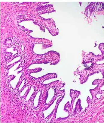
Figure 3. Histopathological apperance of Müllerian duct remnants. Micropapillary wall projections and microtubular spaces which were covered by one layer
of tall columnar epithelium is apparent (H&E; x20). carcinoma, seminoma, yolk sac tumor and teratoma in the testes of patients with PMDS (4). Since a full lit-erature review was not performed formerly, some sur-geons believed that malignancy was not developed
CYSTOSCOPY AND MUCOSECTOMY: ESSENTIALS IN THE MANAGEMENT OF PERSISTENT MÜLLERIAN DUCT SYNDROME... 229 from MDRs. Therefore, they did not recommend the
removal of MDRs due to the difficulty of dissection (2,5,6). The vas deferens is in close proximity with the walls of the Müllerian derivatives. It can be incorpo-rated into the wall of the vagina, as well. The surgeon should bear in mind that excision of these derivatives might damage to the testicular blood supply and vas deferens. Splitting of the Müllerian structures in the midline or microvascular autotransplantation are sug-gested when these derivatives prevented testicular descent (7). However, Farikullah et al performed a thorough search of the literature over the last 40 years and they reported 11 cases of malignancies arising not from the testes but from the retained MDRs, in-cluding squamous cell carcinoma, papillary cystad-enocarcinoma, clear cell carcinoma, adenocarcino-ma, adenosarcoma and clear cell adenocarcinoma. They estimated the malignant transformation of PMDS between 3.1% and 8.4% (8). Furthermore, the MDRs can cause urination discomfort, stones, urinary tract infections or periodic hematuria in the event of es-trogen excess (9,10). Although the priority is orchi-dopexy to preserve fertility and to prevent testicular malignancy in the management of PMDS associated with TTE, the recent report of malignant degeneration of the 11 MDRs led us to remove the these remnants in our case (8). We have excised the MDRs, which were located away from the vas deferens. Considering the reported malignancies of MDRs arising from its muco-sa, the parts which lay close to the vas deferens were opened and the mucosectomy was performed in our patient. Then, resection was completed just above its junction with the urethra. Cystoscopy assistance and placement of a 4 Fr catheter marked at centimeter intervals were essential for the complete excision of mucosa in our patient. To the best of our knowledge, cystoscopy assisted mucosectomy in PMDS has not been reported previously.
Given the long-term follow-up with magnetic resonance imaging is insistently recommended when MDRs have been preserved, cystoscopy assisted mu-cosectomy saved our patient from this imaging all through his life. We have been made the follow-up of testes due to the risk of infertility and testicular malig-nancy by using only physical examination.
Successful laparoscopic removal of MDRs has been reported (8,11). Although laparoscopy pro-vides excellent magnified operative view, we did not prefer laparoscopy for two reasons: First, both testes and MDRs in a hernia sac were detected not intraab-dominally, but in the right inguinal canal in our case, Secondly, mucosectomy in the distal part of MDRs may be difficult laparoscopically.
CONCLUSION
Removal of MDRs is advised in order to re-duce the jeopardy of malignancy, urinary tract in-fections, stones and hematuria. On the other hand, excision down to urethra which can compromise the integrity and vascularity of the vas deferens is diffu-cult, even in experienced surgical hands. Complete excision of these structures by mucosectomy of the dis-tal part of remnant which lay closed to vas deferens is a safe and effective method. Cystoscopy assistance and placement of a catheter into MDRs were essential for the complete excision of this mucosa.
Picard JY, Cate RL, Racine C, Josso N. The Persistent Müllerian Duct Syndrome: An Update Based Upon a Personal Experience of 157 Cases. Sex Dev. 2017; 11:109-125.
Rajesh A, Farooq M. A rare case of male pseudoher-maphroditism-persistent mullerian duct syndrome with transverse testicular ectopia- Case report and review of literature. Int J Surg Case Rep. 2017;37:72-75. Karakuş SC, Kılınçaslan H, Ertaşkın I, Koku N, De-liağa H. Delayed and incidental diagnosis of transverse testicular ectopia. Balkan Med J. 2012;29:447-449. Palanisamy S, Patel ND, Sabnis SC, Palanisamy N, Vi-jay A, Chinnusamy P. Laparoscopic hysterectomy with bilateral orchidectomy for Persistent Mullerian duct syndrome with seminoma testes: Case report. J Minim Access Surg. 2015;11:273-275.
Vandersteen DR, Chaumeton AK, Ireland K, Tank ES. Surgical management of persistent müllerian duct syn-drome. Urology. 1997;49:941–945.
Berkmen F. Persistent mullerian duct syndrome with or without transverse testicular ectopia and testis tumours. Br J Urol. 1997; 79: 122–126.
Brandli DW, Akbal C, Eugsster E, Hadad N, Havlik RJ, Kaefer M. Persistent Mullerian duct syndrome with bi-lateral abdominal testis: surgical approach and review of the literature. J Pediatr Urol. 2005; 1: 423–427. Farikullah J, Ehtisham S, Nappo S, Patel L, Hennayake S. Persistent Mullerian duct syndrome: lessons learned from managing a series of eight patients over a 10-year period and review of literature regarding malignant risk from the Müllerian remnants. BJU Int. 2012;110: E1084-E1089.
Thiel DD, Erhard MJ. Uterine adenosarcoma in a boy with persistent müllerian duct syndrome: first reported case. J Pediatric Surg. 2005; 40: e29-31.
Smith-Harrison LI, Patel MS, Smith RP, Schenkman NS. Persistent Müllerian duct structures presenting as hematuria in an adult: Case report of robotic surgical removal and review of the literature. Urol Ann. 2015; 7:544-546.
Valioulis I, Simaioforidis V, Syllas K. Laparoscopically assisted treatment of transverse testicular ectopia with persistent Müllerian duct syndrome: a case report and review of the literatüre. Hippokratia. 2016; 20: 88-89. **1. *2. 3. *4. 5. 6. 7. *8. 9. 10. 11.
REFERENCES AND RECOMMENDED READINGS (*of special interest, **of outstanding interest)
View publication stats View publication stats