Pediatric Mass Lesions of the Head and Neck Region and
Fine-Needle Aspiration Biopsy Results
Erkan Eşki1, Volkan Akdoğan1, Seda Babakurban Türkoğlu1, Muhammed Furkan Sökmen1, Fatma Çaylaklı1, Cem Özer1, Emine Tuba Canpolat2, İsmail Yılmaz1
1Department of Otorhinolaryngology, Başkent University School of Medicine, Ankara, Turkey 2Department of Pathology, Başkent University School of Medicine, Ankara, Turkey
Original Investigation
Address for Correspondence:
Erkan Eşki,
E-mail: [email protected] Received Date: 15.12.2015 Accepted Date: 29.04.2016
© Copyright 2016 by Official Journal of the Turkish Society of Otorhinolaryngology and Head and Neck Surgery Available online at www.turkarchotorhinolaryngol.org DOI: 10.5152/tao.2016.1371
Abstract Objective: 1. To provide a classification of pediatric
mass of the head and neck region and evaluate their frequency. 2. To examine the findings of fine-needle aspiration biopsy (FNAB) in pediatric patients along with its contribution to diagnosis.
Methods: Totally, 233 pediatric patients (125 boys and
108 girls) operated at Başkent University for head and neck mass were included. Clinical, radiological, and histopathological data were retrieved from medical re-cords.
Results: The mean age was 119±65 months, and the
mean duration of follow-up was 75±49 months. Lo-calization of the masses was as follows: 208 (89%) in the neck, 21 (9%) in the oral cavity, 2 (1%) in the neck and nasopharynx, and two (1%) in the larynx. The most common surgical procedure was open excisional biopsy (n=105, 45%) followed by cystic mass excision (n=72, 31%) and salivary gland excision (n=33, 14%). Based
on histopathological findings, benign cystic lesions were the most common disease group (n=77, 33.1%), whereas reactive lymphadenopathy was the most com-mon condition (n=36, 15%) when a single disease was considered. Infectious/inflammatory diseases, malig-nancies, and benign salivary gland diseases were pres-ent in 49 (21%), 24 (10.3%), and 22 (9.4%) patipres-ents, respectively. FNAB was performed in 29.8% of the pa-tients with an accuracy of 90.3% (95% CI, 80.1–96.4).
Conclusion: The differential diagnosis of head and
neck masses during childhood includes a wide spec-trum with the different conditions being benign cystic diseases of congenital origin and reactive lymphade-nopathies. Owing to its high predictive value, FNAB represents a rapid and reliable method that can be commonly used in both adult and pediatric patients.
Keywords: Pediatric, neck, tumor, histopathology, fine
needle aspiration biopsy
Introduction
Neck masses are common among pediatric pop-ulation. The etiology is wide-spectrum. Inflam-matory, congenital and neoplastic masses must be considered in differential diagnosis (1-3). Al-though the most common cause is conventionally accepted to be congenital lesions, some authors suggest that reactive lymphadenopathies (LAP) that develop following viral and bacterial infec-tions are the most common cause (4, 5). The final diagnosis is established through pathological ex-amination after excision, but fine needle aspiration biopsy (FNAB) from the mass can be performed for pre-diagnosis (6). In the studies conducted in USA over the past 20 years, it has been report-ed that the results of FNAB in preport-ediatric cases are as correct as those in adult patients and FNAB protects patient from negative outcomes such as general anesthesia, pain, scar, wound infection, and delayed diagnosis (7-10).
In this study, it was aimed to classify pediatric masses of the head and neck region in general, to determine their frequency, and to compare with literature, also to examine the findings of FNAB in pediatric patients, and to reveal the contribution of FNAB to the diagnosis.
Methods
This study was approved by Başkent University Medical and Health Sciences Research Com-mittee (Project no: KA 1/290) and supported by the Research Fund of Başkent University. Pedi-atric patients followed due to head-neck masses in Başkent University Ear-Nose-Throat Depart-ments between 1998 and 2015 were evaluated ret-rospectively. The patients older than 204 months were excluded from the study. The location of mass, surgical procedure that was applied, the results of pathology, and the findings of FNAB and radiological examination were obtained from the medical records. Histopathological diagnoses
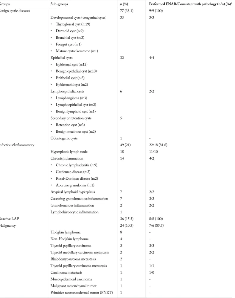
Table 1. Classification of histopathological diagnoses and cases undergoing FNAB
Groups Sub-groups n (%) Performed FNAB/Consistent with pathology (n/n) (%)*
Benign cystic diseases 77 (33.1) 9/9 (100)
Developmental cysts (congenital cysts) 33 3/3 • Thyroglossal cyst (n:19)
• Dermoid cyst (n:9) • Branchial cyst (n:3) • Foregut cyst (n:1)
• Mature cystic keratome (n:1)
Epithelial cysts 32 4/4
• Epidermal cyst (n:12) • Benign epithelial cyst (n:10) • Epithelial cyst (n:8) • Epidermoid cyst (n:2)
Lymphoepithelial cysts 6 2/2
• Lymphangioma (n:3) • Lymphoepithelial cyst (n:2) • Benign lymphoid cyst (n:1)
Secondary or retention cysts 5
-• Retention cyst (n:3) • Benign mucinous cyst (n:2)
Odontogenic cysts 1
-Infectious/Inflammatory 49 (21) 22/18 (81.8)
Hyperplastic lymph node 18 11/10
Chronic inflammation 14 4/2
• Chronic lymphadenitis (n:9) • Castleman disease (n:2) • Rosai-Dorfman disease (n:2) • Abortive granulomas (n:1)
Atypical lymphoid hyperplasia 7 2/2
Caseating granulomatous inflammation 7 3/2
Granulomatous inflammation 2 2/2 Lymphohistiocytic inflammation 1 -Reactive LAP 36 (15.5) 8/8 (100) Malignancy 24 (10.3) 7/6 (85.7) Hodgkin lymphoma 8 -Non-Hodgkin lymphoma 4
-Thyroid papillary carcinoma 3 3/3
Thyroid medullary carcinoma metastasis 2 2/2
Rhabdomyosarcoma metastasis 2
-Thyroid papillary carcinoma metastasis 1 1/1
Carcinoma metastasis 1 1/0
Mucoepidermoid carcinoma 1
-Malignant mesenchymal tumor 1
-of patients were separately evaluated by dividing into sub-age groups, which is a method that is conventionally used in the specialization areas of Pediatrics. According to this method, the sub-age group of 0-2 years was defined as infant; the sub-age group of 3-12 years was defined as child; and the sub-age group of 13-17 years was defined as adolescence.
For the statistical analysis of data, SPSS 17.0 statistical soft-ware (Version 17.0, SPSS Inc.; Chicago, IL, USA) was used. Categorical data were summarized as number and percentage. Continuous variables were presented as mean and standard de-viation (as median and minimum-maximum when needed). By considering the results of pathology as the golden standard, the superiority of the results of FNAB was evaluated by calculating sensitivity, positive predictive value, and accuracy.
Results
A total of 233 pediatric patients (125 boys and 108 girls) oper-ated for head and neck mass were included in the study. The ages of patients at the time of surgery were between 1 month and 204 months (mean=119±65 months) and the mean duration of follow-up was 75±49 months (1-192 months).
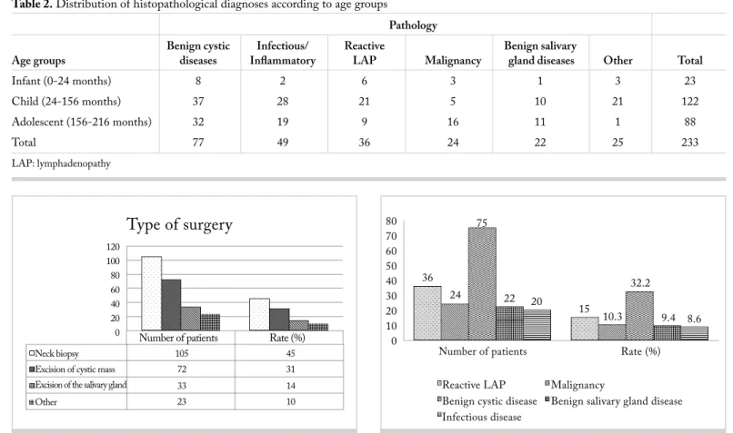
The mass location was in the neck in 208 patients (89%), in the oral cavity in 21 patients (9%), and in both the neck and naso-pharynx in 2 patients (1%), and in the larynx in 2 patients (1%). Neck biopsy expressed open excisional biopsy process and it was the surgical procedure that was applied mostly [in 105 cases (45%)]. It was followed by the excision of cystic mass in 72 cases (31%) and the excision of the salivary gland in 33 cases (14%) (Figure 1).
Considering the histopathological diagnoses separately, the most common was reactive lymphadenopathy at the rate of 15% (n=36). With regard to the groups, the most common diagno-sis was benign cystic diseases, most of which were congenital masses (n=77, 33.1%). Of the patients, 49 (21%) were diag-nosed with infectious/inflammatory disease, 24 (10.3%) were diagnosed with malignancy, and 22 (9.4%) were diagnosed with benign salivary gland disease (Figure 2). All histopathological diagnoses and patients undergoing FNAB are given in Table 1. Histopathological diagnoses of patients according to the sub-age groups (infant: 0-2 years; child: 3-12 years; adolescence: 13-17 years) are presented in Table 2. It was noted that benign
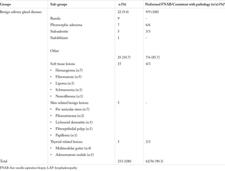
Table 1. Classification of histopathological diagnoses and cases undergoing FNAB (Continue)
Groups Sub-groups n (%) Performed FNAB/Consistent with pathology (n/n) (%)*
Benign salivary gland diseases 22 (9.4) 9/9 (100)
Ranula 9 -Pleomorphic adenoma 7 6/6 Sialoadenitis 5 3/3 Sialolithiasis 1 Other 25 (10.7) 7/6 (85.7)
Soft tissue lesions 15 4/3
• Hemangioma (n:7) • Fibromatosis (n:5) • Lipoma (n:1) • Schwannoma (n:1) • Neurofibroma (n:1)
Skin-related benign lesions 5
-• Pre-auricular sinus (n:7) • Pilomatrixoma (n:2) • Lichenoid dermatitis (n:1) • Fibroepithelial polyp (n:1) • Papilloma (n:1) Thyroid-related lesions 5 3/3 • Multinodular goiter (n:4) • Adenomatosis nodule (n:1) Total 233 (100) 62/56 (90.3)
cystic diseases were mostly seen in all age groups; 58% of re-active LAPs were in the subgroup of child; malignancies were more common among adolescences, and infectious/inflamma-tory diseases were observed mostly in the sub-group of children (Table 2).
Of the patients with neck mass (n=208), 62 (29.8%) underwent FNAB. 56 of FNAB processes matched with the result of his-topathologic examination. Accordingly, the rate of accuracy was found to be 90.3% [sensitivity100 % (CI 93.6%-100%) – positive predictive value 90.3% (CI 80.1%-96.4%)]. According to histo-pathological sub-groups, the accuracy of FNAB was the highest in the groups of benign cystic disease, reactive LAP and benign salivary gland disease with the rate of 100% and the lowest in the group of infectious /inflammatory disease group with the rate of 81.8% (Table 1). The rates of FNAB application were 11.7%, 44.9%, 22.2%, 29.2%, 40.9%, and 28% in benign cystic diseases, infectious/inflammatory diseases, reactive LAP, malignancy, be-nign salivary gland diseases, and other group, respectively. In 264 radiological examinations, the most commonly used was ultrasonography (USG) at the rate of 58% (n=154). It was fol-lowed by computed tomography (CT) (18%, n=47) and mag-netic resonance imaging (MRI) (14%, n=37).
Discussion
The differential diagnosis of pediatric masses of the head and neck region is quite wide. Medical history, physical
examina-tion findings, and imaging techniques are highly important in the diagnosis of congenital, inflammatory and tumoral causes. The mostly preferred imaging technique after physi-cal examination of patients is USG because it is inexpensive, easily accessible, and harmless and it provides rapid differen-tiation of solid-cystic masses (11). Also in our study, the most commonly used technique was found to be USG at the rate of 58%. Particularly for branchial, thyroglossal, and dermoid congenital cystic masses of the neck, USG does not require histopathological evaluation and it is diagnostic at a high rate of accuracy. On the other hand, CT is not preferred as the first choice because of ionized radiation. However, CT and MRI can be used for the masses that have deeper localization and the character of which cannot be defined through USG (12). In our study, while the usage rate of CT was detected to be 18%, MRI was used in 14% of cases. Although radiological examinations are important for the establishment of diagno-sis, histopathological evaluation must definitely be performed in order to make the final diagnosis.
In the approach to pediatric head-neck masses, FNAB must be performed after radiological examinations, especially in lateral neck masses. Fine-needle aspiration biopsy can reduce the number of surgical interventions planned to be performed for diagnostic purpose owing to its high predictive value. In this way, children are kept away from negative outcomes such as general anesthesia, wound infection, pain, scar and delayed diagnosis (7-10). D’anza et al. (13) reported the sensitivity
Table 2. Distribution of histopathological diagnoses according to age groups
Pathology
Benign cystic Infectious/ Reactive Benign salivary
Age groups diseases Inflammatory LAP Malignancy gland diseases Other Total
Infant (0-24 months) 8 2 6 3 1 3 23
Child (24-156 months) 37 28 21 5 10 21 122
Adolescent (156-216 months) 32 19 9 16 11 1 88
Total 77 49 36 24 22 25 233
LAP: lymphadenopathy
Figure 1. The surgeries that were performed
Type of surgery
Number of patients Other Neck biopsy 105 23 33 72 120 100 80 60 40 20 0 45 10 14 31 Excision of cystic massExcision of the salivary gland
Rate (%)
Figure 2. Results of histopathologic examinations (LAP: lymphadenopathy) Infectious disease
Benign cystic disease
Reactive LAP Malignancy
Rate (%) 36 80 70 60 50 40 30 20 10 0 24 75 22 20 15 10.3 32.2 9.4 8.6 Number of patients
of FNAB as 87.5% and the specificity as 100% for revealing malignancy of pediatric neck masses. In our study, 62 patients with neck masses (29.8%) underwent FNAB and the match rate of FNAB result to histopathological diagnosis was gener-ally 90.3% and 85.7% in malignant masses. In the study with the widest series in literature, FNAB sensitivity was detected to be 97.1% and sensitivity was 93.2% (13). In the study of Dilber et al. (14) with 17 pediatric and 110 adult patients, the accuracy of FNAB in all age groups was reported to be 94.3% for congenital-developmental pathologies, 89.4% for infectious-inflammatory pathologies (93.5% for specific in-fections), and 83.7% for neoplastic pathologies (85.9% for malignant neoplasms). They reported no statistically signifi-cant difference between the accuracy of FNAB and age group. Despite that, due to the establishment of clinical diagnosis through medical history, physical examination and radiologi-cal examinations in many cases and FNAB’s requiring pediat-ric cytopathologist and sometimes general anesthesia reduce the rates of FNAB in pediatric neck masses.
The most common cause of pediatric neck masses is known to be congenital lesions in general (4). However, some changes can be observed in the distribution depending to the socio-econom-ic status (15). According to Meier and Grimmer (5) and D’anza (13), the most common pathology is reactive lymphadenopathy following upper respiratory tract infection. On the other hand, the most common cause is the inflammatory group according to Al-Mayoof (15) (57%) and it is attributed to the socio-econom-ic situation of the country where the study was conducted. In our study, general congenital lesions were more common and the most frequent disease was reactive LAP at the rate of 15% when the diseases were considered one by one. Total rate of reactive LAP and infectious/inflammatory groups was found to be 36% in our study and the result was consistent with the socio-eco-nomic situation of our country.
In pediatric population, the rate of malignant neck mass is be-tween 11% and 15% (16). Malignancy was detected in 10% of our cases. The most common malignancy was Hodgkin lympho-ma (8/24=33.3%). Although most of patients were benign, the suspect of malignancy increases when the mass grows rapidly, exists for a long time, and occurs especially in supraclavicular and posterior-cervical regions (4).
Fine-needle aspiration biopsy is a minimal invasive method used for the diagnosis of neoplastic and non-neoplastic lesions in the head and neck region. The procedure is generally with-out complication and it is safe, and well-tolerable by patients. It usually does not require anesthesia and the results are ob-tained rapidly. It serves as a complementary diagnostic method for histopathological examination. It is important that it allows the identification of benign reactive LAP, which is common in childhood, without any need for open biopsy (17). In this study, cytological and histological findings were consistent and the rate of accuracy was quite high. Therefore, it has been concluded that FNAB is an effective diagnostic method in the diagnosis and treatment of pediatric head-neck masses.
Conclusion
As a result, the etiology of pediatric head-neck masses is highly wide-spectrum. The rates of different pathologies in our study were found to be consistent with literature. Moreover, FNAB can be used in pediatric cases with head-neck mass because it can decrease the number of surgical interventions planned to be performed for diagnosis owing to its high predictive value in the establishment of diagnosis. These results display the importance of cytopathological examination for making the final diagnosis, as well as medical history, physical examination and imaging techniques.
Ethics Committee Approval: Ethics committee approval was received
for this study from the ethics committee of Başkent University.
Informed Consent: Informed consent was not obtained from patients
due to the retrospective design of the study.
Peer-review: Externally peer-reviewed.
Author Contributions: Concept - İ.Y.; Design - İ.Y., M.F.S.;
Super-vision - İ.Y.,; Resources - E.E.; Materials - M.F.S., E.E., V.A., S.B., C.Ö., F.Ç., E.T.C.; Data Collection and/or Processing - E.E., V.A., S.B., C.Ö., F.Ç., E.T.C.; Analysis and/or Interpretation - İ.Y.; Litera-ture Search - M.F.S., E.E.; Writing Manuscript - M.F.S., E.E.; Critical Review - İ.Y.
Conflict of Interest: No conflict of interest was declared by the
aut-hors.
Financial Disclosure: This study has received of financial support by
Başkent University research fund.
References
1. Dickson PV, Davidoff AM. Malignant neoplasms of the head and neck. Semin Pediatr Surg 2006; 15: 92-8. [CrossRef]
2. Turkington JR, Paterson A, Sweeney LE, Thornbury GD. Neck masses in children. Br J Radiol 2005; 78: 75-85. [CrossRef]
3. Tracy TF Jr, Muratore CS. Management of common head and neck masses. Semin Pediatr Surg 2007; 16: 3-13. [CrossRef]
4. Karaca İ. Çocuklarda baş boyun patolojileri. Turk Ped Ars 2010; 45: 20-2. [CrossRef]
5. Meier JD, Grimmer JF. Evaluation and management of neck mas-ses in children. Am Fam Physician 2014; 89: 353-8.
6. Niedzielska G, Kotowski M, Niedzielski A, Dybiec E, Wieczorek P. Cervical lymphadenopathy in children – incidence and diagnos-tic management. Int J Pediatr Otorhinolaryngol 2007; 71: 51-6.
[CrossRef]
7. Mobley DL, Wakely PE, Frable MS. Fine-needle aspiration bi-opsy: application to pediatric head and neck masses. Laryngoscope 1991; 101: 469-72. [CrossRef]
8. Cohen MB, Bottles K, Ablin AR, Miller TR. The use of fine need-le aspiration biopsy in children, Western J. Med. 1989; 150: 665-7. 9. Derias NW, Chong WH, O’Conner AF. Fine needle aspiration
cytology of a head and neck swelling in a child: a non-inva-sive approach to diagnosis. J Laryngol Otol 1992; 106: 755-7.
[CrossRef ]
10. Liu E, Bernstein J, Sculateri N, Wu H. Fine needle aspiration bi-opsy of pediatric head and neck masses. Int J Pediatr Otorhinolar-yngol 2001; 60: 135-40. [CrossRef]
11. Rooks VJ, Cable BB. Head and neck ultrasound in the pedi-atric population. Otolaryngol Clin N Am 2010; 43: 1255-66.
[CrossRef ]
12. Shekdar KV, Mirsky DM, Kazahaya K, Bilaniuk LT. Magnetic resonance imaging of the pediatric neck: an overview. Magn Reso-nance Imaging Clin N Am 2012; 20: 573-603. [CrossRef]
13. D’anza B, Kraseman SJ, Helwig CC, Grene JS, Wood WE. FNA biopsy of pediatric cervicofacial masses and validation of clini-cal characteristics of malignancy. Int J Pediatr Otorhinolaryngol 2015;79: 1196-200. [CrossRef]
14. Dilber M, Erişen L, Yerci Ö, Coşkun H, Basut O, Onart S, et al. Tiroid dışı baş-boyun kitlelerinde ince iğne aspirasyon
sitolo-jisi sonuçlarımız: Uludağ deneyimi. Turk Otorhinolaryngol Arch 2005; 45: 86-93.
15. Al-Mayoof AF. Neck masses in paediatric population: An expe-rience with children attended the Central Teaching Hospital of Pediatrics in Baghdat 2008-2009. African J Paediatr Surg 2015; 12: 136-9. [CrossRef]
16. Rosenberg HK. Sonography of pediatric neck masses. Ultrasound Q 2009; 25: 111-27. [CrossRef]
17. Anne S, Teot LA, Mandell DL. Fine needle aspiration biopsy: Role in diagnosis of pediatric head and neck masses. Int J Pediatr Otorhinolaryngol 2008;72: 1547-53. [CrossRef]