I had presented the abstract of this manuscript in 25th National Congress of Cardiology, İstanbul, 2009.
Address for Correspondence: Dr. Didem Oğuz, Başkent Üniversitesi Tıp Fakültesi, Oymacı Sokak No:7 Altunizade/İstanbul-Türkiye Phone: +90 216 554 15 00 Fax: +90 216 651 98 58 E-mail: [email protected]
Accepted Date: 14.11.2013 Available Online Date: 26.02.2014
©Copyright 2014 by Turkish Society of Cardiology - Available online at www.anakarder.com DOI:10.5152/akd.2014.4612
A
BSTRACTObjective: This study aims to show the effect of myeloperoxidase (MPO), hsCRP, TNF-alpha values and leukocyte count on the development of coronary collateral arteries in patients with severely diseased coronary arteries.
Methods: Current study is an observational cross-sectional study. In the study, 295 patients who had functional obstruction or total coronary occlusion at least 1 month on their angiograms were included. We divided the study population into two groups according to their collateral grade as good collateral (Group 1) (169 patients) and poor collateral (Group 2) (126 patients). Multiple logistic regression analysis was used for independent variables associated with the coronary collateral grade.
Results: History of stable angina pectoris was statistically more prevalent in good collateral group (61.5% and 48.4%, p=0.025). Furthermore, MPO activation was higher in good collateral group and the difference was statistically significant (3.7 U/mL and 3.0 U/mL p=0.001). In multiple logistic regression analysis, stable angina pectoris [OR 1.7, 95% CI (1.05-2.8), p=0.03] and high MPO levels [OR 2.7, 95% CI (1.7-4.3), p<0.001] were found to be independent predictors of good collateral development.
Conclusion: We think that proinflammatory enzymes and cytokines released from these cells rather than inflammatory cells themselves may play an important role on the collateral development. (Anadolu Kardiyol Derg 2014; 14: 336-41)
Key words: collateral arteries, inflammatory markers, stable angina pectoris, angiogenesis, regression analysis.
Didem Oğuz, Yusuf Atmaca
1, Çağdaş Özdöl
1, Ayhan Ongun Özdemir
1, Cansın Tulunay Kaya
1, Çetin Erol
1 Department of Cardiology, Faculty of Medicine, Başkent University; İstanbul-Turkey1Department of Cardiology, Faculty of Medicine, Ankara University; Ankara-Turkey
The relationship between coronary collateral artery development and
inflammatory markers
Introduction
Atherosclerotic heart disease is the most frequent cause of death in adults. Recent advances in the modernization of coronary intensive care units, the use of beta blockers, coronary bypass surgery, fibrinolytic therapy and primary coronary angioplasty and similar treatment methods has resulted in important declines in mortality rates due to coronary heart disease (1, 2). A well-developed collateral artery also restricts the infarction area during acute myocardial infarction (MI) and prevents the occurrence of Q wave MI, left ventricular aneurysm and coronary failure. As is known, inflammation has a significant role in the progression of atherosclerosis. For this reason, an examination of inflammatory markers that can affect the development of collateral arteries is important (2-7).
The imbalance between oxygen requirements as a result of coronary artery stenosis or coronary artery occlusion lead to an increase in coronary collateral development in humans, dogs
and pigs. The formation of coronary collaterals takes the form of de novo “angiogenesis” with the budding of new capillaries in existing blood vessels or as “arteriogenesis” that is present as a result of the development and maturing of anastomosis vessels in existing arteries since birth (6). In both cases endothelial cells and the growth factors secreted from inflammatory cells such as thrombocyte and monocyte that attack the ischemic region play an important role (6-8).
The tumor necrosis factor b (TNF-alfa) is a proinflammatory cytokine that is produced from leading macrophages. It increases the expression of adhesion molecules (such as VCAM-1 and ICAM-1). And also by increasing the production of nitric oxide (NO) and prostacycline from endothelium, it increases blood flow and creates local vasodilatation (9-14).
CRP is one of the marker of vascular inflammation. Some studies show inverse graded association between CRP and the presence of coronary collaterals in patients with stable angina pectoris (15-18). In our study there is no relation between hsCRP
and collateral development. White blood cells are one of the important cells that give response to inflammation. Widely used, stable and with better standardization compared to other inflammatory indicators, white blood cell measurement is considered as an independent predictor of smoking, age etc. in cardiovascular mortality (19).
Myeloperoxidase (MPO) is stored inside azurophilic granules within polymorphic nuclei neutrophils and macrophages (20). MPO, through hydrogen peroxide reaction, forms permeable oxidative substances that contains free radicals, antimicrobial activities and causes oxidative damage in tissues (20-23). It is elevated in coronary artery disease and higher levels of MPO is found with progression of coronary artery disease (CAD) (21).
It has been shown that TNF-alfa and through nitric acid oxide synthesis of high sensitive C-reactive protein (hsCRP), cytokines and development factors regulates angiogenesis (12). But there is no sufficient data in the relationship between MPO, white cells and collateral development. There are interindividual differences in the number and extent of collateral vessels among patients with a similiar degree of coronary atherosclerosis (4). This is due to environmental and genetic disparities. In this study, we have aimed in identifying the inflammatory markers such as MPO serum, TNF-alfa, hsCRP and white blood cell count which we consider to effect the collateral development.
Methods
Study design
This study has a cross-sectional and observational design. Study population
The study population consisted of 295 patients who underwent clinically indicated coronary angiography in our department and were found to have at least one major coronary occlusion, or a stenosis of ≥95% with Thrombolysis In Myocardial Infarction (TIMI) grade ≤1 anterograde flow between October 2008 and April 2009. All patients had stable anginal symptoms and/or positive stress test results or electrocardiographic changes indicating ischemia.
Study protocol
Clinical characteristics of the patients including age, weight, gender and any data known to influence development of collaterals such as current medications, history of hypertension, diabetes mellitus, complete blood count and fasting glucose levels were documented. Patients with a history of coronary bypass surgery were excluded if the operation has been done within the past 30 days and the distal aspect of the qualifying severly stenosed or occluded artery is supplied by the patent bypass greft. Patients who had percutaneous coronary intervention within 30 days, acute or chronic infectious disease and malignancy were also excluded. All patients gave written informed consent and local Ethics Commitee approved the study protocol.
Baseline clinical examination
Routine blood samples were taken from the patients who were admitted to hospital for coronary angiography after overnight fasting. Patients with low density lipoprotein (LDL) levels greater than 100 mg/dL or under lipid lowering drug therapy were considered to have hyperlipidemia. Patients with systolic or diastolic blood pressure ≥140 mm Hg or 90 mm Hg or who were under antihypertensive treatment were considered to be hypertensive (based on the mean of the two readings). Patients who were being treated for diabetes mellitus (DM) or who had a fasting glucose concentration ≥126 mg/dL were considered to have DM. Smoking habit was recorded as one that has lasted for more than 1 year. The Friedewald’s formula was used for LDL cholesterol measurement.
Laboratory analyses
5 mL of blood samples were collected from the arterial sheaths immediately following coronary angiography from each patient and vacutiner tubes centrifuged at 5000 rpm for 15 minutes within 1 hour. Then plasma collected and stored at -20˚C. MPO was measured with enzyme linked immunosorbent assay (ELISA) (Imtec MPO ANCA. Wiesbaden-Germany) and TNF-alpha was measured with ELISA (Biosource, Carlsbad, California, USA) in immunology laboratuary. hsCRP was measured with nephelometry (CardioPhase hsCRP, BN II nefelometre, Dade- Behring-Germany) and white blood cell counts were measured in the Beckman Coulter LH 750 automotic blood count analyzer by the VSC prinsible in Ankara University İbni-Sina hospital central laboratuary.
Coronary angiography and grading of coronary collaterals Coronary angiography was completed by two experienced cardiologist blinded to the study (Philips Integris 3000 system Philips, Holland) according to the Judkins technique using automated quantitative coronary artery stenosis assessment program of same system. The diseased vessel was identified as stenosis of the major coronary artery of at least 75%. Collateral flow was graded according to the Rentrop classification (6) and based on the injection that best opacified the occluded vessel: grade 0, no visible filling of any collateral vessels; grade 1, filling of side branches of the artery to be perfused by collateral vessels without visualization of the epicardial segment; grade 2, partial filling of the epicardial segment by collateral vessels; and grade 3, complete filling of the epicardial segment by collateral vessels. According to this classification we divided 295 patients into two groups. Grade 0 and 1 were named as poor collateral (Group 2), grade 2 and 3 were named as good collateral (Group 1). In subjects with >1 collateral supplying the distal aspect of the diseased artery, the higher collateral grade was used. In subjects with >1 qualifying severly diseased vessel, the vessel with the higher collateral grade was chosen for analysis.
Statistical analysis
Categorical variables were defined as percentage, continuous variables were presented as mean±standard error. Comparisons
among groups were tested for normal distribution with student-t test, for abnormal distribution with Mann-Whitney U test. Relationship between continuous variables that were not normally distributed and collateral grade was tested with Spearman correlation test. History of stable angina pectoris and serum MPO level were found to be significantly correlated with collateral grade. Therefore, these variables were used in a multiple logistic regression analysis as independent variables to determine their effect on collateral grade. Statistical significance was defined as p<0.05. The SPSS statistical software (SPSS for windows 11.5, SPSS Inc, Chicago, IL) was used for all statistical calculations.
Results
Clinical characteristics of the population
The patient population consisted of 218 males (74%) and 77 females (26%) (mean age, 63±10 years). The prevalence of
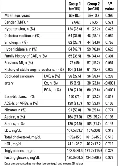
various demographic, angiographic and therapy related characteristics of the study subjects at the time of coronary angiography according to collateral classification are shown in Table 1. Of the 295 patients, 169 had good collaterals (grade 3 and grade 2), whereas 126 had poor collaterals (grade 1 and grade 0). Subjects with good collaterals were significantly more likely to have stable angina pectoris history (p=0.025) and the prevalance of RCA (right coronary artery) lesion was higher in the good collateral group (p=0.02) whereas CX (circumflex artery) lesion was higher in the poor collateral group (p<0.0001). The prevalence of all other cardiovascular risk factors and use of medications were similar at different levels of collaterals.
The relationship of collateral grade and TNF-alpha, MPO and hsCRP
TNF-alpha, MPO, hsCRP levels, white blood cell counts and subgroup levels were shown in Table 2. hsCRP, TNF-alpha levels, white blood cell counts and subgroup levels were not significantly different among groups (p>0.05) (Table 2). Furthermore MPO levels were significantly higher in the good collateral group than poor collateral group (3.7 U/mL vs. 3.0 U/mL, p=0.001) (Table 2, Fig. 1). We also found very weak correlation between the MPO levels and collateral grade albeit p value was significant (p=0.009, r=0.15) (Fig. 2). In multiple logistic regression analysis, stable angina pectoris [odds ratio 1.7, 95% confidence interval (1.05-2.8), p=0.03] and high MPO levels [odds ratio 2.7, 95% confidence interval (1.7-4.3), p<0.001] were found to be independent predictors of good collateral development (Table 3).
Group 1 Group 2 *P (n=169) (n=126) value
Mean age, years 63±10.6 63±10.2 0.996
Gender (M/F), n 127/42 91/35 0.571
Hypertension, n (%) 124 (73.4) 91 (72.2) 0.826 Diabetes mellitus, n (%) 64 (37.9) 48 (38.1) 0.969
Smoking, n (%) 62 (36.7) 44 (34.9) 0.755
Hyperlipidemia, n (%) 84 (49.7) 59 (46.8) 0.625 Family history of CAD, n (%) 65 (38.5) 56 (44.4) 0.301
Previous MI, n (%) 76 (45) 57 (45.2) 0.964
History of stable angina pectoris, n (%) 104 (61.5) 61 (48.4) 0.025 Occluded coronary LAD, n (%) 38 (22.5) 36 (28.6) 0.233 artery Cx, n (%) 15 (8.9) 30 (23.8) <0.0001 RCA, n (%) 120 (71.0) 60 (47.6) <0.0001 Beta-blockers, n (%) 120 (71) 91 (72.2) 0.819 ACE-Is or ARBs, n (%) 138 (81.7) 93 (73.8) 0.106 Nitrates, n (%) 91 (53.8) 70 (55.6) 0.771 Aspirin, n (%) 164 (97.0) 125 (99.2) 0.193 Statins, n (%) 126 (74.6) 103 (81.7) 0.143 LDL, mg/dL 107.5±39.7 107±36.8 0.912 Total cholesterol, mg/dL 178±45.5 181.5±45.8 0.515 HDL, mg/dL 41.1±26.7 40.2±12.2 0.719 Triglycerides, mg/dL 153.6±80.4 171.2±115.6 0.328 Fasting glucose, mg/dL 120.6±60.5 124.5±66.9 0.979
Data are presented as number (percentage) and mean±SD values Group 1; good collateral, Group 2; poor collateral
* Student-t test and Mann-Whitney U test
ACE-I - angiotension converting enzyme inhibitor; ARB - angiotension receptor blocker; CAD - coronary artery disease; Cx - circumflex artery; F - female; Gender- Male/Female HDL - high density lipoprotein; LAD - left anterior descending artery; LDL - low density lipoprotein; MI - myocardial infarction M/F; RCA - right coronary artery; SD - standard deviation
Table 1. Characteristics of the subjects according to collateral groups
Group 1 (n=169) Group 2 (n=126) mean±SD mean±SD *P TNF-alpha, pg/mL 37.9±117.2 25.8±47.4 0.940 MPO, U/mL 3.7±2.0 3.0±2.0 0.001* hsCRP, mg/L 13.7±19.5 13.7±21.2 0.990 WBC, x109cells/L 8400±3000 8500±2700 0.622 Neutrophils, % 64.9±11.2 63.4±9.6 0.235 Lymphocytes, % 24.4±9.4 24.5±7.9 0.873 Monocytes, % 8.0±2.7 7.8±2.5 0.438 *significant
*Student-t test and Mann-Whitney U test
HsCRP - high sensitive C-reactive protein; MPO - myeloperoxidase; SD - standard deviation; TNF-alpha - tumor necrosis factor alpha; WBC - white blood cell
Table 2. Relationship between inflammatory markers and collateral growth
*P value OR CI 95%
History of stable angina pectoris 0.03 1.7 1.05-2.8
MPO, >2.91 U/mL <0.001 2.7 1.7-4.3
*Multiple logistic regression analysis
Dependent variable collateral grade, independent variables-history of stable angina pectoris, serum MPO level
MPO - myeloperoxidase
Discussion
In this study, we aimed to indentify possible determinants of coronary collaterals in patients with severe coronary artery disease. We found that stable angina pectoris and MPO levels were independent predictors of development of coronary collaterals.
The presence of collateral vessels protects the heart from myocardial ischemia and infarction, and following an infarction by enabling the development of less Q wave MI, left ventricular aneurysm and coronary failure. So they provide a positive effect in in-hospital and long-term prognosis (1). An increased shear stress following serious arterial blockage arteriogenesis is
stimulated. In this mechanism it is thought that MCP-1 (monocyte chemotactic protein-1) is effective. While shear stress is at a maximum at the beginning, this stress gradually decreases as the diameter of collateral arteries expands (5, 7, 8). Assuming that hypertension plays a facilitating role in this mechanism, we can consider it to have a positive effect on collateral development. In our study, hypertension in the good collateral group was also detected more but this difference was not found to be statistically significant.
We know that coronary collateral growth is impaired in type 2 diabetes mellitus and metabolic syndrome (24). In our study, 112 diabetic patients were present and in terms of DM frequency, no significant difference was determined among the two groups. This may due to insufficient number of patients and undetectable arteries formed by angiogenesis with coronary angiography. Additionally, a comparison of the number of diseased arteries between the two groups was not carried out in this study.
Although many studies have suggested that statins and nitrates may promote vessel growth, clinical study results are generally lacking (8, 25). We did not find any relation between coronary colllateral and use of this group of drugs.
In the latest studies conducted, it has been suggest that CRP results in differentiating endothelial precursor cells, that it badly effects survival and that it disrupts the angiogenic and arteriogenic functions of the cells (15, 18). Whereas in the study carried out by Turu et al. (16) CRP stimulated in angiogenesis. Even though there are studies that show an inverse relationship between collateral artery development and high CRP levels (15), we did not determine a meaningful relationship between collateral development and hsCRP in our study.
Barron et al. (26) in a study conducted on 975 patients found a correlation between high blood cell counts, increased thrombus load, increased coronary failure and incidences of death after acute myocardial infarction. Even though there is no study available that indicates the relationship between coronary collateral development and white cell counts, we compared white cell counts between good and bad collateral arteries and found no significant difference.
TNF-alfa is an important cytokine that plays a role in inflammation. Its effects on the cardiovascular system are currently being researched. Numerous studies have shown that TNF-alfa reduces eNOS production in various arteries thus leading to a decline in NO production (12-14). In a study carried out by Luo et al. (10) TNF-alfa through p55 receptors affected arteriogenesis in a positive way. In our study we considered TNF-alfa having an effect on angiogenesis. So we compared TNF-alfa levels with patients that have good and bad collateral but could not determine a significant difference.
Carrão et al. (27) found that granulocyte-colony stimulating factor (G-CSF) directly stimulates cardiomyocytes and promote angiogenesis. Besides this, there are many studies that indicate the important role played by monocytes and macrophages in collateral development where MPO is stored (20, 21, 28). Kocaman et al. (29) divided 210 patients, where coronary artery
Figure 2. Correlation between myeloperoxidase level and collateral grade
15.00 12.00 9.00 6.00 3.00 0.00 Myeloperoxidase (U/mL) r=0.15 p=0.009 0.00 1.00 2.00 3.00 Collateral Grade
Figure 1. Comparasion of myeloperoxidase level between good and poor collateral groups
15.00 10.00 5.00 0.00 Myeloperoxidase (U/mL) Poor collateral p=0.001 Collateral Grade Good collateral
stenosis of these patients was 95% or above and did not have diabetes, into two groups of good and bad collateral and found that the group with good collateral had a high monocyte count and that the monocyte count was an independent predictor in the development of good collateral. In our study it was determined that while in terms of white cell count, a noticeable difference was not found among the two groups, the MPO level was found to be high among patients with good collateral and that this difference was statistically meaningful. We interpreted the high level of MPO in particular as due to the possibility of an increase in MPO activity in neutrophiles and monocytes. And G-CSF may also increase MPO activity but in our study we did not measure the serum G-CSF level. Additionally, after a multiple logistic regression analysis is carried out, a high MPO level was determined to be the independent predictor of good collateral artery development. Even though there are no head to head studies that show the relationship between collateral development and MPO levels in the literature, we consider that in collateral development rather than inflammatory cells, the proinflammatory enzymes and cytokines secreted from these cells can play a more important role.
Study limitations
We consider that low number of subject and unknown duration of the medication were the two limitations of this study. That’s why having diabetes mellitus and use of medication were not significantly different among groups. Additionally we performed all measurements at one point in time. HsCRP, MPO, blood cell count and TNF-alpha are inflammatory markers and we tried to find out the predictive value of them measured at one point in time to detect the relation of the presence of collateral circulation. Design of our study reflected routine daily practice in the majority of cardiology clinics. Repeated angiography assesments may be needed but due to the invasive nature of coronary angiography we did not perform.
Conclusion
The findings from our study leave us to think that while TNF-alfa, hsCRP and white cell count does not have a meaningful effect on collateral development, MPO levels has a positive effect on collateral development. But randomized clinical studies with follow up are required to evaluate the role of them on cardiovascular outcomes.
Conflict of interest: None declared. Peer-review: Externally peer-reviewed.
Authorship contributions: Concept - Y.A., D.O.; Design - Y.A., D.O.; Supervision - Ç.E., Y.A.; Resource - Ç.E., Y.A.; Materials - A.O.Ö., C.T.K.; Data collection &/or processing - Ç.Ö., C.T.K., A.O.Ö., D.O.; Analysis &/or interpretation - Ç.Ö., D.O.; Literature search - D.O.; Writing - D.O.; Critical review - Y.A., D.O.
References
1. Gonçalves LM. Angiogenic growth factors: potential new treatment for acute myocardial infarction? Cardiovasc Res 2000; 45: 294-302. [CrossRef] 2. Berry C, Balachandran KP, L’Allier PL, Lesperance J, Bonan R,
Oldroyd KG. Importance of collateral circulation in coronary heart disease. Eur Heart J 2007; 28: 278-91. [CrossRef]
3. van Royen N, Piek JJ, Buschmann I, Hoefer I, Voskuil M, Schaper W. Stimulation of arteriogenesis; a new concept for the treatment of arterial occlusive disease. Cardiovasc Res 2001; 49: 543-53. [CrossRef] 4. Heil M, Schaper W. Pathophysiology of collateral development.
Coron Artery Dis 2004; 15: 373-8. [CrossRef]
5. Ganz P, Braunwald E. Coronary blood flow and myocardial ischemia. In: Braunwald E, editor. Braundwald’s Heart Disease: A Textbook of Cardiovascular Medicine. 6 th ed. Philadelphia: WB Saunders Company; 2001.p.1087-108.
6. Çelik T, Çelik M, İyisoy A. Coronary collateral circulation. Arch Turk Soc Cardiol 2010; 38: 505-14.
7. Schaper W. Collateral Circulation: past and present. Basic Res Cardiol 2009; 104: 5-21. [CrossRef]
8. Werner GS, Figulla HR. Direct assessment of coronary steal and associated changes of collateral hemodynamics in chronic total coronary occlusions. Circulation 2002; 106: 435-40. [CrossRef] 9. Hoefer IE, van Royen N, Rectenwald JE, Bray EJ, Abouhamze Z,
Moldawer LL, et al. Direct evidence for tumor necrosis factor alfa signaling in arteriogenesis. Circulation 2002; 105: 1639-41. [CrossRef] 10. Luo D, Luo Y, He Y, Zhang H, Zhang R, Li X, et al. Differential functions of tumor
necrosis factor receptor 1 and 2 signaling in ischemia-mediated arteriogenesis and angiogenesis. Am J Pathol 2006; 169: 1886-98. [CrossRef] 11. Zhang H, Park Y, Wu J, Chen X, Lee S, Yang J, et al. Role of TNF-alpha
in vascular dysfunction. Clin Sci (London) 2009; 116: 219-30. [CrossRef] 12. Seidel M, Billert H, Kurpisz M. Regulation of eNOS expression in
HCAEC cell line treated with opioids and proinflammatory cytokines. Kardiol Pol 2006; 64: 153-8.
13. Goodwin BL, Pendleton LC, Levy MM, Solomonson LP, Eichler DC. Tumor necrosis factor alpha reduces argininosuccinate synthase expression and nitric oxide production in aortic endothelial cells. Am J Physiol Heart Circ Physiol 2007; 293: H1115-21. [CrossRef] 14. Picchi A, Gao X, Belmadani S, Potter BJ, Focardi M, Chilian WM, et al.
Tumor necrosis factor alpha induces endothelial dysfunction in the prediabetic metabolic syndrome. Circ Res 2006; 99: 69-77. [CrossRef] 15. Kerner A, Gruberg L, Goldberg A, Roguin A, Lavie P, Lavie L, et al.
Relation of C-reactive protein to coronary collaterals in patients with stable angina pectoris and coronary artery disease. Am J Cardiol 2007; 99: 509-12. [CrossRef]
16. Turu MM, Slevin M, Matou S, West D, Rodriguez C, Luque A, et al. C-reactive protein exerts angiogenic effects on vascular endothelial cells and modulates associated signalling pathways and gene expression. BMC Cell Biol 2008; 9: 47. [CrossRef] 17. Casas JP, Shah T, Hingorani AD, Danesh J, Pepys MB. C-reactive
protein and coronary heart disease: a critical review. J Intern Med 2008; 264: 295-314. [CrossRef]
18. Clearfield MB. C-reactive protein: A new risk assessment tool for cardiovascular disease. J Am Osteopath Assoc 2005; 105: 409-16. 19. Hoffman M, Blum A, Baruch R, Kaplan E, Benjamin M. Leukocytes and
coronary heart disease. Atherosclerosis 2004; 172: 1-6. [CrossRef] 20. Roman RM, Wendland AE, Polanczyk CA. Myeloperoxidase and
coronary arterial disease: from research to clinical practice. Arq Bras Cardiol 2008; 91: 11-9.
21. Ndrepepa G, Braun S, Mehilli J, Von Beckerath N, Schömig A, Kastrati A. Myeloperoxidase level in patients with stable coronary artery disease and acute coronary syndromes. Eur J Clin Invest 2008; 38: 90-6. [CrossRef]
22. Lau D, Baldus S. Myeloperoxidase and its contributory role in inflammatory vascular disease. Pharmacol Ther 2006; 111: 16-26. [CrossRef]
23. Mocatta TJ, Pilbrow AP, Cameron VA, Senthilmohan R, Frampton CM, Richards AM, et al. Plasma concentrations of myeloperoxidase predict mortality after myocardial infarction. J Am Coll Cardiol 2007; 49: 1993-2000. [CrossRef]
24. Rocic P. Why is coronary collateral growth impaired in type 2 diabetes and the metabolic syndrome? Vascular Pharmacology 2012; 57: 179-86. [CrossRef]
25. Landmesser U, Engberding N, Bahlmann FH, Schaefer A, Wiencke A, Heineke A, et al. Statin-induced improvement of endothelial progenitor cell mobilization, myocardial neovascularization, left ventricular function and survival after experimental myocardial
infarction requires endothelial nitric oxide synthase. Circulation 2004; 110: 1933-9. [CrossRef]
26. Barron HV, Cannon CP, Murphy SA, Braunwald E, Gibson CM. Association between white blood cell count, epicardial blood flow, myocardial perfusion and clinical outcomes in the setting of acute myocardial infarction: a thrombolysis in myocardial infarction 10 substudy. Circulation 2000; 102: 2329-34. [CrossRef]
27. Carrão AC, Chilian WM, Yun J, Kolz C, Rocic P, Lehmann K, et al. Stimulation of coronary collateral growth by granulocyte stimulating factor: role of reactive oxygen species. Arterioscler Thromb Vasc Biol 2009; 29: 1817-22. [CrossRef]
28. Lavine KJ, Kovacs A, Weinheimer C, Mann DL. Repetitive myocardial ischemia promotes coronary growth in the adult mammalian heart. J Am Heart Assoc 2013; 2: e000343. [CrossRef] 29. Kocaman SA, Arslan U, Tavil Y, Okuyan H, Abacı A, Çengel A. Increased
circulating monocyte count is related to good collateral development in coronary artery disease. Atherosclerosis 2008; 197: 753-6. [CrossRef]